Quantcast
Channel: AGINGSCIENCES™ – Anti-Aging Firewalls™
Viewing all 149 articles
Browse latest View live

The pivotal role of Nrf2. Part 3 – Part 3 – Is promotion of Nrf2 expression a viable strategy for human human healthspan and lifespan extension?

$
0
0

By Vince Giuliano

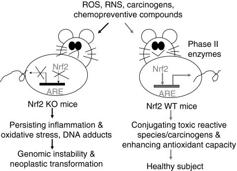
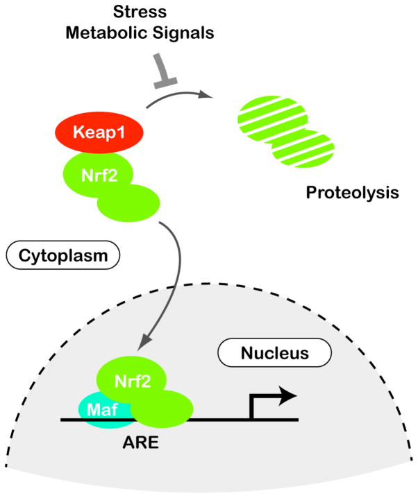
This is the third of three blog entries relating to the Nrf2 pathway.  The first blog entry The pivotal role of Nrf2. Part 1 – a new view on the control of oxidative damage and generation of hormetic effects dealt with the general mechanisms of operation of Nrf2, and with positive effects of Nrf2 in preventing or treating a number of pathological conditions via its ability to turn on genes for the body’s own antioxidant defense system and genes for combating stress – hundreds of such genes.

The second blog entry The pivotal role of Nrf2. Part 2 – foods, phyto-substances and other substances that turn on Nrf2 deals with how a number of substances that are incidentally antioxidants, plant-derived phyto-substances in particular, actually exercise their benefits through promoting the expression of Nrf2 which in turn activates the body’s own antioxidant and hormetic defense systems.  I have added several new items to that blog entry after first publishing it.

This third blog entry explores whether supplementation with substances that promote Nrf2 might be life-extending.  Taken together the three blog entries answer a question that has frequently been addressed to me: “How can you simultaneously warn against indiscriminant antioxidant supplementation and at the same time so enthusiastically endorse consuming foods which have strong antioxidant capabilities, ones like broccoli, coffee, olive oil, chocolate, garlic, green tea and blueberries?  And, if antioxidants are bad for you, how can you continue to advocate taking so many antioxidant supplements like curcumin, alpha-lipoic acid, ashwagandha, boswellia, ginger and resveratrol?”  I explore the question from two viewpoints: first in terms of what the research evidence says, and second from the viewpoint of my personal opinion.

Many researchers see the Nrf2 pathway as being intrinsically involved with aging and longevity.

The November 2010 publication Nrf2, a guardian of healthspan and gatekeeper of species longevity states the case for the Nrf2 pathway being critical for longevity.  (Italics are mine)  Although aging is a ubiquitous process that prevails in all organisms, the mechanisms governing both the rate of decline in functionality and the age of onset remain elusive. A profound constitutively upregulated cytoprotective response is commonly observed in naturally long-lived species and experimental models of extensions to lifespan (e.g., genetically-altered and/or experimentally manipulated organisms), as indicated by enhanced resistance to stress and upregulated downstream components of the cytoprotective nuclear factor erythroid 2-related factor 2 (Nrf2)-signaling pathway. The transcription factor Nrf2 is constitutively expressed in all tissues, although levels may vary among organs, with the key detoxification organs (kidney and liver) exhibiting highest levels. Nrf2 may be further induced by cellular stressors including endogenous reactive-oxygen species or exogenous electrophiles. The Nrf2-signaling pathway mediates multiple avenues of cytoprotection by activating the transcription of more than 200 genes that are crucial in the metabolism of drugs and toxins, protection against oxidative stress and inflammation, as well as playing an integral role in stability of proteins and in the removal of damaged proteins via proteasomal degradation or autophagy. Nrf2 interacts with other important cell regulators such as tumor suppressor protein 53 (p53) and nuclear factor-kappa beta (NF-κB) and through their combined interactions is the guardian of healthspan, protecting against many age-related diseases including cancer and neurodegeneration. We hypothesize that this signaling pathway plays a critical role in the determination of species longevity and that this pathway may indeed be the master regulator of the aging process.” – “Increased cytoprotection is a key contributor to longevity. We predict that marked differences among species in Nrf2 protein levels and concomitant signaling exist and that they are commensurate with species longevity. In this regard we predict that naturally or genetically-altered long-lived species have more free Nrf2 that is localized in the nucleus, resulting in greater Nrf2 binding to the ARE and thus in an increase in the transcription of cytoprotective target genes such as GST and NQO1, which show greater activity and that this enhanced cellular protection contributes to species longevity. We hypothesize that shorter-lived species, in contrast, show low constitutive levels of Nrf2 with concomitant lower primed defenses against cellular insults and are thus more susceptible to toxins and accumulated damage.

Note that this publication discusses the role of the Nrf2 pathway in terms of species longevity and does not discuss whether deliberate Nrf2 activation might enhance the longevity of members of a specific species such as us humans.

Animal models provide clues as to the role of Nrf2 in controlling longevity.  Prime among these is the naked mole rat.

The naked mole rat is a good example: it lives eight times as long as comparable-size mice and has six times the amount of endogenous expression of Nrf2.

I discussed the naked mole rat in the blog entries Animal models of aging – the African naked mole rat and Revisiting the naked mole rat, making the points that naked mole rats have a powerful long-lived antioxidant defense system which mice do not have, and that naked mole rats never get cancers.  The November 2011 publication RNA Sequencing Reveals Differential Expression of Mitochondrial and Oxidation Reduction Genes in the Long-Lived Naked Mole-Rat When Compared to Mice reports: “The naked mole-rat (Heterocephalus glaber) is a long-lived, cancer resistant rodent and there is a great interest in identifying the adaptations responsible for these and other of its unique traits. We employed RNA sequencing to compare liver gene expression profiles between naked mole-rats and wild-derived mice. Our results indicate that genes associated with oxidoreduction and mitochondria were expressed at higher relative levels in naked mole-rats. The largest effect is nearly 300-fold higher expression of epithelial cell adhesion molecule (Epcam), a tumour-associated protein. Also of interest are the protease inhibitor, alpha2-macroglobulin (A2m), and the mitochondrial complex II subunit Sdhc, both ageing-related genes found strongly over-expressed in the naked mole-rat. These results hint at possible candidates for specifying species differences in ageing and cancer, and in particular suggest complex alterations in mitochondrial and oxidation reduction pathways in the naked mole-rat.” –  “Lastly, one gene recently hypothesized to be important in species longevity is Nrf2 [31], also known as Nfe2l2, which we interestingly found over 6-fold over-expressed in the naked mole-rat (Dataset S1).”  My sense is that the higher Nrf2 expression is probably a necessary factor in enabling the naked mole rat’s extraordinary longevity though other characteristics of the naked mole rat’s biochemical systems are also important(ref).

Other long-lived species also appear to have strong antioxidant defense systems in which Nrf2 expression plays a key role, for example turtles(ref)(ref).

The 2008 publication Keap1/Nrf2 signaling regulates oxidative stress tolerance and lifespan in Drosophila reported: “Keap1/Nrf2 signaling defends organisms against the detrimental effects of oxidative stress and has been suggested to abate its consequences, including aging-associated diseases like neurodegeneration, chronic inflammation, and cancer. Nrf2 is a prominent target for drug discovery, and Nrf2-activating agents are in clinical trials for cancer chemoprevention. However, aberrant activation of Nrf2 by keap1 somatic mutations may contribute to carcinogenesis and promote resistance to chemotherapy. To evaluate potential functions of Keap1 and Nrf2 for organismal homeostasis, we characterized the pathway in Drosophila. We demonstrate that Keap1/Nrf2 signaling in the fruit fly is activated by oxidants, induces antioxidant and detoxification responses, and confers increased tolerance to oxidative stress. Importantly, keap1 loss-of-function mutations extend the lifespan of Drosophila males, supporting a role for Nrf2 signaling in the regulation of longevity. Interestingly, cancer chemopreventive drugs potently stimulate Drosophila Nrf2 activity, suggesting the fruit fly as an experimental system to identify and characterize such agents.”

One view of aging is that it is associated with and partially driven by vascular oxidative stress which is directly linked with Nrf2 expression.

The 2010 publication Vascular oxidative stress in aging: a homeostatic failure due to dysregulation of NRF2-mediated antioxidant response expounds this view: “There is strong evidence showing that aging is associated with vascular oxidative stress, which has been causally linked to the development of cardiovascular diseases. NF-E2-related factor-2 (Nrf2) is a transcription factor, which is activated by reactive oxygen species in the vasculature of young animals leading to the upregulation of various antioxidant genes. The present study was designed to elucidate age-related changes in the homeostatic role of Nrf2-driven free radical detoxification mechanisms in the vasculature. We found that in the aorta of Fischer 344 × Brown Norway rats, aging results in a progressive increase in O2·− production, and downregulates protein and mRNA expression of Nrf2, which is associated with a decreased nuclear Nrf2 activity and a decrease in the Nrf2 target genes NAD(P)H:quinone oxidoreductase 1, γ-glutamylcysteine synthetase, and heme oxygenase-1. There was an inverse relationship between vascular expression of Nrf2 target genes and age-related increases in the expression of the NF-κB target genes ICAM-1 and IL-6, which was significant by regression analysis. In cultured aorta segments of young (3 mo old) rats treatment with H2O2 and high glucose significantly increases nuclear translocation of Nrf2 and upregulates the expression of Nrf2 target genes. In contrast, in cultured aorta segments of aged (24 mo old) rats, the induction of Nrf2-dependent responses by H2O2 and high glucose are blunted. High glucose-induced vascular oxidative stress was more severe in aortas of aged rats, as shown by the significantly increased H2O2 production in these vessels, compared with responses obtained in aortas from young rats. Moreover, we found that aging progressively increases vascular sensitivity to the proapoptotic effects of H2O2 and high glucose treatments. Taken together, aging is associated with Nrf2 dysfunction in the vasculature, which likely exacerbates age-related cellular oxidative stress and increases sensitivity of aged vessels to oxidative stress-induced cellular damage.”  I agree with the authors although aging clearly has to do with a lot more than oxidative vascular stress.

Contrarian research suggests that overexpression of Nrf2 could lead to aortic atherosclerosis, creating more harm than good.

The October 2010 publication NF-E2–Related Factor 2 Promotes Atherosclerosis by Effects on Plasma Lipoproteins and Cholesterol Transport That Overshadow Antioxidant Protectionreports:  ObjectiveTo test the hypothesis that NF-E2–related factor 2 (Nrf2) expression plays an antiatherogenic role by its vascular antioxidant and anti-inflammatory properties.   Methods and ResultsNrf2 is an important transcription factor that regulates the expression of phase 2 detoxifying enzymes and antioxidant genes. Its expression in vascular cells appears to be an important factor in the protection against vascular oxidative stress and inflammation. We developed Nrf2 heterozygous (HET) and homozygous knockout (KO) mice on an apolipoprotein (apo) E–null background by sequential breeding, resulting in Nrf2−/−, apoE−/− (KO), Nrf2−/+, apoE−/− (HET) and Nrf2+/+, and apoE−/− wild-type littermates. KO mice exhibited decreased levels of antioxidant genes with evidence of increased reactive oxygen species generation compared with wild-type controls. Surprisingly, KO males exhibited 47% and 53% reductions in the degree of aortic atherosclerosis compared with HET or wild-type littermates, respectively. Decreased atherosclerosis in KO mice correlated with lower plasma total cholesterol in a sex-dependent manner. KO mice also had a decreased hepatic cholesterol content and a lower expression of lipogenic genes, suggesting that hepatic lipogenesis could be reduced. In addition, KO mice exhibited atherosclerotic plaques characterized by a lesser macrophage component and decreased foam cell formation in an in vitro lipid-loading assay. This was associated with a lower rate of cholesterol influx, mediated in part by decreased expression of the scavenger receptor CD36.  ConclusionNrf2 expression unexpectedly promotes atherosclerotic lesion formation in a sex-dependent manner, most likely by a combination of systemic metabolic and local vascular effects.”

According to a Science Daily article on this research: “We were very surprised at the finding,” said principal investigator Dr. Jesus Araujo, director of environmental cardiology at the David Geffen School of Medicine at UCLA. “In fact, the atherosclerosis-producing factors were greater than the antioxidant benefits. The development of novel antioxidant therapies is quite important, and this research may help shed light on why treatments affecting this protein may not be as effective as we thought.” – “For the study, the team was able to isolate and identify Nrf2′s actions by looking at what would happen in mice that were specially bred without the protein. — Researchers found that male mice without Nrf2 had decreased levels of antioxidants, as would be expected, but also exhibited a 53 percent reduction in atherosclerotic plaques in the aorta, compared with normal animals. Mice with only half the gene expression for Nrf2 exhibited the same degree of plaque formation as normal animals.”  The research describes reduced atherosclerotic plaque reduction in Nrf2 knockout mice.  It does not investigate whether induced overexpression of Nrf2 would lead to additional atherosclerosis although it raises this as a disturbing possibility.

Other contrarian research suggests that quality of antioxidant defense and longevity within a species may not be correlated.

See the blog entries Consistency – “The hobgoblin of small minds?”, Oxidative damage – cause or effect? and The free radical theory of aging. Is it really a theory of aging?It does not appear to apply to mice living in luxury mouse resorts.”

AMPK expression appears to control the aging process.

The December 2011 e-publication AMP-activated protein kinase (AMPK) controls the aging process via an integrated signaling network makes the point that control of Nrf2 along with control of other key pathways by AMPK drives aging.  “Efficient control of energy metabolic homeostasis, enhanced stress resistance, and qualified cellular housekeeping are the hallmarks of improved healthspan and extended lifespan. AMPK signaling is involved in the regulation of all these characteristics via an integrated signaling network. Many studies with lower organisms have revealed that increased AMPK activity can extend the lifespan. Experiments in mammals have demonstrated that AMPK controls autophagy through mTOR and ULK1 signaling which augment the quality of cellular housekeeping. Moreover, AMPK-induced stimulation of FoxO/DAF-16, Nrf2/SKN-1, and SIRT1 signaling pathways improves cellular stress resistance.  Furthermore, inhibition of NF-κB signaling by AMPK suppresses inflammatory responses. Emerging studies indicate that the responsiveness of AMPK signaling clearly declines with aging.  The loss of sensitivity of AMPK activation to cellular stress impairs metabolic regulation, increases oxidative stress and reduces autophagic clearance. These age-related changes activate innate immunity defence, triggering a low-grade inflammation and metabolic disorders. We will review in detail the signaling pathways of this integrated network through which AMPK controls energy metabolism, autophagic degradation and stress resistance and ultimately the aging process.”

Also, see the June 2010 blog entry AMPK and longevity.

The July 2011 publication AMP-activated protein kinase inhibits NF-κB signaling and inflammation: impact on healthspan and lifespan reported: “Adenosine monophosphate-activated protein kinase (AMPK) is a crucial regulator of energy metabolic homeostasis and thus a major survival factor in a variety of metabolic stresses and also in the aging process. Metabolic syndrome is associated with a low-grade, chronic inflammation, primarily in adipose tissue. A low-level of inflammation is also present in the aging process. There are emerging results indicating that AMPK signaling can inhibit the inflammatory responses induced by the nuclear factor-κB (NF-κB) system. The NF-κB subunits are not direct phosphorylation targets of AMPK, but the inhibition of NF-κB signaling is mediated by several downstream targets of AMPK, e.g., SIRT1, PGC-1α, p53, and Forkhead box O (FoxO) factors. AMPK signaling seems to enhance energy metabolism while it can repress inflammatory responses linked to chronic stress, e.g., in nutritional overload and during the aging process. AMPK can inhibit endoplasmic reticulum and oxidative stresses which are involved in metabolic disorders and the aging process. Interestingly, many target proteins of AMPK are so-called longevity factors, e.g., SIRT1, p53, and FoxOs, which not only can increase the stress resistance and extend the lifespan of many organisms but also inhibit the inflammatory responses. The activation capacity of AMPK declines in metabolic stress and with aging which could augment the metabolic diseases and accelerate the aging process. We will review the AMPK pathways involved in the inhibition of NF-κB signaling and suppression of inflammation. We also emphasize that the capacity of AMPK to repress inflammatory responses can have a significant impact on both healthspan and lifespan.”

The 2008 publication NF-kappa B and Nrf2 as potential chemopreventive targets of some anti-inflammatory and antioxidative phytonutrients with anti-inflammatory and antioxidative activities reports: “Chemoprevention refers to the use of defined non-toxic chemical regimens to inhibit, reverse or retard the process of multi-stage carcinogenesis that involves multiple signal transduction events. A new horizon in chemoprevention research is the recent discovery of molecular links between inflammation and cancer. Components of the cell signaling network, especially those that converge on the ubiquitous eukaryotic redox-sensitive transcription factor, nuclear factor-kappa B (NF-kappa B), have been implicated in the pathogenesis of many inflammation-associated disorders. A wide variety of chemopreventive and chemoprotective phytochemicals and phytonutrients can alter or correct undesired cellular functions caused by abnormal pro-inflammatory signal transmissions, mediated by NF-kappaB. Modulation of cellular signaling involved in chronic inflammatory responses, induced by anti-inflammatory agents, hence provides a rational and pragmatic strategy in molecular target-based chemoprevention and cytoprotection. Induction of phase-2 detoxifying or antioxidant genes represents an important cellular defence in response to oxidative and electrophilic insults. Nuclear transcription factor erythroid 2p45 (NF-E2)-related factor 2 (Nrf2) plays a crucial role in regulating phase-2 detoxifying/antioxidant gene induction.  Many antioxidants derived from dietary and medicinal plants have been found to activate this particular redox-sensitive transcription factor, thereby potentiating the cellular antioxidant or detoxification capacity.”

The January 2012 publication Phytochemicals suppress nuclear factor-κB signaling: impact on health span and the aging process reports consistently: “Purpose of review: We will briefly review the current knowledge on the major molecular targets of plant-derived phytochemicals, particularly their connections to nuclear factor-κB (NF-κB) signaling, and accordingly link these observations to their anti-inflammatory properties and beneficial effects on the aging process and age-related degenerative diseases.  Recent findings: Many of the major phytochemicals, for example, flavonoids and terpenoids, possess significant therapeutic properties including anti-inflammatory and anticancer effects. Although phytochemicals have multiple molecular targets, recent studies have indicated that many of them can activate signaling pathways driven by AMP-activated protein kinase and nuclear factor-erythroid 2-related factor 2. These pathways are potent inhibitors of NF-κB signaling, a crucial inducer of inflammatory responses and cancer formation. Current opinion suggests that inflammation has a critical role in the aging process and in the pathogenesis of age-related degenerative diseases and, thus, anti-inflammatory properties of phytochemicals could explain their beneficial effects on health span and lifespan.  Summary: Plant-derived phytochemicals are promising lead compounds helping modern drug discovery to develop potent and safe inhibitors for age-related inflammatory disorders driven by NF-κB signaling.”

WRAPPING IT UP

These four last-mentioned publications make what I believe are very telling points:

  • AMPK signaling has positive impacts on both lifespan and healthspan, and this appears to apply across  a wide variety of species.
  • Multiple phytosubstances are promoters of AMPK and simultaneously are inhibitors of NF-kappaB and are activators of the Nrf2 pathway. 
  • Health benefits associated with those substancesn are conferred by activation of antioxidant and hormetic response genes by Nrf2, by limitation of inflammation due to inhibition of NF-kappaB expression and by a variety of associated mechanisms.

Another key point is that:

  • The redox control pathways in which Nrf2 plays a key role and the NF-kappaB pathways controlling inflammation and driving many disease processes are in close interaction.

From the March 2011 publication From Normal Cells to Malignancy: Distinct Role of Pro-inflammatory Factors and Cellular Redox Mechanisms: “The genesis of many solid cancers is a complex, multistep process that includes cellular neoplastic transformation, resistance to apoptosis, loss of control of cell cycle, angiogenesis and acquisition of invasive properties. Among a number of factors, the simultaneous existence of chronic inflammatory mechanism and downregulation of antioxidant defense mechanism of cells are emerging major causes of neoplastic transformation and the progression of many solid cancers. The Longer the inflammation persists, higher the risk of developing many age related malignancies such as organs of colon, stomach and prostate etc. Concurrent occurrence of these processes that may affects DNA mutations in cells by excessive generation of free radicals, Reactive Oxygen Species (ROS) and other active intermediates that exceed the limit of the cells ability to neutralize it. Chronic inflammatory conditions may activate a variety of pro-tumorigenic activities, such as stimulation of proliferative pathway, chemotactic activity, increased invasive potential of cells and angiogenesis. However, many of the antioxidant or cellular redox molecules play a crucial role in maintaining cellular homeostasis and response to oxidative damage but down-regulation of some of the cellular molecules of this process may leads to malignant transformation.” –Rel/NF-κB is a family of dimeric transcription factors that control the expression of numerous genes involved in the inflammatory process and other physiological responses of cells [57]. Many genes including TNFα, IL-1β, IL-6, IL-8, IL-12 cell adhesion molecules (vascular cell adhesion molecule-1 and intercellular cell adhesion molecule-1), iNOS, and COX-2 have been known to regulated via the NF-κb transcription factor [58-59]. Activation of many of these genes are involved in cell growth, differentiation, regulation of apoptosis, chemotaxis and metastasis progression in prostate and other solid cancers (Figure 2) [60-61]. These factors become activated by exposure to pro-inflammatory stimuli or activation of inflammatory responsive gene element that controls cellular redox balance mechanism [62-63], such as activation of oxygen-evoked sensitive HIF-1α and NF-κB. These two factors closely coupled with intracellular redox state or redox equilibrium and responsive to expression and trans-activation of many genes. So the differential regulation of HIF-1 α and NF-κB by oxygen-sensitive and redox-dependent pathways may play a role in inflammation related diseases [64] (Figure 3).”

Back to the central question: Is promotion of Nrf2 expression a viable strategy for human healthspan and lifespan extension?

I think the answer is probably YES based on the preponderance of research evidence as presented in these three blog entries.  But it may be some time before this can be definitively proven.

Existing research such as cited in these three blog entries does establish:

  1. Increasing Nrf2 expression can help prevent, ameliorate or clear up numerous age-related health problems in a variety of species, problems ranging from Type 2 diabetes to ischemic stroke, to cancers to cardiovascular problems to recovery from physical injuries.  To the extent diseases or conditions of infirmity can be prevented or cured people will live longer.  “Longevity is the art of not dying.”
  2. Nrf2 expression is clearly associated with control of aging in a number of species.  It is particularly high in certain long-lived species.
  3. Nrf2 expression can be practically increased by consuming a number of food substances, drugs and, particularly phyto-substance supplements such as those I have discussed in many other blog entries.
  4. Again, increasing expression of Nrf2 cannot be considered independently from acting on tightly linked pathways such as inhibition of expression of ND-kappaB and its inflammatory concomitants.
  5. If additional research establishes that induced expression of Nrf2 in humans leads to serious atherosclerosis or unacceptably raises plasma cholesterol levels and cholesterol content in the liver, then the YES answer I gave above might be switched to either NO or to researcher’s favorite fudbge answer which is “We don’t know yet.  It will take more research to find out.”

So, the above is my answer to the question ““How can you simultaneously warn against indiscriminant antioxidant supplementation and at the same time so enthusiastically endorse consuming foods which have strong antioxidant capabilities, ones like broccoli, coffee, olive oil, chocolate, garlic, green tea and blueberries?  And, if antioxidants are bad for you, how can you continue to advocate taking so many antioxidant supplements like curcumin, alpha-lipoic acid, ashwagandha, boswellia, ginger and resveratrol?”  These substances primarily function via Nrf2 to promote the body’s own antioxidant and hormetic defenses and inhibit NF-kappaB and inflammation.”  Their direct chemical antioxidant properties are of completely secondary importance.

Finally, I need emphasize that Nrf2 and NF-kappaB are only aspects of the ultimate conversation having to do with phytosubstances and aging.  There are several important pathways that have to do with longevity and many of these are tightly linked.  It is worth repeating some of the concluding observations from previous blog entries.

  • Phytosubstances like curcumin, green tea, garlic and resveratrol are very complex and simultaneously affect multiple pathways.  This makes the precise attribution of their health benefits a difficult task.  For example, a large number of phytosubstances downregulate the expression of NF-kappaB (limiting the expression of inflammatory cytokines) and simultaneously promote the expression of Nrf2 (causing the expression of antioxidant and hormetic-protective genes).  We often can identify a net positive health result but are unable to pinpoint the genes that cause them.
  • Activation of pathways involved with metabolism and longevity such as Keap1-Nrf2, AMPK, PI3-kinase, AKT, mTOR, MAPK, PPAR-gamma, FoxO/DAF-16, the GH-IGF axis, P16(Ink4a), SIRT1, telomerase, Klotho and NF-kappaB seems to be more important in assessing the impacts of phytosubstances and their health-producing effects than activation of single genes.
  • Dozens or hundreds of genes in pathways may be involved when something biologically important is going on, be that development of a cancer or Alzheimer’s disease or diabetes or be that a protective program that protects against such diseases.
  • Complicated natural substances, phytochemicals, have evolved to address such pathways while traditional pharmaceuticals, simpler substances that address a single protein or gene or inhibit a single kinase, do not. That is one reason why pharmaceutical approaches to the age related diseases such as cancers, diabetes and Alzheimer’s disease have achieved only limited successes. And why the phytosubstances seem to offer so many possibilities
  • Keap-1 – Nrf2 appears to be such a critical pathway. It appears that once the Keap1-Nrf2 pathway is activated it remains so for up to 24 hours, suggesting daily consumption of phytosubstances could be beneficial.
  • New ways are coming to the fore for looking at health-producing effects of phytosubstances, such as epigenetic actions and effects of and on stem cells.  These ways will yield new insights.
  • There is much more to be written in the Antioxidant Chapter of the Great Book of Life before it is complete.  But, a Chapter it remains.
Gut microbiota, probiotics, prebiotics and synbiotics – keys to health and longevity

$
0
0

By Vince Giuliano

To take care of your own health you have to take care of about a hundred trillion other living entities.  Each of us is in close relationship with that many micro-organisms belonging to hundreds of different species that live in our intestines.  Our own health and longevity is closely tied up to the balance among these species and their vitality.  Recent research indicates that signals sent out by colon microbiota directly affect our immune systems, states of health, and can either avert or lead to and promote disease conditions.  It is thought that new and effective therapies for many disease conditions might be based on working with gut microbiota.  And new research shows that promoting an optimal balance of gut microbiota species in mice can extend their lifespans.  Probiotics and prebiotics could be life-saving and life-prolonging. 

There is strong and widespread current interest in gut microbiota and I have found thousands of relevant research publications related to gut microbiota.  My purpose here it to introduce the topic in this blog, to establish its relevance, and to identify some recent key research findings.

Introduction

Aging of a human happens in a meta-organism consisting not only of that person’s body and cells but also of the 100 trillion organisms that exist in that persons gut.

This is a mind-numbing proposition, as if aging were not complicated enough just considering the person.  To introduce the topic I quote from the just-published (Feb 2012) article Ageing of the human metaorganism: the microbial counterpart: “Human beings have been recently reviewed as ‘metaorganisms’ as a result of a close symbiotic relationship with the intestinal microbiota. This assumption imposes a more holistic view of the ageing process where dynamics of the interaction between environment, intestinal microbiota and host must be taken into consideration. — Age-related physiological changes in the gastrointestinal tract, as well as modification in lifestyle, nutritional behaviour, and functionality of the host immune system, inevitably affect the gut microbial ecosystem. Here we review the current knowledge of the changes occurring in the gut microbiota of old people, especially in the light of the most recent applications of the modern molecular characterisation techniques. The hypothetical involvement of the age-related gut microbiota unbalances in the inflamm-aging, and immunosenescence processes will also be discussed. Increasing evidence of the importance of the gut microbiota homeostasis for the host health has led to the consideration of medical/nutritional applications of this knowledge through the development of probiotic and prebiotic preparations specific for the aged population. The results of the few intervention trials reporting the use of pro/prebiotics in clinical conditions typical of the elderly will be critically reviewed.”

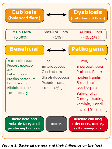
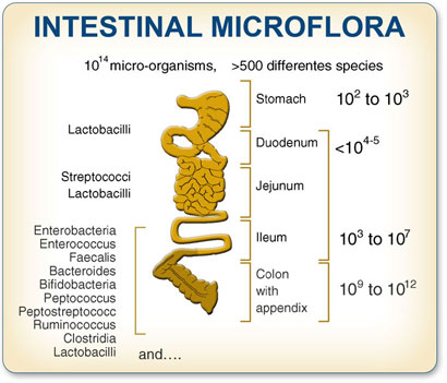
Image from customprobiotics.com

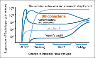
The typical mix of microbiota in the gut changes from infancy to adulthood, remains fairly stable, and then changes again in old age. 

Every time a human baby is born, a rich and dynamic microbial ecosystem develops from a sterile environment. During the first year of life, until weaning, the gut ecosystem is prevalently colonized by opportunistic microorganisms to which a baby is exposed in its environment (Palmer et al. 2007). The earliest colonizers are often aerobes such as Staphylococcus, Streptococcus and Enterobacteria, followed by anaerobic later colonizers such as Eubacteria and Clostridia. After these earlier stages, it is generally though that the microbiota of breast-fed infants is largely dominated by Bifidobacterium. After weaning, the developmental changes in the gut mucosa and in the intestinal IS, together the introduction of a solid diet, drive to the transition to a resilience adult-like profile of the human gut microbiota, characterised by a remarkable microbial biodiversity. The ageing of the gut microbiota starts after a subject-specific ‘threshold age’ which depends on individual characteristics such as diet, country and, eventually, frailty. In any case, changes of diet, lifestyle and the immunosenescence of the intestinal IS dramatically impact the microbial ecology of the human GI tract. Symmetrically to what happens in the early stage of our life, the aged-type microbiota shows a low microbial biodiversity, and it is characterised by an increase in opportunistic environmental facultative aerobes Staphylococcus, Streptococcus, Enterobacteriaceae, as well as a reduction in anaerobes, such as Clostridium clusters IV and XIVa and Bacteroidetes. However, differently from the infant-type microbiota, the aged type is characterised by a low abundance of Bifidobacterium(ref).”

The 2009 publication Acquisition, evolution and maintenance of the normal gut microbiota reports: “The gut is sterile at birth, but is rapidly colonised by faecal and vaginal bacteria of maternal origin. Over the succeeding weeks, months and years, a complex microbiota develops that plays a major role in host physiology. While the digestive tract is colonised to varying degrees by micro-organisms throughout its length, due to acid pH and the short retention time of gastric contents, bacterial numbers in the stomach are usually low. The rapid passage of digestive materials through the upper gut does not provide time for significant bacterial growth to occur, but cell numbers increase considerably in the distal ileum. The rate of movement of intestinal contents slows in the colon, which facilitates the development of complex bacterial communities. The large intestine is an intricate ecosystem that contains a complex microbiota composed of several hundred different types of bacteria. The growth and metabolism of microbial communities in the large intestine are determined by many factors, such as diet, environment and host physiological processes, as well as the anatomic structure of the digestive tract, disease, immunity, host genetics, drugs and ageing. Modifications in diet and host immune system activity, as well as physiological changes in the digestive tract affect microbiota composition in older people. The elderly have fewer bifidobacteria and higher numbers of enterobacteria and clostridia than young adults. Increased antibiotic use in older people and simply going into hospital have been shown to change bacterial community structure in the colonic microbiota, although the metabolic significance of this is unclear.”

From Genomic Insights into Bifidobacteria:The human large intestine is a very complex ecosystem that is still not fully understood, and while its microbial composition consists primarily of just four bacterial phyla, Bacteroidetes, Firmicutes, Actinobacteria, and Proteobacteria (as well as some Archaea, Eukarya, and viruses), it is highly variable at the genus level between individuals, likely due to factors such as age, health, diet, etc. (25, 99, 127, 244). Therefore, the adaptation capabilities of the intestinal bacteria corresponding to these variable factors likely influence the overall composition of intestinal microflora in the intestine (89).”

The 2007 publication Intestinal bacteria and ageing reported: “Advancements in science and medicine, as well as improved living standards, have led to a steady increase in life expectancy, and subsequently a rise in the elderly population. The intestinal microbiota is important for maintenance of host health, providing energy, nutrients and protection against invading organisms. Although the colonic microbiota is relatively stable throughout adult life, age-related changes in the gastrointestinal (GI) tract, as well as changes in diet and host immune system reactivity, inevitably affect population composition. Recent studies indicate shifts in the composition of the intestinal microbiota, which may lead to detrimental effects for the elderly host. Increased numbers of facultative anaerobes, in conjunction with a decrease in beneficial organisms such as the anaerobic lactobacilli and bifidobacteria, amongst other anaerobes, have been reported. These changes, along with a general reduction in species diversity in most bacterial groups, and changes to diet and digestive physiology such as intestinal transit time, may result in increased putrefaction in the colon and a greater susceptibility to disease. — Therapeutic strategies to counteract these changes have been suggested in ageing people. These include dietary supplements containing prebiotics, probiotics and a combination of both of these, synbiotics. Limited feeding trials show promising results with these supplements, although further longer-term investigations are required to substantiate their use in elderly healthcare fields.”

The 2010 publication Role of the gut microbiota in defining human health reported: “The human superorganism is a conglomerate of mammalian and microbial cells, with the latter estimated to outnumber the former by ten to one and the microbial genetic repertoire (microbiome) to be approximately 100-times greater than that of the human host. Given the ability of the immune response to rapidly counter infectious agents, it is striking that such a large density of microbes can exist in a state of synergy within the human host. This is particularly true of the distal gastrointestinal (GI) tract, which houses up to 1000 distinct bacterial species and an estimated excess of 1 x 10(14) microorganisms. An ever-increasing body of evidence implicates the GI microbiota in defining states of health and disease. Here, we review the literature in adult and pediatric GI microbiome studies, the emerging links between microbial community structure, function, infection and disease, and the approaches to manipulate this crucial ecosystem to improve host health.”

While the study of gut bacteria has been going on for more than 100 years, many new insights are now flowing from massive genomic sequencing techniques.

Bifidobacteria appear to be an important set of species of gut bacteria from a health viewpoint.

Study of the relationships of gut bacteria to health are very far from new. Identification of Bifidobacteria goes back to 1900.  Also from the September 2010 publication Genomic Insights into Bifidobacteria “Tissier (315) suggested that the large number of bifidobacteria in the feces of healthy breast-fed infants was likely the reason for their lower incidence of infantile diarrhea. In his pediatric work, he used bifidobacteria for the treatment of this intestinal diarrhea, and this likely represents the first example of the oral administration of a live microorganism for the treatment of a disease (316). The abundance of bifidobacteria in the feces of breast-fed infants was thought to be due to the Bifidobacterium-stimulating properties of human breast milk (38, 46, 60, 96, 196, 321). Numerous studies have substantiated the higher bifidobacterial counts and lower incidences of gastroenteritis in breast-fed infants than in formula-fed infants (3, 40, 51, 98).”  The Summary of the publication relates: “Since the discovery in 1899 of bifidobacteria as numerically dominant microbes in the feces of breast-fed infants, there have been numerous studies addressing their role in modulating gut microflora as well as their other potential health benefits. Because of this, they are frequently incorporated into foods as probiotic cultures. An understanding of their full interactions with intestinal microbes and the host is needed to scientifically validate any health benefits they may afford. Recently, the genome sequences of nine strains representing four species of Bifidobacterium became available. A comparative genome analysis of these genomes reveals a likely efficient capacity to adapt to their habitats, with B. longum subsp. infantis exhibiting more genomic potential to utilize human milk oligosaccharides, consistent with its habitat in the infant gut. Conversely, B. longum subsp. longum exhibits a higher genomic potential for utilization of plant-derived complex carbohydrates and polyols, consistent with its habitat in an adult gut. An intriguing observation is the loss of much of this genome potential when strains are adapted to pure culture environments, as highlighted by the genomes of B. animalis subsp. lactis strains, which exhibit the least potential for a gut habitat and are believed to have evolved from the B. animalis species during adaptation to dairy fermentation environments.”

“The composition of the microflora in the human large intestine, as estimated using culturing techniques, is usually dominated by the genera Bacteroides, Eubacterium, and Bifidobacterium, with several other predominant genera, such as Clostridium, Peptostreptococcus, Enterococcus, Lactobacillus, and members of the family Enterobacteriaceae (117, 118). The number of species estimated by culturing techniques is approximately 400. Nonculturing analysis of the gut microflora was greatly facilitated by the direct isolation of DNA from feces and by amplification of the 16S rRNA genes representing the entire microflora. Cloning and sequencing of individual rRNA genes enabled the numerically dominant genera of bacteria to be identified. This molecular analysis of the intestinal microflora in fecal and colonic samples initially suggested that the human large intestine contains more than 500 different bacterial species and that about 75% of them are nonculturable (81, 83). However, a recent extensive metagenomic analysis revealed that this is an overestimation, with most individuals harboring approximately 160 different bacterial species, the majority of which are “known” (239). While the total number of species present in the human gut is not known, recent molecular studies indicate that it is in excess of 1,000 (239, 241), with an upper estimate of 1,150 suggested by an extensive metagenomic analysis of 124 individuals (239)(ref).”

Gut microflora may play an even more important role in maintaining human health than previously thought.  Specifically it appears they are involved in the control of energy and metabolic homeostasis.

This theme has been repeated in a substantial number of publications for over five years now. The 2007 reviewpublication Gut microflora as a target for energy and metabolic homeostasis reported “Purpose of review: Gut microbiota plays an important role in health and disease, but this ecosystem remains incompletely characterized and shows a wide diversity. This review discusses new findings that may explain how gut microbiota can be involved in the control of energy and metabolic homeostasis.  Recent findings: Over the past 5 years studies have highlighted some key aspects of the mammalian host-gut microbial relationship. Gut microbiota could now be considered a ‘microbial organ’ placed within a host organ. Recent data suggest that the modulation of gut microbiota affects host metabolism and has an impact on energy storage. Several mechanisms are proposed that link events occurring in the colon and the regulation of energy metabolism.  Summary: Gut microflora may play an even more important role in maintaining human health than previously thought. The literature provides new evidence that the increased prevalence of obesity and type 2 diabetes cannot be attributed solely to changes in the human genome, nutritional habits, or reduction of physical activity in our daily lives. One must also consider this important new environmental factor, namely gut microbiota. Scientists may take into consideration a key question: could we manipulate the microbiotic environment to treat or prevent obesity and type 2 diabetes? This opens up a new area in nutrition research.”

The August 2011 publication Programming of host metabolism by the gut microbiota reports: “The human gut harbors a vast ensemble of bacteria that has co-evolved with the human host and performs several important functions that affect our physiology and metabolism. The human gut is sterile at birth and is subsequently colonized with bacteria from the mother and the environment. The complexity of the gut microbiota is increased during childhood, and adult humans contain 150-fold more bacterial genes than human genes. Recent advances in next-generation sequencing technology and mechanistic testing in gnotobiotic mice have identified the gut microbiota as an environmental factor that contributes to obesity. Germ-free mice are protected against developing diet-induced obesity and the underlying mechanisms whereby the gut microbiota contributes to host metabolism are beginning to be clarified. The obese phenotype is associated with increased microbial fermentation and energy extraction; however, other microbially modulated mechanisms contribute to disease progression as well. The gut microbiota has profound effects on host gene expression in the enterohepatic system, including genes involved in immunity and metabolism. For example, the gut microbiota affects expression of secreted proteins in the gut, which modulate lipid metabolism in peripheral organs. In addition, the gut microbiota is also a source of proinflammatory molecules that augment adipose inflammation and macrophage recruitment by signaling through the innate immune system. TLRs (Toll-like receptors) are integral parts of the innate immune system and are expressed by both macrophages and epithelial cells. Activation of TLRs in macrophages dramatically impairs glucose homeostasis, whereas TLRs in the gut may alter the gut microbial composition that may have profound effects on host metabolism. Accordingly, reprogramming the gut microbiota, or its function, in early life may have beneficial effects on host metabolism later in life.”

The mixture of kinds of bacteria in the gut may contribute to metabolic disorders such as obesity, diabetes, and cardiovascular diseases or can contribute strongly to maintenance of health homeostasis.

The November 2010 publication Ecology and Physiology of the Intestinal Tract reports: “The number of microorganisms inhabiting the human digestive tract exceeds the number of body cells by a factor of ten. This microbial community affects host physiology and host health. The metabolic potential of the gut microbiota is immense affording the extraction of energy from otherwise indigestible carbohydrates (dietary fiber) and the conversion of host-derived substances, non-nutritive dietary components and drugs. Recognized functions of the gut microbiota include provision of colonization resistance against pathogens and priming of both the innate and the acquired immune systems. However, the intestinal microbiota may also contribute to the development of diseases such as ulcerative colitis and colorectal cancer. Culture-dependent studies provided basic knowledge on the gut microbiota, but only the advent of culture-independent molecular methods led to a better understanding of host-microbe interactions. The application of metagenomics to the gut microbial ecosystem revealed truly remarkable correlations between certain diseases and the gut microbiome. It also led to the suggestion of the existence of a ‘core microbiome’ that encompasses key functions shared by each individual. However, the mechanisms underlying host-microbe interactions have not yet been unraveled.””

Image from website of engormix.com.

The January 2012 publication Is the gut microbiota a new factor contributing to obesity and its metabolic disorders? Relates: “The gut microbiota refers to the trillions of microorganisms residing in the intestine and is integral in multiple physiological processes of the host. Recent research has shown that gut bacteria play a role in metabolic disorders such as obesity, diabetes, and cardiovascular diseases. The mechanisms by which the gut microbiota affects metabolic diseases are by two major routes: (1) the innate immune response to the structural components of bacteria (e.g., lipopolysaccharide) resulting in inflammation and (2) bacterial metabolites of dietary compounds (e.g., SCFA from fiber), which have biological activities that regulate host functions. Gut microbiota has evolved with humans as a mutualistic partner, but dysbiosis in a form of altered gut metagenome and collected microbial activities, in combination with classic genetic and environmental factors, may promote the development of metabolic disorders. This paper reviews the available literature about the gut microbiota and aforementioned metabolic disorders and reveals the gaps in knowledge for future study.”

Other relevant publications include:

The environment within: how gut microbiota may influence metabolism and body composition (2010)

Changes in gut microbiota control inflammation in obese mice through a mechanism involving GLP-2-driven improvement of gut permeability (2009)

Diet-induced metabolic improvements in a hamster model of hypercholesterolemia are strongly linked to alterations of the gut microbiota (2009)

A discussion of the role of gut flora in autism and inflammatory bowel disease can be found on this webpage of science-autism.org which is about published scientific data concerning autism.   The following diagram is also from that web page.  It illustrates the role microbiota can play in breaching the intestinal barrier wall and generating an inflammatory reaction.

Image from Gut Flora in Autism:  “A well considered pathway for the effect of bacterial flora on the inflammatory response of the gut wall in IBD. The inflammation response starts when the gut barrier is breeched and this causes a further inflammatory response.”

With advanced aging, changes typically happen in the ecology of the gut microorganism, system resulting in increased susceptibility to infectious diseases and related infirmities.

The 2010 publication Through ageing, and beyond: gut microbiota and inflammatory status in seniors and centenarians reports: “Age-related physiological changes in the gastrointestinal tract, as well as modifications in lifestyle, nutritional behaviour, and functionality of the host immune system, inevitably affect the gut microbiota, resulting in a greater susceptibility to infections.  Methodology/Principal Findings: By using the Human Intestinal Tract Chip (HITChip) and quantitative PCR of 16S rRNA genes of Bacteria and Archaea, we explored the age-related differences in the gut microbiota composition among young adults, elderly, and centenarians, i.e subjects who reached the extreme limits of the human lifespan, living for over 100 years. We observed that the microbial composition and diversity of the gut ecosystem of young adults and seventy-years old people is highly similar but differs significantly from that of the centenarians. After 100 years of symbiotic association with the human host, the microbiota is characterized by a rearrangement in the Firmicutes population and an enrichment in facultative anaerobes, notably pathobionts. The presence of such a compromised microbiota in the centenarians is associated with an increased inflammatory status, also known as inflammageing, as determined by a range of peripheral blood inflammatory markers.  This may be explained by a remodelling of the centenarians’ microbiota, with a marked decrease in Faecalibacterium prauznitzii and relatives, symbiotic species with reported anti-inflammatory properties. As signature bacteria of the long life we identified specifically Eubacterium limosum and relatives that were more than ten-fold increased in the centenarians.  Conclusions/Significance: We provide evidence for the fact that the ageing process deeply affects the structure of the human gut microbiota, as well as its homeostasis with the host’s immune system. Because of its crucial role in the host physiology and health status, age-related differences in the gut microbiota composition may be related to the progression of diseases and frailty in the elderly population.”

Image from the website of Les Laboratoires Nicar, Inc.

On the other hand, the 2010 e-publication Human intestinal microbiota and healthy ageing review publication reports: “Earlier studies have indicated a decrease in anaerobes and bifidobacteria and a concomitant increase in enterobacteria in the intestinal microbiota with ageing. However, new data obtained with molecular techniques suggests decreased stability and increased diversity of the gut microbiota with advancing age. Further, no simple marker change in microbiota composition can be identified. Except for the reduced immune function, ageing itself may have relatively little effect on overall gastrointestinal function. Concomitant changes in nutrition, increased incidence of disease and corresponding use of medication with advancing age modify the composition of the microbial community of the gastrointestinal tract. This mini-review will focus on the recent findings on the gut microbiota of the elderly and on the potential benefits of probiotics, prebiotics and synbiotics.”

TLR and NLR signaling in intestinal epithelial cells triggered by gut microbiota contribute to a range of homeostatic mechanisms including proliferation, wound healing, epithelial integrity, and regulation of mucosal immune functions.

The 2010 publication The role of innate signaling in the homeostasis of tolerance and immunity in the intestine reports: “In the intestine innate recognition of microbes is achieved through pattern recognition receptor (PRR) families expressed in immune cells and different cell lineages of the intestinal epithelium. Toll-like receptor (TLR) and nucleotide-binding and oligomerization domain-like receptor (NLR) families are emerging as key mediators of immunity through their role as maturation factors of immune cells and triggers for the production of cytokines and chemokines and antimicrobial factors. At the mucosal surface chronic activation of the immune system is avoided through the epithelial production of a glycocalyx, steady-state production of antimicrobial factors as well as the selective expression and localization of PRRs. Additionally, the polarization of epithelial TLR signaling and suppression of NF-kappaB activation by luminal commensals appears to contribute to the homeostasis of tolerance and immunity. Several studies have demonstrated that TLR signaling in epithelial cells contributes to a range of homeostatic mechanisms including proliferation, wound healing, epithelial integrity, and regulation of mucosal immune functions. The intestinal epithelium appears to have uniquely evolved to maintain mucosal tolerance and immunity, and future efforts to further understand the molecular mechanisms of intestinal homeostasis may have a major impact on human health.”

Our old friend NF-kappaB can be activated by TLR and NOD signaling by gut microbiota, resulting in possible positive or detrimental effects.

The 2010 publication Role of NF-kappaB activation in intestinal immune homeostasis reports: “Inflammatory bowel diseases (IBD) are characterised by a disturbance of intestinal immune homeostasis, either caused by or followed by inappropriate responses to the resident commensal bacteria. Although the transcription factor NF-kappaB actively participates in the excessive inflammatory response observed in IBD, recent studies with mice defective in NF-kappaB activation have revealed that NF-kappaB also serves an essential protective function in the intestinal immune system. The enormous amount of commensal bacteria in the intestine might play a role in the distinct functions of NF-kappaB in the intestine, as they can initiate signalling to NF-kappaB through both Toll-like receptors and NOD-like receptors in intestinal epithelial cells as well as mucosal immune cells. However, the exact individual contributions of different NF-kappaB-activating stimuli as well as the target cells that mediate the detrimental or beneficial functions of NF-kappaB in the intestine are still elusive. In this review, I will summarise and discuss the current knowledge on the role of different NF-kappaB-activating pathways in preserving intestinal immune homeostasis and the development of intestinal inflammation.”

Gut microbiota have a strong impact on immune system responses.

The May 2011 publication Influence of gastrointestinal commensal bacteria on the immune responses that mediate allergy and asthma reports: “The human intestine contains more than 100 trillion microorganisms that maintain a symbiotic relationship with the host. Under normal conditions, these bacteria are not pathogenic and in fact confer health benefits to the host. The microbiota interacts with the innate and adaptive arms of the host’s intestinal mucosal immune system and through these mechanisms drives regulatory cell differentiation in the gut that is critically involved in maintaining immune tolerance. Specifically, the microbiota can activate distinct tolerogenic dendritic cells in the gut and through this interaction can drive regulatory T-cell differentiation. In addition, the microbiota is important in driving T(H)1 cell differentiation, which corrects the T(H)2 immune skewing that is thought to occur at birth. If appropriate immune tolerance is not established in early life and maintained throughout life, this represents a risk factor for the development of inflammatory, autoimmune, and allergic diseases. Early-life events are instrumental in establishing the microbiota, the composition of which throughout life is influenced by various environmental and lifestyle pressures. Significant efforts are now being made to establish interventional approaches that can create a healthy microbiota that confers maximum tolerogenic immunomodulatory effects in the gut and that will protect against systemic inflammatory disease pathologies.”

Regarding probiotics, prebiotics and synbiotics

Practical interventions in gut bacteria mixes are primarily based on taking probiotic, prebiotic or symbiotic supplements.  In a nutshell, a probiotic supplement contains one or more strains of live bacteria known to be health-promoting.  A prebiotic is a dietary supplement designed to promote the proliferation and sustainability in the gut of health-producing bacterial strains.  And a symbiotic is blend of a probiotic and a probiotic.  The distinctions are discussed in the 2008 publication Probiotics, prebiotics, and synbiotics: “.According to the German definition, probiotics are defined viable microorganisms, sufficient amounts of which reach the intestine in an active state and thus exert positive health effects. Numerous probiotic microorganisms (e.g. Lactobacillus rhamnosus GG, L. reuteri, bifidobacteria and certain strains of L. casei or the L. acidophilus-group) are used in probiotic food, particularly fermented milk products, or have been investigated–as well as Escherichia coli strain Nissle 1917, certain enterococci (Enterococcus faecium SF68) and the probiotic yeast Saccharomyces boulardii–with regard to their medicinal use. Among the numerous purported health benefits attributed to probiotic bacteria, the (transient) modulation of the intestinal microflora of the host and the capacity to interact with the immune system directly or mediated by the autochthonous microflora, are basic mechanisms. They are supported by an increasing number of in vitro and in vivo experiments using conventional and molecular biologic methods. In addition to these, a limited number of randomized, well-controlled human intervention trials have been reported. Well-established probiotic effects are: 1. Prevention and/or reduction of duration and complaints of rotavirus-induced or antibiotic-associated diarrhea as well as alleviation of complaints due to lactose intolerance. 2. Reduction of the concentration of cancer-promoting enzymes and/or putrefactive (bacterial) metabolites in the gut. 3. Prevention and alleviation of unspecific and irregular complaints of the gastrointestinal tracts in healthy people. 4. Beneficial effects on microbial aberrancies, inflammation and other complaints in connection with: inflammatory diseases of the gastrointestinal tract, Helicobacter pylori infection or bacterial overgrowth. 5. Normalization of passing stool and stool consistency in subjects suffering from obstipation or an irritable colon. 6. Prevention or alleviation of allergies and atopic diseases in infants. 7. Prevention of respiratory tract infections (common cold, influenza) and other infectious diseases as well as treatment of urogenital infections. Insufficient or at most preliminary evidence exists with respect to cancer prevention, a so-called hypocholesterolemic effect, improvement of the mouth flora and caries prevention or prevention or therapy of ischemic heart diseases or amelioration of autoimmune diseases (e.g. arthritis). A prebiotic is “a selectively fermented ingredient that allows specific changes, both in the composition and/or activity in the gastrointestinal microflora that confers benefits upon host well being and health”, whereas synergistic combinations of pro- and prebiotics are called synbiotics. Today, only bifidogenic, non-digestible oligosaccharides (particularly inulin, its hydrolysis product oligofructose, and (trans)galactooligosaccharides), fulfill all the criteria for prebiotic classification. They are dietary fibers with a well-established positive impact on the intestinal microflora. Other health effects of prebiotics (prevention of diarrhoea or obstipation, modulation of the metabolism of the intestinal flora, cancer prevention, positive effects on lipid metabolism, stimulation of mineral adsorption and immunomodulatory properties) are indirect, i.e. mediated by the intestinal microflora, and therefore less-well proven. In the last years, successful attempts have been reported to make infant formula more breast milk-like by the addition of fructo- and (primarily) galactooligosaccharides.”

The 2010 publication Prebiotic effects: metabolic and health benefits relates general experience with probiotics over a 15 year time frame: “The different compartments of the gastrointestinal tract are inhabited by populations of micro-organisms. By far the most important predominant populations are in the colon where a true symbiosis with the host exists that is a key for well-being and health. For such a microbiota, ‘normobiosis’ characterises a composition of the gut ‘ecosystem’ in which micro-organisms with potential health benefits predominate in number over potentially harmful ones, in contrast to ‘dysbiosis’, in which one or a few potentially harmful micro-organisms are dominant, thus creating a disease-prone situation. The present document has been written by a group of both academic and industry experts (in the ILSI Europe Prebiotic Expert Group and Prebiotic Task Force, respectively). It does not aim to propose a new definition of a prebiotic nor to identify which food products are classified as prebiotic but rather to validate and expand the original idea of the prebiotic concept (that can be translated in ‘prebiotic effects’), defined as: ‘The selective stimulation of growth and/or activity(ies) of one or a limited number of microbial genus(era)/species in the gut microbiota that confer(s) health benefits to the host.’ Thanks to the methodological and fundamental research of microbiologists, immense progress has very recently been made in our understanding of the gut microbiota. A large number of human intervention studies have been performed that have demonstrated that dietary consumption of certain food products can result in statistically significant changes in the composition of the gut microbiota in line with the prebiotic concept. Thus the prebiotic effect is now a well-established scientific fact. The more data are accumulating, the more it will be recognised that such changes in the microbiota’s composition, especially increase in bifidobacteria, can be regarded as a marker of intestinal health. The review is divided in chapters that cover the major areas of nutrition research where a prebiotic effect has tentatively been investigated for potential health benefits. The prebiotic effect has been shown to associate with modulation of biomarkers and activity(ies) of the immune system. — Confirming the studies in adults, it has been demonstrated that, in infant nutrition, the prebiotic effect includes a significant change of gut microbiota composition, especially an increase of faecal concentrations of bifidobacteria. This concomitantly improves stool quality (pH, SCFA, frequency and consistency), reduces the risk of gastroenteritis and infections, improves general well-being and reduces the incidence of allergic symptoms such as atopic eczema. Changes in the gut microbiota composition are classically considered as one of the many factors involved in the pathogenesis of either inflammatory bowel disease or irritable bowel syndrome. — The use of particular food products with a prebiotic effect has thus been tested in clinical trials with the objective to improve the clinical activity and well-being of patients with such disorders. Promising beneficial effects have been demonstrated in some preliminary studies, including changes in gut microbiota composition (especially increase in bifidobacteria concentration). Often associated with toxic load and/or miscellaneous risk factors, colon cancer is another pathology for which a possible role of gut microbiota composition has been hypothesised. — Numerous experimental studies have reported reduction in incidence of tumours and cancers after feeding specific food products with a prebiotic effect. Some of these studies (including one human trial) have also reported that, in such conditions, gut microbiota composition was modified (especially due to increased concentration of bifidobacteria). Dietary intake of particular food products with a prebiotic effect has been shown, especially in adolescents, but also tentatively in postmenopausal women, to increase Ca absorption as well as bone Ca accretion and bone mineral density. Recent data, both from experimental models and from human studies, support the beneficial effects of particular food products with prebiotic properties on energy homaeostasis, satiety regulation and body weight gain. Together, with data in obese animals and patients, these studies support the hypothesis that gut microbiota composition (especially the number of bifidobacteria) may contribute to modulate metabolic processes associated with syndrome X, especially obesity and diabetes type 2. It is plausible, even though not exclusive, that these effects are linked to the microbiota-induced changes and it is feasible to conclude that their mechanisms fit into the prebiotic effect. However, the role of such changes in these health benefits remains to be definitively proven. As a result of the research activity that followed the publication of the prebiotic concept 15 years ago, it has become clear that products that cause a selective modification in the gut microbiota’s composition and/or activity(ies) and thus strengthens normobiosis could either induce beneficial physiological effects in the colon and also in extra-intestinal compartments or contribute towards reducing the risk of dysbiosis and associated intestinal and systemic pathologies.”

Raising bifidobacteria levels by the use of probiotics and prebiotics appears to be the most-established strategy for modifying microbial balance in human colons to promote health.  However, much remains to be learned.

The December 2009 publication Food-based strategies to modulate the composition of the intestinal microbiota and their associated health effects reports: “The most well known food-based strategies to modulate the composition of the intestinal microbiota are the dietary use of prebiotics, probiotics and their combination, synbiotics. Currently established prebiotic compounds are mainly targeting the bifidobacteria population of the colon microbiota.  A good illustration of the importance of high colonic bifidobacteria levels is the observation that breast milk creates an environment in the colon (because of its high amount in galacto-oligosaccharides with prebiotic activity) favouring the development of a simple flora, dominated by bifidobacteria to which various health benefits have been ascribed.  – Currently, high colonic bifidobacteria levels has been considered favourably at all ages and strategies to augment their presence have been demonstrated in placebo-controlled intervention studies; e.g. in toddlers to reduce sickness events, in adults to reduce the risk for developing gastrointestinal diseases and in the elderly to re-enhance their declining immune activity. The intestinal microbiota can be considered as a metabolically adaptable and rapidly renewable organ of the body. However, unbalances in its microbial community and activities are found to be implicated in disease initiation and progression, such as chronic inflammatory bowel diseases and colonic cancers. Restoration of this balance by increasing bifidobacteria levels has demonstrated to reduce disease severity of patients and to improve well-being in healtly volunteers. New emerging evidence on the difference in the composition of the colonic microbiota between obese and lean volunteers has opened new areas for pre-, pro- and synbiotic research. Additionally, as knowledge will increase about the microbial bio-conversion of polyphenolic compounds into bioactive metabolites in the colon and whether food-based strategies can augment such bioconversion into more potent compounds with anti-oxidant and/or anti-inflammatory activity new areas of research will be discovered. This paper provides an up-to-date review of the health benefits associated to the induction of high bifidobacteria levels in the colon by the use of prebiotics (inulin and oligofructose).”

Prebiotics may be useful for the prevention or treatment of insulin resistance, diabetes and obesity.

The 2011 publication Gut microbiota and the pathogenesis of insulin resistance reports: “Several reviews recently explored how the gut microbiota was able to control host energy metabolism, and thereby the development of adiposity. In this review, we focused on the state of the art that supports a link between the gut microbiota composition and activity, and the management of glycemia associated with overweight and diabetes. Several microbial-derived compounds are related to disturbances of glucose homeostasis including the gram-negative-derived lipopolysaccharides. Some nutrients with prebiotic properties, which escape the digestion in the upper part of the gut, modify the composition of the gut microbiota in favor of bacteria that could play a beneficial role on glucose homeostasis, namely by modulating the endocrine function of the gut, and by reinforcing the gut barrier. Adequate intervention studies in diabetic patients are required to assess the relevance of those experimental data for human health.”

The August 2011 publication Modulation of the gut microbiota by nutrients with prebiotic properties: consequences for host health in the context of obesity and metabolic syndrome reports: “The gut microbiota is increasingly considered as a symbiotic partner for the maintenance of health. The homeostasis of the gut microbiota is dependent on host characteristics (age, gender, genetic background…), environmental conditions (stress, drugs, gastrointestinal surgery, infectious and toxic agents…). Moreover, it is dependent on the day-to-day dietary changes. — Experimental data in animals, but also observational studies in obese patients, suggest that the composition of the gut microbiota is a factor characterizing obese versus lean individuals, diabetic versus non diabetic patients, or patients presenting hepatic diseases such as non alcoholic steatohepatitis. Interestingly, the changes in the gut microbes can be reversed by dieting and related weight loss. The qualitative and quantitative changes in the intake of specific food components (fatty acids, carbohydrates, micronutrients, prebiotics, probiotics), have not only consequences on the gut microbiota composition, but may modulate the expression of genes in host tissues such as the liver, adipose tissue, intestine, muscle. This in turn may drive or lessen the development of fat mass and metabolic disturbances associated with the gut barrier function and the systemic immunity. The relevance of the prebiotic or probiotic approaches in the management of obesity in humans is supported by few intervention studies in humans up to now, but the experimental data obtained with those compounds help to elucidate novel potential molecular targets relating diet with gut microbes. The metagenomic and integrative metabolomic approaches could help elucidate which bacteria, among the trillions in human gut, or more specifically which activities/genes, could participate to the control of host energy metabolism, and could be relevant for future therapeutic developments.” – “Improvement of obesity and related metabolic disorders by the prebiotic approach. Nutrients with prebiotic properties allows, by changing the gut microbiota, to promote the endocrine function of the gut (increase in GLP-1, and GLP-2 producing cells), and to modulate the activation of the endocannabinoid system in the intestine and in the adipose tissue. All those effects contribute to lessen gut permeability (improved distribution of the tight junction proteins ZO-1 and Occludin), thereby decreasing endotoxemia, and systemic inflammation. Changes in GLP-1 contribute to decrease food intake, fat mass, glycemia and insulin resistance. eCB, endocannabinoid; GLP-1, glucagon-like peptide 1; GLP-2, glucagon-like peptide 2; LPS, lipopolysaccharides; ZO-1, zonula occludens 1.”

The LKM512 strain of yogurt bacteria has attracted particular attention as a probiotic for increasing polyamines levels and inhibiting inflammation.

“Chronic low-grade inflammation is recognized as an important factor contributing to senescence and age-related diseases. In mammals, levels of polyamines (PAs) decrease during the ageing process; PAs are known to decrease systemic inflammation by inhibiting inflammatory cytokine synthesis in macrophages. Reductions in intestinal luminal PAs levels have been associated with intestinal barrier dysfunction. The probiotic strain Bifidobacterium animalis subsp. lactis LKM512 is known to increase intestinal luminal PA concentrations.” (ref)

The 2009 publication Polyamine-rich food decreases age-associated pathology and mortality in aged micerelates polyamine consumption to senescence markers in mice: “The purpose of this study was to test whether oral intake of foods rich in polyamines (spermine and spermidine) suppresses age-associated pathology in aged mice. Synthetic polyamines were mixed into experimental chows, and 24-week-old Jc1:ICR male mice were fed one of three chows containing differing polyamine concentrations. The spermine and spermidine concentrations in the low, normal, and high polyamine chows were 143 and 224 nmol/g, 160 and 434 nmol/g, and 374 and 1540 nmol/g, respectively. An increase in concentration of polyamine in the blood was found only in mice fed the high polyamine chow at 50 weeks of age. While the body weights of mice in all three groups were similar, the survival rate of mice fed high polyamine chow was significantly higher than those in the other two groups (p=0.011). Mice fed the high polyamine chow analyzed at 88 weeks of age, corresponding to the end of the study, demonstrated lower incidence of glomerulosclerosis and increased expression of senescence marker protein-30 in both kidney and liver compared to those fed the low polyamine chow. As these pathological changes are associated with senescence, oral polyamine appears to inhibit the progression of age-associated pathologies.”

The 2009 publication Dynamics of fecal microbiota in hospitalized elderly fed probiotic LKM512 yogurt reports: “The comprehensive dynamics of intestinal microbiota including uncultured bacteria in response to probiotic consumption have not been well studied. The aims of this study were twofold: firstly to analyze the impact on intestinal microbiota of yogurt fermented by Bifidobacterium animalis subsp. lactis LKM512, Lactobacillus delbrueckii subsp. bulgaricus LKM1759, and Streptococcus thermophilus LKM1742 (LKM512 yogurt) and placebo fermented by these lactic acid bacterial strains without LKM512; and secondly to investigate the changes in intestinal microbiota that influence the concentration of PA, one of the beneficial metabolites produced by bacteria in the intestine. The LKM512 yogurt/placebo trial was performed in six hospitalized elderly patients (three men and three women with an average age of 80.3 years) and lasted seven weeks with the following schedule: pre-consumption for one week, LKM512 yogurt consumption for two weeks, washout period for two weeks, and placebo consumption for two weeks. The amount of ingested LKM512 yogurt or placebo was 100 g/day/individual. Fecal samples were analyzed using T-RFLP and real-time PCR. The T-RFLP patterns in five of the six volunteers were changed in a similar fashion by LKM512 yogurt consumption, although these patterns were individually changed following consumption of placebo. It was confirmed that B. animalis subsp. lactis was increased dramatically and Lactobacillus spp. tended to be decreased by LKM512 yogurt consumption. Some indigenous uncultured bacteria were increased and some decreased by LKM512 yogurt/placebo consumption. The similar changes in the intestinal microbiota of the elderly caused by consumption of the LKM512 yogurt were found to be influenced by the LKM512 strain itself, and not by the lactic acid bacteria in the yogurt. Moreover, this study suggests that the increase in intestinal PA concentrations caused by LKM512 yogurt consumption is probably dependent on the LKM512 strain colonizing the intestine.”

Some researchers think targeting the gut microbiota may be an avenue for human life extension.

The December 2011 publication Gut microbiota as a candidate for lifespan extension: an ecological/evolutionary perspective targeted on living organisms as metaorganisms speculates on this possibility: “An emerging central concept in evolutionary biology suggests that symbiosis is a universal characteristic of living organisms that can help in understanding complex traits and phenotypes. During evolution, an integrative circuitry fundamental for survival has been established between commensal gut microbiota and host. On the basis of recent knowledge in worms, flies, and humans, an important role of the gut microbiota in aging and longevity is emerging. The complex bacterial community that populates the gut and that represents an evolutionary adapted ecosystem correlated with nutrition appears to limit the accumulation of pathobionts and infections in all taxa, being able of affecting the efficiency of the host immune system and exerting systemic metabolic effects. There is an urgent need to disentangle the underpinning molecular mechanisms, which could shed light on the basic mechanisms of aging in an ecological perspective. Thus, it appears possible to extend healthy aging and lifespan by targeting the host as a metaorganism by manipulating the complex symbiotic ecosystem of gut microbiota, as well as other possible ecosystems of the body.”

While this discussion is theoretical, it does appear that life extension in mice is possible by feeding them probiotics.

Administration of probiotics can extend the lives of mice.

I was motivated to research and generate this blog entry by coming across the August 2011 publication Longevity in mice is promoted by probiotic-induced suppression of colonic senescence dependent on upregulation of gut bacterial polyamine production.  Background: Chronic low-grade inflammation is recognized as an important factor contributing to senescence and age-related diseases. In mammals, levels of polyamines (PAs) decrease during the ageing process; PAs are known to decrease systemic inflammation by inhibiting inflammatory cytokine synthesis in macrophages. Reductions in intestinal luminal PAs levels have been associated with intestinal barrier dysfunction. The probiotic strain Bifidobacterium animalis subsp. lactis LKM512 is known to increase intestinal luminal PA concentrations. Methodology/Principal findings: We supplemented the diet of 10-month-old Crj:CD-1 female mice with LKM512 for 11 months, while the controls received no supplementation. Survival rates were compared using Kaplan-Meier survival curves. LKM512-treated mice survived significantly longer than controls (P<0.001); moreover, skin ulcers and tumors were more common in the control mice. We then analyzed inflammatory and intestinal conditions by measuring several markers using HPLC, ELISA, reverse transcription-quantitative PCR, and histological slices. LKM512 mice showed altered 16S rRNA gene expression of several predominant intestinal bacterial groups. The fecal concentrations of PAs, but not of short-chain fatty acids, were significantly higher in LKM512-treated mice (P<0.05). Colonic mucosal function was also better in LKM512 mice, with increased mucus secretion and better maintenance of tight junctions. Changes in gene expression levels were evaluated using the NimbleGen mouse DNA microarray. LKM512 administration also downregulated the expression of ageing-associated and inflammation-associated genes and gene expression levels in 21-month-old LKM512-treated mice resembled those in 10-month-old untreated (younger) mice.  Conclusion/Significance: Our study demonstrated increased longevity in mice following probiotic treatment with LKM512, possibly due to the suppression of chronic low-grade inflammation in the colon induced by higher PA levels. This indicates that ingestion of specific probiotics may be an easy approach for improving intestinal health and increasing lifespan. Further studies are required to clarify its effectiveness in humans.” – “When the mice aged or were administered LKM512 treatments, 55 of 93 gene pathways were altered significantly (Z-score >1.98), while the gene expression for 78 of these 93 gene pathways was similar between younger mice and mice receiving LKM512 (Fig. 4C, Fig. S2). Pathways that were downregulated by ageing were upregulated by LKM512 administration and vice versa. In other words, LKM512 administration suppressed ageing-associated change in gene pathways. When the mice aged or were administered LKM512 treatments, 55 of 93 gene pathways were altered significantly (Z-score >1.98), while the gene expression for 78 of these 93 gene pathways was similar between younger mice and mice receiving LKM512 (Fig. 4C, Fig. S2). Pathways that were downregulated by ageing were upregulated by LKM512 administration and vice versa. In other words, LKM512 administration suppressed ageing-associated change in gene pathways.” – “The anti-inflammatory effects of LKM512 administration were also revealed by a DNA microarray. Expression levels of genes in the TNF-NFκB, IL-1, IL-2, and IL-6 pathways were higher in the control group than in LKM512 and younger mice; additionally, gene expression levels in LKM512 mice were similar to those in younger mice (Fig. 5D and Fig. S3).” – “At 25 weeks, 8-OHdG concentrations, which indicate oxidative DNA damage [32], tended to be lower in LKM512 mice than in control mice (P=0.09) (Fig. S4A). Furthermore, pathway analysis using a DNA microarray indicated that the oxidative stress pathway was more active in the control mice than in LKM512 mice (Fig. S4B).”

Final comments

This blog entry provides a general introduction to gut microbiota, their importance to health and to probiotics, prebiotics and synbiotics.  And it provides samples from a large and hopeful body of research that might impact significantly on health and longevity.  I am impressed that a probiotic diet regimen can extend the lives of mice via a gene activation pattern.  However, those of you who follow this blog know that there are a number of other substances that can also do that.  In my perception gut microbiota is an important field of research to be followed. 

Finally, I want to disclose that for several years I have been taking a symbiotic as part of my personal regimen, one consisting of ten strains of probiotic organisms plus 200 mg of fructooligosaccharides, a prebiotic.  And barely a day goes by without my eating a couple of yogurt snacks, ones containing live cultures.

Blue light, sleep, mental alertness and health

$
0
0

By Vince Giuliano

We are being exposed to a lot more blue and ultraviolet light in recent years, especially at night.  This is due to 1. fluorescent bulbs replacing incandescent bulbs in homes and workspaces, especially “daytime” spectrum bulbs, 2. Flat LED TV screens replacing the old big-box tube fluorescent screens, 3. flat LED computer screens replacing the old fluorescent ones, and 4.  White-light LED streetlamps replacing yellow-light sodium vapor lamps.  In each case the light emitted is significantly shifted to the blue and even to the ultraviolet.  And giant TV and big computer screens pre-empt larger and larger parts of the total field of vision.  The result of greater exposure to blue light is inhibition of expression of melatonin, changes in our circadian sleep rhythms, changes in alertness and possible psychological and disease-susceptibility disturbances.  These “blue light” effects have received significant research attention recently and have been discussed in the popular press.  Some have even opined that lighting has become a public health issue. 

Further, with aging, natural human lenses tend to be yellow and less sensitive to blue and UV.  Having such lenses may inhibit melatonin suppression and the natural circadian rhythms.  How do these factors interact?  The purpose of this blog is to review the relevant research and to summarize the major issues and findings to date.

There is much more that meets the eye than what we see.

Besides providing visual images, inputs from the eye to other body systems impact on mood, alertness, sense of wellbeing and on the integrity of circadian rhythms related to health.  Essentially all vertebrates are subject to a circadian regulatory system where the body adjusts dynamically to the daily cycle of light and dark as well as to how this cycle is affected by season.  Key to this process is a small group of hypothalamic nerve cells in the brain, the suprachiasmatic nucleus (SCN), which serves as a master circadian pacemaker.  The SCN controls  the timing of the sleep-wake cycle and coordinates this with other circadian rhythms to enhance behavioral and environmental adaptation.

 “Diagram of various neurological structures, including the suprachiasmatic nucleus (SCN), that are involved in human circadian rhythm control.”  Image credit: http://thebrain.mcgill.ca

A starting orientation for this blog is provided by the 2004 publication Human pineal physiology and functional significance of melatonin.  “Descriptions of the pineal gland date back to antiquity, but its functions in humans are still poorly understood. In both diurnal and nocturnal vertebrates, its main product, the hormone melatonin, is synthesized and released in rhythmic fashion, during the dark portion of the day-night cycle. Melatonin production is controlled by an endogenous circadian timing system and is also suppressed by light. In lower vertebrates, the pineal gland is photosensitive, and is the site of a self-sustaining circadian clock. In mammals, including humans, the gland has lost direct photosensitivity, but responds to light via a multisynaptic pathway that includes a subset of retinal ganglion cells containing the newly discovered photopigment, melanopsin. The mammalian pineal also shows circadian oscillations, but these damp out within a few days in the absence of input from the primary circadian pacemaker in the suprachiasmatic nuclei (SCN). The duration of the nocturnal melatonin secretory episode increases with nighttime duration, thereby providing an internal calendar that regulates seasonal cycles in reproduction and other functions in photoperiodic species. Although humans are not considered photoperiodic, the occurrence of seasonal affective disorder (SAD) and its successful treatment with light suggest that they have retained some photoperiodic responsiveness. In humans, exogenous melatonin has a soporific effect, but only when administered during the day or early evening, when endogenous levels are low. Some types of primary insomnia have been attributed to diminished melatonin production, particularly in the elderly, but evidence of a causal link is still inconclusive. Melatonin administration also has mild hypothermic and hypotensive effects. A role for the pineal in human reproduction was initially hypothesized on the basis of clinical observations on the effects of pineal tumors on sexual development. More recent data showing an association between endogenous melatonin levels and the onset of puberty, as well as observations of elevated melatonin levels in both men and women with hypogonadism and/or infertility are consistent with such a hypothesis, but a regulatory role of melatonin has yet to be established conclusively. A rapidly expanding literature attests to the involvement of melatonin in immune function, with high levels promoting and low levels suppressing a number of immune system parameters. The detection of melatonin receptors in various lymphoid organs and in lymphocytes suggests multiple mechanisms of action. Melatonin has been shown to be a powerful antioxidant, and has oncostatic properties as well, both direct and indirect, the latter mediated by its effects on reproductive hormones. Finally, there are reports of abnormal daily melatonin profiles in a number of psychiatric and neurological disorders, but the significance of such abnormalities is far from clear.”

The role of melatonin in the process is further explained in the 2006 publication Melatonin: Nature’s most versatile biological signal?  Melatonin is a ubiquitous molecule and widely distributed in nature, with functional activity occurring in unicellular organisms, plants, fungi and animals. In most vertebrates, including humans, melatonin is synthesized primarily in the pineal gland and is regulated by the environmental light/dark cycle via the suprachiasmatic nucleus. Pinealocytes function as ‘neuroendocrine transducers’ to secrete melatonin during the dark phase of the light/dark cycle and, consequently, melatonin is often called the ‘hormone of darkness’. Melatonin is principally secreted at night and is centrally involved in sleep regulation, as well as in a number of other cyclical bodily activities. Melatonin is exclusively involved in signaling the ‘time of day’ and ‘time of year’ (hence considered to help both clock and calendar functions) to all tissues and is thus considered to be the body’s chronological pacemaker or ‘Zeitgeber’. Synthesis of melatonin also occurs in other areas of the body, including the retina, the gastrointestinal tract, skin, bone marrow and in lymphocytes, from which it may influence other physiological functions through paracrine signaling. Melatonin has also been extracted from the seeds and leaves of a number of plants and its concentration in some of this material is several orders of magnitude higher than its night-time plasma value in humans. Melatonin participates in diverse physiological functions. In addition to its timekeeping functions, melatonin is an effective antioxidant which scavenges free radicals and up-regulates several antioxidant enzymes. It also has a strong antiapoptotic signaling function, an effect which it exerts even during ischemia. Melatonin‘s cytoprotective properties have practical implications in the treatment of neurodegenerative diseases. Melatonin also has immune-enhancing and oncostatic properties. Its ‘chronobiotic’ properties have been shown to have value in treating various circadian rhythm sleep disorders, such as jet lag or shift-work sleep disorder. Melatonin acting as an ‘internal sleep facilitator’ promotes sleep, and melatonin‘s sleep-facilitating properties have been found to be useful for treating insomnia symptoms in elderly and depressive patients. A recently introduced melatonin analog, agomelatine, is also efficient for the treatment of major depressive disorder and bipolar affective disorder. Melatonin‘s role as a ‘photoperiodic molecule’ in seasonal reproduction has been established in photoperiodic species, although its regulatory influence in humans remains under investigation. Taken together, this evidence implicates melatonin in a broad range of effects with a significant regulatory influence over many of the body’s physiological functions.”

Image source: http://en.wikipedia.org/wiki/Circadian_rhythm

The natural changes in blue light in day and night drives an important aspect of the 24 hour circadian cycle involving the suprachiasmatic nuclei in regulating pineal melatonin synthesis and secretion.  In the evenings there is no blue light to inhibit expression of melatonin.  Melatonin secretion starts.  Melatonin promotes sleep and performs a number of natural antioxidant and other restorative functions(ref)(ref)(ref).  Comes daytime, blue light leads to melatonin secretion stopping.  Clearing of melatonin resulting in sharper cognitive and memory functions.  Artificial blue light at night, possibly generated by LEDs and especially not on a consistent basis, disrupts this cycle. Further, total daily melatonin production may be decreased due to the night work.

There are multiple body clocks.  Light at night inhibits melatonin, getting the SCN out of synchronization with the other clocks leading to negative or possibly pathological consequences.

The 2010 publication Circadian dysfunction in disease reports: “The classic view of circadian timing in mammals emphasizes a light-responsive ‘master clock’ within the hypothalamus which imparts temporal information to the organism. Recent work indicates that such a unicentric model of the clock is inadequate. Autonomous circadian timers have now been demonstrated in numerous brain regions and peripheral tissues in which molecular-clock machinery drives rhythmic transcriptional cascades in a tissue-specific manner. Clock genes also participate in reciprocal regulatory feedback with key signalling pathways (including many nuclear hormone receptors), thereby rendering the clock responsive to the internal environment of the body. This implies that circadian-clock genes can directly affect previously unforeseen physiological processes, and that amid such a network of body clocks, internal desynchronisation may be a key aspect to circadian dysfunction in humans. Here we consider the implications of decentralised and internally responsive clockwork to disease, with a focus on energy metabolism and the immune response.” 

Quoting from Victor’s recent blog entery Circadian Regulation, NMN, Preventing Diabetes, and Longevity:  “Circadian clock functions are ubiquitous and impacted by aging, dietary, lifestyle and environmental conditions. They play important roles with respect to metabolism, health and disease susceptibilities. A key link between circadian regulation and metabolism appears to be the sirtuin SIRT1. Age or disease related-dysregulation of circadian metabolic control can lead to multiple kinds of havoc including type 2 diabetes.” 

Blue light seems to be particularly important in driving the daily SCN clock by inhibiting the expression of melatonin. 

The fact that the spectral composition of light can impact on the expression of melatonin has been studied since the mod 80s.  The effect was discussed in the 1984 publication The influence of different light spectra on the suppression of pineal melatonin content in the Syrian hamster, the 1985 publication Photoperiodic and light spectral conditions which inhibit circulating concentrations of thyroxine in the male hamster, and the 1986 publication [Effect of UVA on biosynthesis of melatonin in the retina].  From the latter: “Although exposure to UVA light affects the visual process only slightly, melatonin biosynthesis was shown to be influenced significantly. Since melatonin is said to play an important role in light adaptation processes, it may be suggested that an imbalance between visual process and light adaptation may occur, predominantly in the aphakic eye.”

The 2001 publication An action spectrum for melatonin suppression: evidence for a novel non-rod, non-cone photoreceptor system in humans provided a more detailed report:

  1. “Using the ability of light to suppress nocturnal melatonin production, we aimed to investigate its spectral sensitivity and produce an action spectrum. Melatonin suppression was quantified in 22 volunteers in 215 light exposure trials using monochromatic light (30 min pulse administered at circadian time (CT) 16-18) of different wavelengths (λmax 424, 456, 472, 496, 520 and 548 nm) and irradiances (0.7-65.0 μW cm−2).
  2. At each wavelength, suppression of plasma melatonin increased with increasing irradiance. Irradiance-response curves (IRCs) were fitted and the generated half-maximal responses (IR50) were corrected for lens filtering and used to construct an action spectrum.
  3. The resulting action spectrum showed unique short-wavelength sensitivity very different from the classical scotopic and photopic visual systems. The lack of fit (r2 < 0.1) of our action spectrum with the published rod and cone absorption spectra precluded these photoreceptors from having a major role. Cryptochromes 1 and 2 also had a poor fit to the data. Fitting a series of Dartnall nomograms generated for rhodopsin-based photopigments over the λmax range 420-480 nm showed that rhodopsin templates between λmax 457 and 462 nm fitted the data well (r2≥ 0.73). Of these, the best fit was to the rhodopsin template with λmax 459 nm (r2 = 0.74).
  4. Our data strongly support a primary role for a novel short-wavelength photopigment in light-induced melatonin suppression and provide the first direct evidence of a non-rod, non-cone photoreceptive system in humans.”

From the 2011 publication Non-Visual Effects of Light on Melatonin, Alertness and Cognitive Performance: Can Blue-Enriched Light Keep Us Alert?  “The differential spectral sensitivity of non-image forming responses to visual responses [6], [7] has challenged the classical involvement of rod and cone photopigments in responses to light. Since Berson and co-workers [1] detected intrinsic photosensitive retinal ganglion cell (ipRGC) in the mammalian retina, it began to emerge that the eye plays a dual role in detecting light for a range of behavioral and physiological responses apart from the classical visual responses. Melanopsin-containing ipRGCs have a specialized non-visual retino-hypothalamic tract which provides direct neuronal connections to the suprachiasmatic nucleus (SCN), as well as direct and indirect (via SCN) projections to brain areas implicated in the regulation of arousal [32]. Furthermore, the SCN has connections to the pineal gland, which is responsible for the regulation of melatonin, as well as with many areas that share input from the visual photoreceptor system, such as the lateral geniculate nucleus, pretectum, and superior colliculus [33]. However, very recent findings suggest that cone photoreceptors also contribute substantially to non-visual responses at the beginning of a light exposure and at low irradiances, whereas melanopsin may be the primary circadian photopigment in response to long-duration light exposure and at high irradiances [8].”  And yes, it does appear that blue light can keep us alert.

The non-visual impacts of light received by eyes is mediated by retinal ganglion cells which express melanopsin.

These cells are different than the rods and cones which enable us to see objects, and they respond to differently to light spectra, with response peaking in the blue.  Discovered in 2000 and as reported in A novel human opsin in the inner retina, “Here we report the of a novel human opsin, melanopsin, that is expressed in cells of the mammalian inner retina. The human melanopsin gene consists of 10 exons and is mapped to chromosome 10q22. This chromosomal localization and gene structure differs significantly from that of other human opsins that typically have four to seven exons. A survey of 26 anatomical sites indicates that, in humans, melanopsin is expressed only in the eye. In situ hybridization histochemistry shows that melanopsin expression is restricted to cells within the ganglion and amacrine cell layers of the primate and murine retinas. Notably, expression is not observed in retinal photoreceptor cells, the opsin-containing cells of the outer retina that initiate vision. The unique inner retinal localization of melanopsin suggests that it is not involved in image formation but rather may mediate nonvisual photoreceptive tasks, such as the regulation of circadian rhythms and the acute suppression of pineal melatonin. The anatomical distribution of melanopsin-positive retinal cells is similar to the pattern of cells known to project from the retina to the suprachiasmatic nuclei of the hypothalamus, a primary circadian pacemaker.”

Also from the 2011 publication Non-visual effects of light on melatonin, alertness and cognitive performance: can blue-enriched light keep us alert?  “The non-visual effects of ocular light at short wavelengths strongly impinge on the human circadian timing system [1], [2], most probably via novel photoreceptors with the photopigment melanopsin [3], [4], [5]. Maximal response of this non-image-forming (NIF) system to light occurs between 446 and 483 nm for melatonin suppression [6], [7]. Furthermore, circadian phase shifts seem to be more sensitive to 460-nm light compared to 555-nm light at high irradiances [8]. Repercussions on human physiology include increased heart rate and core body temperature after blue (460 nm) but not after green light (550 nm) of equal photon density when administered in the evening [9], together with decreased electroencephalographic (EEG) slow-wave activity in the first cycle of non- rapid eye (NREM) sleep and shortened rapid eye movement (REM) sleep duration in the first two cycles [10].”

Light peaked at 6500K appears to have the highest bioactive effect for melatonin suppression.

The same publication reports “Comparison of salivary melatonin levels across different light conditions indicated that light at 6500K, 3000K and 2500K resulted, respectively, in an increase of 29.5±5%, 49±7.6% and 42±8.6% in comparison to pre-light exposure (1-way ANOVA, F2,17=2.1, p=0.03). –Here we demonstrate that the alerting response to polychromatic light in the evening is wavelength-dependent, such that light at 6500K is more effective than light at 2500K and 3000K in reducing subjective sleepiness and enhancing cognitive performance, specifically associated with tasks of sustained attention. — In our study, light exposure caused a wavelength-dependent suppression of salivary melatonin, such that light at 6500K resulted in a significant attenuated melatonin secretion, particularly after 90 minutes of light exposure, and which persisted during post-light exposure. This stands in agreement with recent findings suggesting that the human circadian pacemaker is highly sensitive to short wavelength light [6], [7], as indexed by action spectra for human melatonin suppression and assessment of human circadian phase resetting [30], [31].”

A trend in street lighting is replacing sodium vapor bulbs with more energy-efficient LED lighting, shifting the spectrum of such light from the yellow toward the blue.

This is happening throughout the country.  For example on Feb 22, 2012, the Dothan Eagle reported Energy-efficient street lights being installed reported: “New energy-efficient streetlights are going up on West Main Street, reducing energy consumption and greenhouse gas emissions. — Richard Ash, electric operations supervisor for Dothan Utilities, said the street lights were purchased for the city through a grant from the U.S. Department of Energy, aimed at pursuing energy efficiency to reduce greenhouse gas emissions. The grant was for $600,000. About $100,000 of that is being used for the streetlights. — The new streetlights are 157-watt LED lights and replace 250-watt high pressure sodium bulbs. Ash said the new lights use less electricity and will last longer than the old sodium bulbs. According to Lighting Orient, an LED light manufacturer, LED lights provide 50 to 60 percent energy savings over traditional sodium bulbs. They may also appear brighter than the old bulbs because they emit white, rather than yellow light. — Dothan Utilities workers are currently installing about 68 lights from the intersection of West Main Street at Ross Clark Circle to Montana Street. Lights have also been installed on Westgate Parkway and Honeysuckle Road. Installation should be complete in about two weeks.”

Flat-screen LED TV sets and backlit LED computer monitors have already largely displaced the older fluorescent CRT TV sets and monitors.  These new displays often display light whose frequency peak is more in the blue.  Blue light from light-emitting diodes at night can cause significant suppression of expression of melatonin in humans with consequent disruption of the daily circadian rhthym with possible negative health consequences.

The May 2011 publication Evening exposure to a light-emitting diodes (LED)-backlit computer screen affects circadian physiology and cognitive performance reports: “Many people spend an increasing amount of time in front of computer screens equipped with light-emitting diodes (LED) with a short wavelength (blue range). Thus we investigated the repercussions on melatonin (a marker of the circadian clock), alertness, and cognitive performance levels in 13 young male volunteers under controlled laboratory conditions in a balanced crossover design. A 5-h evening exposure to a white LED-backlit screen with more than twice as much 464 nm light emission {irradiance of 0,241 Watt/(steradian × m(2)) [W/(sr × m(2))], 2.1 × 10(13) photons/(cm(2) × s), in the wavelength range of 454 and 474 nm} than a white non-LED-backlit screen [irradiance of 0,099 W/(sr × m(2)), 0.7 × 10(13) photons/(cm(2) × s), in the wavelength range of 454 and 474 nm] elicited a significant suppression of the evening rise in endogenous melatonin and subjective as well as objective sleepiness, as indexed by a reduced incidence of slow eye movements and EEG low-frequency activity (1-7 Hz) in frontal brain regions. Concomitantly, sustained attention, as determined by the GO/NOGO task; working memory/attention, as assessed by “explicit timing”; and declarative memory performance in a word-learning paradigm were significantly enhanced in the LED-backlit screen compared with the non-LED condition. Screen quality and visual comfort were rated the same in both screen conditions, whereas the non-LED screen tended to be considered brighter. –  Our data indicate that the spectral profile of light emitted by computer screens impacts on circadian physiology, alertness, and cognitive performance levels. The challenge will be to design a computer screen with a spectral profile that can be individually programmed to add timed, essential light information to the circadian system in humans.”

So, for people like myself who tend to work at night in front of a large bluish LED screen, compared to working on an old smaller and yellower screen:  1.On the one hand, alertness, cognitive and memory performance and feeling of wellbeing are improved, sleepiness is diminished and productivity is improved, and 2. On the other hand evening expression of melatonin is inhibited, the nighttime circadian sleep cycle is delayed, depth and quality of sleep may be impaired, and there could be additional negative consequences. 

The December 2011 publication Blue light from light-emitting diodes elicits a dose-dependent suppression of melatonin in humans points out the importance of light frequency on LED inhibition of melatonin.  This document reports: “Light suppresses  melatonin in humans, with the strongest response occurring in the short-wavelength portion of the spectrum between 446 and 477 nm that appears blue. Blue monochromatic light has also been shown to be more effective than longer-wavelength light for enhancing alertness. Disturbed circadian rhythms and sleep loss have been described as risk factors for astronauts and NASA ground control workers, as well as civilians. Such disturbances can result in impaired alertness and diminished performance. Prior to exposing subjects to short-wavelength light from light-emitting diodes (LEDs) (peak λ = 469 nm; 1/2 peak bandwidth = 26 nm), the ocular safety exposure to the blue LED light was confirmed by an independent hazard analysis using the American Conference of Governmental Industrial Hygienists exposure limits. Subsequently, a fluence-response curve was developed for plasma melatonin suppression in healthy subjects (n = 8; mean age of 23.9 ± 0.5 years) exposed to a range of irradiances of blue LED light. Subjects with freely reactive pupils were exposed to light between 2:00 and 3:30 AM. Blood samples were collected before and after light exposures and quantified for melatonin. The results demonstrate that increasing irradiances of narrowband blue-appearing light can elicit increasing plasma melatonin suppression in healthy subjects (P < 0.0001). The data were fit to a sigmoidal fluence-response curve (R(2) = 0.99; ED(50) = 14.19 μW/cm(2)). A comparison of mean melatonin suppression with 40 μW/cm(2) from 4,000 K broadband white fluorescent light, currently used in most general lighting fixtures, suggests that narrow bandwidth blue LED light may be stronger than 4,000 K white fluorescent light for suppressing melatonin.”

One negative consequence of inhibition of melatonin production by exposure to blue LED light at night could be elevated risk of breast cancer.

The October 2011 publication Circadian regulation of molecular, dietary, and metabolic signaling mechanisms of human breast cancer growth by the nocturnal melatonin signal and the consequences of its disruption by light at night reports; “This review article discusses recent work on the melatonin-mediated circadian regulation and integration of molecular, dietary, and metabolic signaling mechanisms involved in human breast cancer growth and the consequences of circadian disruption by exposure to light at night (LAN). The antiproliferative effects of the circadian melatonin signal are mediated through a major mechanism involving the activation of MT(1) melatonin receptors expressed in human breast cancer cell lines and xenografts. In estrogen receptor (ERα+) human breast cancer cells, melatonin suppresses both ERα mRNA expression and estrogen-induced transcriptional activity of the ERα via MT(1) -induced activation of G(αi2) signaling and reduction of 3′,5′-cyclic adenosine monophosphate (cAMP) levels. Melatonin also regulates the transactivation of additional members of the steroid hormone/nuclear receptor super-family, enzymes involved in estrogen metabolism, expression/activation of telomerase, and the expression of core clock and clock-related genes. The anti-invasive/anti-metastatic actions of melatonin involve the blockade of p38 phosphorylation and the expression of matrix metalloproteinases. Melatonin also inhibits the growth of human breast cancer xenografts via another critical pathway involving MT(1) -mediated suppression of cAMP leading to blockade of linoleic acid uptake and its metabolism to the mitogenic signaling molecule 13-hydroxyoctadecadienoic acid (13-HODE). Down-regulation of 13-HODE reduces the activation of growth factor pathways supporting cell proliferation and survival. Experimental evidence in rats and humans indicating that LAN-induced circadian disruption of the nocturnal melatonin signal activates human breast cancer growth, metabolism, and signaling provides the strongest mechanistic support, thus far, for population and ecological studies demonstrating elevated breast cancer risk in night shift workers and other individuals increasingly exposed to LAN.”

Photo-triggering of circadian rhythms may be impaired in older people because of yellowing of their natural lenses which blocks blue light.

The December 2011 publication Short wavelength light filtering by the natural human lens and IOLs – implications for entrainment of circadian rhythm reports: “PurposePhotoentrainment of circadian rhythm begins with the stimulation of melanopsin containing retinal ganglion cells that respond directly to bluelight. With age, the human lens becomes a strong colour filter attenuating transmission of short wavelengths. — The purpose of the study was to examine the effect the ageing human lens may have for the photoentrainment of circadian rhythm and to compare with intraocular implant lenses (IOLs) designed to block UV radiation, violet or bluelight.   Methods: The potential for photoentrainment of circadian rhythm was computed for 29 human donor lenses (18-76years) and five IOLs (one UV, two violet and two bluelight blocking) based on the transmission properties of the lenses and the spectral characteristics of melanopsin activation and two of it’s physiological outcomes; melanopsin-driven pupillary light reponse and light-induced melatonin suppression. Results: The potential for melanopsin stimulation and melatonin suppression was reduced by 0.6-0.7 percentage point per year of life because of yellowing of the natural lens. The computed effects were small for the IOLs and did not exceed that of a 22.2-year-old natural lens for the blue-blocking IOLs. Conclusion: The results show that photoentrainment of circadian rhythm may be significantly impaired in older subjects because of the colour filtering characteristics of the human lens, whereas the effects were small for all three types of IOLs studied. Consequently, the ageing process of the natural lens is expected to influence the photoentrainment of circadian rhythm, whereas IOLs are not expected to be detrimental to circadian rhythm.”

So, one would think that yellowed lenses would protect older people against LED-originated blue light at night as well as inhibiting light-activated clearance of melatonin in the morning.  This is not necessarily the case for people who have had cataract surgery since artificial replacement lenses may readily transmit bluish light.  I have had cataract surgery and have an implanted replacement lense only in my left eye.  When I look at things with only one eye open, scenes and objects are brighter and bluish seen from the left eye, darker and yellowish when seen from the right eye.

Research has been conducted on intraocular implants with different spectral characteristics.  Some are blue-blocking.  See these publications.  Some researchers feel there are detriments but no health benefits to blue blocking.  See the 2010 publication Blue-blocking IOLs decrease photoreception without providing significant photoprotection.

On a personal note

Relevant to the above, typically: 1.  I work until 9-10:30 PM on a 26” backlit LED computer monitor or watch TV on a 47” flat LED screen, 2.  I take 2mg of melatonin together with other supplements about 10:45PM, 3.  I go to bed at 11:30PM, 4.  I have no trouble going to sleep or sleeping, 5. When I get up at 7:30 or 8:00 AM, I am often still quite sleepy.  Exposing myself to bright light and exercising help shake this off as well as does going back to work in front of the LED monitor.  It seems my melatonin supplement has a 10-hour impact.

Focus on phytosubstances – amazing properties of epimedium and icariin

$
0
0

By Vince Giuliano

Icariin is the active flavinoid substance in the traditional Chinese medicinal herb Epimedium brevicornum Maxim.  Icariin can be derived from several species of plants in the Epimedium family.  These plants are known most popularly as Horny Goat Weed or Yin Yang Huo.[1]  Although known and marketed widely in the United States as an aphrodisiac(ref), icariin and its sister epimedium-derived flavinoids have been subect to extensive research, mainly in China and elsewhere in Asia, and have been shown to exhibit amazing health-producing properties. A series of in-vitro and animal model studies have shown that icariin can promote the differentiation and proliferation of cardiomyocyte s and otherstem cells in multiple organ systems,  act as an antidepressant, be protective of neural cells, inhibit the breakdown of bone tissue, stimulate the development of new bone tissue, inhibit the actions of several toxic substances, attenuate unwanted microglial activation, stimulate angiogenesis, have a powerful effect in regulating the immune response, inhibit the inflammatory response in arthritis and other inflammatory disease conditions, and reduce or reverse bone loss due to injury or arthritis.  Icariin administration extends the lifespan and healthspan of nematodes.  It affects expression in numerous signaling pathways including MAPK, IGF-1, BMP, AMPK, NF-kappaB, MEK/ERK- and PI3K/Akt/eNOS, and, potentially could be the basis for new treatments addressing cancers, arthritis, osteoarthritis, asthma, acne, Alzheimer’s disease — and the list goes on.  Finally, yes: older rats systematically administered icariin do exhibit accelerated sexual activity.

Image source Northern Shade Gardening

Other than for its sexual effects, icariin and epimedium are not well known in the US.  The Pumbed datbase shows 241 research publications related to icariin and 344 related to epimedium, some of which overlap.  Almost all of this research is conducted in China, a little elsewhere in Asia and Europe and almost none in the US.  My purpose in this blog entry is to review this research and highlight what is known about icariin. 

I am grateful to my reader Louis who pointed me in the direction of epimedium in several comments to the blog entry In-vivo cell reprogramming for longer lives.

“Epimedium, also known as Rowdy Lamb Herb, Barrenwort, Bishop’s Hat, Fairy Wings, Horny Goat Weed, or Yin Yang Huo (Chinese: 淫羊藿), is a genus of about 60 or more species of herbaceousflowering plants in the family Berberidaceae. The large majority are endemic to southern China, with further outposts in Europe,[1] and central, southern and eastern Asia(ref).”

Icariin is neuroprotective due to several different actions.

Perhaps a good way to start is with the 2010 publication Icariin attenuates lipopolysaccharide-induced microglial activation and resultant death of neurons by inhibiting TAK1/IKK/NF-kappaB and JNK/p38 MAPK pathways.  Several important recurrent themes are introduced: 1. icariin inhibition of the NF-kappaB and P38 MAPK pathways, 2. icariin promotion of release of nitric oxide, 3. Neuroprotection due to icariin, and 3.  icariin-induced reduction of microglial activation.  “Microglia in the central nervous system (CNS) play an important role in the initiation of neuroinflammatory response. Icariin, a compound from Epimedium brevicornum Maxim, has been reported to have anti-inflammatory effect on the macrophage cell line RAW264.7. However, it is currently unknown what anti-inflammatory role icariin may play in the CNS. Here, we reported the discovery that icariin significantly inhibited the release of nitric oxide (NO), prostaglandin E (PGE)-2, reactive oxygen species (ROS) and mRNA expression of proinflammatory cytokines such as tumor necrosis factor (TNF)-alpha, interleukin (IL)-1beta and IL-6 in lipopolysaccharide (LPS)-activated microglia. — Icariin also inhibited the protein expression of inducible nitric oxide synthase (iNOS) and cyclooxygenase (COX)-2 in a dose-dependent manner. Further mechanism studies revealed that icariin blocked TAK1/IKK/NF-kappaB and JNK/p38 MAPK pathways. It was also found that icariin reduced the degeneration of cortical neurons induced by LPS-activated microglia in neuron-microglia co-culture system. Taken together these findings provide mechanistic insights into the suppressive effect of icariin on LPS-induced neuroinflammatory response in microglia, and emphasize the neuroprotective effect and therapeutic potential of icariin in neuroinflammatory diseases.”

The 2010 publication way Neuroprotective effects of icariin on corticosterone-induced apoptosis in primary cultured rat hippocampal neurons expands on another aspect of neuroprotection:  Neurons are damaged following prolonged

tissue showed that IL-4 expression was significantly reduced (P < 0.05), while the exposure to high concentrations of corticosterone, particularly during chronic inflammatory and immune diseases. One of the main mechanisms underlying neuronal injury is apoptosis. In the present study the neuroprotective effects of icariin, an active natural ingredient from the Chinese plant Epimedium sagittatum maxim against corticosterone-induced apoptosis were examined in primary cultured rat hippocampal neuronal cells. Pre-treatment of neuronal cells with icariin suppressed corticosterone-induced cytotoxicity in a dose-dependent manner. Terminal deoxynucleotidyl transferase-mediated deoxyuridine triphosphate (dUTP) nick-end-labeling assay (TUNEL) labeling demonstrated that icariin significantly reduced TUNEL-positive cell numbers induced by exposure of cultured neurons to corticosterone. Moreover, icariin markedly inhibited corticosterone-induced mitochondrial dysfunction, including improved mitochondrial membrane potential and inhibition of caspase-3 activation. Using western blot analysis, corticosterone activated p38MAPK, extracellular regulated kinase 1/2(ERK1/2) ,and c-jun N-terminal protein kinase 1(JNK1) ,while icariin blocked p38 MAPK, but not JNK1 or ERK1/2. Pharmacological approaches showed that the activation of p38 MAPK plays a critical role in corticosterone-induced mitochondrial dysfunction and apoptosis. Taken together, the present results suggest that the protective effects of icariin on apoptosis in hippocampal neuronal cells are potentially mediated through blockade of p38 MAPK phosphorylation.”  Again, the role of inhibiting the P38 MAPK pathway is emphasized.

The March 2011 publication Icariin inhibits hydrogen peroxide-induced toxicity through inhibition of phosphorylation of JNK/p38 MAPK and p53 activity reports: “Oxidative stress caused by hydrogen peroxide (H(2)O(2)) plays an important role in the pathogenesis of Alzheimer’s disease (AD). The prominent damages caused by H(2)O(2) include the ruin of membrane integrity, loss of intracellular neuronal glutathione (GSH), oxidative damage to DNA as well as the subsequent caspase-3 and p53 activation. — Icariin is a flavonoid extracted from the traditional Chinese herb Epimedium brevicornum Maxim. We have previously reported that icariin has a good curative effect on patients with mild cognitive impairment (MCI), AD animal and cell models. However, the molecular mechanism of how icariin exerts neuroprotective effects is still not well understood. To address this question, we exposed undifferentiated neuronal cell lines (PC12 cells) to hydrogen peroxide (H(2)O(2)) and investigated the possible neuroprotective mechanisms of icariin. Vitamin E was used as a positive control. We observed that H(2)O(2) activated the JNK/p38 mitogen-activated protein kinase (MAPK) and induced PC12 cells apoptosis in a concentration-dependent manner. More over, we demonstrated that icariin protected PC12 cells by attenuating LDH leakage, reducing GSH depletion, preventing DNA oxidation damage and inhibiting subsequent activation of caspase-3 and p53, which are the main targets of H(2)O(2)-induced cell damage. In addition, we also found that icariin’s neuroprotective effect may partly correlate with its inhibitory effect on JNK/p38 MAPK pathways. Therefore, our findings suggest that icariin is a candidate for a novel neuroprotective drug to against oxidative-stress induced neurodegeneration.”

The 2009 publication Icariin enhances neuronal survival after oxygen and glucose deprivation by increasing SIRT1reports:It has been reported that icariin protects neurons against ischemia/reperfusion injury. In this study, we found that icariin could enhance neuronal viability and suppress neuronal death after oxygen and glucose deprivation (OGD). Further study showed that neuroprotection by icariin was through the induction of Sirtuin type 1 (SIRT1), an effect that was reversed by SIRT1 inhibitor III and P38 inhibitor SB203580. SIRT1 is an endogenous gene of longevity, which increased neuronal viability and could be activated by stimulating the mitogen-activated protein kinase (MAPK) pathway. However, this study found that icariin activated the MAPK/P38 pathway, not the extracellular signal-regulated kinase (MAPK/ERK) or c-Jun N-terminal protein kinase (MAPK/JNK) to regulate SIRT1 expression. The results suggest that icariin may be developed into a neuroprotectant for ischemia-related brain injury.”

Besides providing neuroprotection, icariin may promote neurogenesis in humans.

The 2010 publication Effects of Epimedium flavonoids on proliferation and differentiation of neural stem cells in vitro reported: “Objective: The purpose of this study is to investigate the effects of Epimedium flavonoids (EF), which is extracted from a traditional Chinese Epimedium herb, and its effect on the proliferation and differentiation of neural stem cells (NSCs) in vitro.  Methods: The single cells isolated from the hippocampi of 1 day old neonatal rats were cultured in a serum-free condition medium DMEM/F12 (1 : 1) with different concentrations of EF or 20 ng/ml epidermal growth factor (EGF) and 10 ng/ml basic fibroblast growth factor (bFGF). After 7 and 28 days, the neurospheres’ diameters were measured. The formed neurospheres were cultured in the differentiation medium containing EF or 10% fetal bovine serum (FBS). After 12 hours and 7 days, immunofluorescent studies for nestin, Musashi-1, BrdU, beta-III-tubulin, NF-200 and GFAP were performed. The number and lengths of 10-15 axons of NF-200 immunopositive cells were measured.  Results: The results showed that the isolated cells had the ability to propagate as neurospheres in the medium with 200 and 400 m g/ml EF, but without any EGF or bFGF, and the volume of neurospheres increase gradually from 7 to 28 days. In comparison with FBS control, the number of NF-200 positive neurons had significantly increased in the EF groups where the newborn neurons were morphologically more mature and able to migrate farther away from neurospheres than in the FBS control.  Discussion: The results demonstrate that EF effectively promotes the proliferation and differentiation of NSCs in vitro, suggesting that EF may have new properties of regulating central nervous system function by neurogenesis.”

Icariin has an effect on regulating the innate imune system.

The November 2011 publication Icariin induces the expression of toll-like receptor 9 in ana-1 murine macrophages reports: “Icariin is the major pharmacologically active compound of Herba epimedii which has been used as a tonic, aphrodisiac and an antirheumatic in traditional Chinese medicine. This study analysed the effect of icariin on the expression of Toll-like receptor 9 (TLR9) which plays an important role in regulation of the innate immune response. Stimulation of Ana-1 murine macrophages with icariin induced a significant dose-dependent expression of TLR9, and its mRNA expression which increased from 3h post-treatment was approximately five-fold that of DMSO-treated cells. Several molecules, such as myeloid differentiation factor 88, tumor necrosis factor-α and interleukin 6, which are involved in the TLR9 downstream signaling pathway, were also significantly up-regulated in response to icariin stimulation. Our findings demonstrated that icariin is able to induce the expression of TLR9.”

Icariin may be immunoregulatory for patients with allergic rhinitis.

The December 2011 publication [Immunoregulatory mechanisms of an optimal Chinese herbal monomer compound in mice with allergic rhinitis] concluded as a result of a mouse model study: “The Chinese herbal monomer compound can inhibit the proliferation of cultured splenic lymphocytes of mice with allergic rhinitis. The effects of the compound of lowering intracellular calcium concentration and arresting cell cycle at G(0)/G(1) phases from entering into S and G(2)/M phases are responsible for its antiproliferation activity.”

Icariin limits inflammation.

The 2010 publication Icariin attenuates LPS-induced acute inflammatory responses: involvement of PI3K/Akt and NF-kappaB signaling pathway reports: “This study aimed to investigate the mechanism underlying the attenuation of LPS-induced lung inflammation by icariin in vivo and in vitro. The anti-inflammatory effects of icariin on LPS-induced acute inflammatory and the molecular mechanism were investigated. Pretreatment with icarrin (20mg/kg) could attenuate acute lung inflammation by inhibiting mRNA expressions of tumor necrosis factor alpha (TNF-alpha), interleukin-6 (IL-6), metalloproteinase cycloxygenase-2 (COX-2), and inducible nitric oxide synthase (iNOS) in the lung of LPS-treated mice.  In addition, icariin suppressed the secretion of TNF-alpha, prostaglandin E2 (PGE(2)) and nitric oxide (NO) as well as NF-kappaB p65 activation. Furthermore, decreased myeloperoxidase (MPO) activity was observed in the lung tissue and LPS-induced cytotoxicity in the RAW 264.7 macrophages cells was also markedly attenuated by icariin. Western blotting analysis and confocal microscopy showed that icariin pretreatment reduced the nucleus transportation and constant level of NF-kappaB p65 in the RAW 264.7 macrophage cells. However, the protective effects of icariin were reversed by a PI3K/Akt inhibitor (wortmannin). Our in vitro and in vivo results suggested that activation of the PI3K/Akt pathway and the inhibition of NF-kappaB were involved in the protective effects of icariin on LPS-induced acute inflammatory responses.”

The January 2012 publication Attenuation of LPS-induced inflammation by ICT, a derivate of icariin, via inhibition of the CD14/TLR4 signaling pathway in human monocytes reports: “Objective: To evaluate the anti-inflammatory potential of ICT in LPS stimulated human innate immune cells.  Background: 3, 5, 7-Trihydroxy-4′-methoxy-8-(3-hydroxy-3- methylbutyl)-flavone (ICT) is a novel derivative of icariin, the major active ingredient of Herba Epimedii, an herb used in traditional Chinese medicine. We previously demonstrated its anti-inflammatory potential in a murine macrophage cell line as well as in mouse models.  Methods: We measured TNF-α production by ELISA, TLR4/CD14 expression by flow cytometry, and NF-κB and MAPK activation by western blot all in LPS-stimulated PBMC, human monocytes, or THP-1 cells after treatment with ICT.  Results: ICT inhibited LPS-induced TNF-α production in THP-1 cells, PBMCs and human monocytes in a dose-dependent manner. ICT treatment resulted in down-regulation of the expression of CD14/TLR4 and attenuated NF-κB and MAPK activation induced by LPS.  Conclusion: We illustrate the anti-inflammatory property of ICT in human immune cells, especially in monocytes. These effects were mediated, at least partially, via inhibition of the CD14/TLR4 signaling pathway.”

Inflammation of airways is an important aspect of asthma pathology.  The September 2011 publication Molecular mechanism of icariin on rat asthmatic model reports: “Background: Effects of icariin on airway inflammation in asthmatic rats and the intervention of LPS induced inflammation are interfered with the machanism of icariin. Our study aimed to observe the effect of icariin on ovalbumin-induced imbalance of Th1/Th2 cytokine expression and its mechanism.  Methods: Sixty male SD rats were randomly divided into control group (PBS), asthma group (ovalbumin (OVA)-induced), dexamethasone group, and OVA+icariin low, medium and high dose groups (5, 10, 20 mg/kg, respectively). Each group had ten rats. The model of OVA sensitization was a rat asthma model. Enzyme-linked immunosorbent assay (ELISA) method was used to observe the effects of icariin on interleukin-4 (IL-4) and inerferon γ (IFN-γ) in rats’ lung tissue. Immunohistochemical staining was applied to detect the intervention effects of icariin on T cells (T-bet) and gatabinding protein 3 (GATA-3) in rat pulmonary tissue. Realtime RT-PCR was used to observe the intervention effects of icariin on T-bet and GATA-3 mRNA expression in rat pulmonary tissue and spleen lymphocytes. Western blotting was used to observe the icariin intervention effects on T-bet, GATA-3 and nuclear factor-Kappa B (NF-κB) p65 protein expressions in rat pulmonary tissue.  Results: The ELISA results from pulmonary IFN-γ expression increased but not significantly when we compared OVA+icariin medium and high dose groups with the asthma group. Immunohistochemical staining of pulmonary tissue showed that the GATA-3 decreased significantly while the T-bet staining did not change in the OVA+icariin high dose group. In pulmonary tissue and spleen lymphocytes T-bet and GATA-3 mRNA expressions were significantly reduced (P < 0.05) in icariin treatment groups compared with the asthma model group. GATA-3 and T-bet mRNA in rat spleen lymphocytes in the asthma group were higher than in the control group. GATA-3 mRNA expression in pulmonary tissue significantly decreased (P < 0.05) while T-bet mRNA expression decreased but not significantly in the icariin treatment group compared with the asthma group. T-bet and GATA-3 protein expressions in pulmonary tissue increased significantly compared with the asthma group, which meant that icariin could inhibit the increase of GATA-3 protein, but not of T-bet. The bronchus, blood vessels and periphery pulmonary tissue had infiltration of inflammatory cells in the OVA+icariin high dose group while NF-κB p65 cells were reduced, and expression of NF-κB p65 in this group was less than in the asthma group. The expression of total p65 protein decreased with icariin treatment while the expression of cytoplasmic p65 protein increased.  Conclusions: Icariin could regulate the imbalance of Th1/Th2 cytokines in asthmatic rat pulmonary tissue. Icariin could regulate the imbalance of Th1/Th2 associated transcription factors T-bet and GATA-3 in asthmatic rat pulmonary tissue and spleen lymphocytes. Icariin could inhibit the activation of NF-κB p65 protein in asthmatic rat pulmonary tissue.”

Icariin may delay aging or postpone the onset of age-related diseases.

At least, the substance appears capable of doing that in nematode C.-elegans worms.  The December 2011 publication Icariin and its derivative icariside II extend healthspan via insulin/IGF-1 pathway in C. elegansreported: “Compounds that delay aging might also postpone age-related diseases and extend healthspan in humans. Icariin is a flavonol extracted from several plant species of the Epimedium family. The icariin and its metabolic derivatives have been shown to exert wide protective effects in age-related diseases. However, whether icariin and its derivatives have the potency of delaying aging remains unclear. Here, we report that icariin and its derivative icariside II extend C. elegans lifespan. Using HPLC, we found high level of icariside II in the animals treated with icariin, suggesting icariside II is the bioactive form in vivo of icariin. Icariside II also increased the thermo and oxidative stress tolerance, slowed locomotion decline in late adulthood and delayed the onset of paralysis mediated by polyQ and Aβ(1-42) proteotoxicity. The lifespan extension effect of icariside II is dependent on the insulin/IGF-1 signaling (IIS) since the daf-16(mu86) and daf-2(e1370) failed to show any lifespan extension upon icariside II treatment. Consistently, icariside II treatment upregulates the expression of DAF-16 targets in the wild-type. Moreover, our data suggests that the heat shock transcription factor HSF-1 has a role in icariside II-dependent lifespan extension further implicating the IIS pathway. In conclusion, we demonstrate a novel natural compound, icariside II as the bioactive form of icariin, extends the healthspan via IIS pathway in C. elegans.”—“ Icariside II increases the mRNA expression of FOXO/DAF-16 targets hsp12.3 and sod3 significantly.”—“ Icariside II ameliorates protein aggregation and protetoxicity-mediated paralysis phenotype.” – “Icariside II promotes stress resistance and slows age related decline in movement in C. elegans.” 

So, the IGF1, IIS and FOXO/DAF-16, pathways are also affected by icariin administration,.  This is not surprising given the known relationships of these pathways to longevity.

Icariin promotes the directed differentiation of embryonic stem cells into cardiomyocytes.

The 2005 publication Inducible effects of icariin, icaritin, and desmethylicaritin on directional differentiation of embryonic stem cells into cardiomyocytes in vitroreported:  Aim: To investigate the possible inducible effects of icariin, icaritin, and desmethylicaritin on the directional differentiation of embryonic stem (ES) cells into cardiomyocytes in vitro.  Results: The total percentage of beating EBs treated with 10(-7) mol/L icariin, icaritin, or desmethylicaritin was 87% (P<0.01), 59% (P<0.01), and 49%, respectively. All the beating cardiomyocytes derived from the ES cells expressed cardiac-specific proteins for a-actinin and troponin T. Among them, 10(-7) mol/L icariin treatment resulted in a significantly advanced and increased mRNA level of a-cardiac major histocompatibility complex (MHC) and myosin light chain 2v (MLC-2v) in EBs in the early cardiac developmental stage. Before shifting to the cardiomyocyte phenotype, icariin could evoke the accumulation of ES cells in G0/G1 and accelerate apoptosis of the cell population (P<0.05).  Conclusion: Icariin facilitated the directional differentiation of ES cells into cardiomyocytes at a concentration of 10(-7) mol/L. The promoting effect of icariin on cardiac differentiation was related to increasing and accelerating gene expression of a-cardiac MHC and MLC-2v, as well as regulating the cell cycles and inducing apoptosis.”

The 2007 publication Icariin-mediated expression of cardiac genes and modulation of nitric oxide signaling pathway during differentiation of mouse embryonic stem cells into cardiomyocytes in vitro reports: Aim: To investigate effects of icariin on cardiac gene expression and the modulation of nitric oxide (NO) signal transduction during the differentiation of embryonic stem (ES) cells into cardiomyocytes in vitro.  Methods: The expression levels of cardiac developmental-dependent genes were measured using reverse transcription-polymerase chain reaction (RT-PCR). The chronotropic responses of cardiomyocytes to b-adrenoceptor stimulation were determined. The levels of cAMP and cGMP in ES cells were measured using radioimmunoassay. Endogenous NO levels were measured by using the Griess reaction. Aminoguanidine (AG) was used to confirm the influence of icariin on the endogenous NO signal pathway.  Results: Icariin significantly elevated mRNA levels of cardiac transcription factors GATA4 and Nkx2.5, and cardiac-specific alpha-MHC, MLC-2v and beta-AR genes in a concentration- and time-dependent manner (P<0.05). Cardiomyocytes derived from embryoid body (EB) treated with icariin were more sensitive to isoprenaline (P<0.01). Treatment of ES cells with icariin resulted in a continued elevation in the cAMP/cGMP ratio before a shift to the cardiomyocyte phenotype (P<0.05). AG decreased the NO level, and delayed and decreased the incidence of contracting EB to only approximately 35% on d 5+11, an effect that could be rescued by icariin. When cells were cocultured with icariin and AG, the percentage of beating EB reached a peak level of 73% on d 5+11 (P<0.05).  Conclusion: The inducible effects of icariin were partly related to increase in the expression of cardiac developmental-dependent genes, and elevation of the cAMP/cGMP ratio in ES cells, as well as upregulation of endogenous NO generation during the early stages of cardiac development.”

The March 2010 publication Icariin induces mouse embryonic stem cell differentiation into beating functional cardiomyocytes reports: “Icariin, the primary active component of Epimedium extracts, has recently been shown to induce cardiomyocyte differentiation of murine embryonic stem (mES) cells in vitro. However, as these cardiomyocytes were not functionally characterized, the potential application of icariin-induced cardiomyocytes in clinical practice remains unclear. Therefore, in this study, we characterized the structure and function of icariin-induced cardiomyocytes to evaluate their potential application in transplantation for cardiac failure treatment. mES cells were cultured as embryoid bodies (EBs) via the direct suspension method in the presence of icariin. The protein expression profiles and ultrastructural characteristics of mES cell-derived cardiomyocytes were then characterized by immunofluorescence and transmission electron microscopy, respectively. In addition, the expression of cardiac-specific and calcium handling genes was detected by semi-quantitative reverse transcription-polymerase chain reaction (RT-PCR). Cardiomyocytes induced by icariin treatment expressed the cardiac-specific proteins myosin light chain-1v (MLC1v), atrial natriuretic polypeptide (ANP), and cardiac troponin I (cTnI). Furthermore, these cells appeared to possess myofibrils organized into mature sarcomeres that had formed A and I bands. In addition, icariin treatment upregulated the mRNA levels of MLC1v, ANP, cTnI, calsequestrin (CSQ), and sodium-calcium exchanger (NCX) in these cells. Icariin induces the differentiation of mES cells into beating cardiomyocytes with normal structure and function. Therefore, these cells may have promising applications in cardiac cell therapy or tissue engineering.”

The November 2010 publication Icariin-mediated differentiation of mouse adipose-derived stem cells into cardiomyocytes reported: “In this study, we investigated the ability of mouse adipose-derived stem cells (ADSCs) to differentiate into a cardiac phenotype in vitro. Icariin (ICA) has previously been shown to induce cardiomyocyte (CM) differentiation of murine embryonic stem cells in vitro, but its effect on ADSCs remains unclear. We isolated ADSCs from white adipose tissue and analyzed selected surface antigens using flow cytometry. ADSCs and CMs were co-cultured in transwell plates, with or without the addition of either ICA or ICA plus the extracellular signal-regulated kinase (ERK) inhibitor PD98059. Cardiac-specific gene expression was examined by reverse transcription-polymerase chain reaction and western blotting. ICA facilitated differentiation of ADSCs into CMs that expressed cardiac-specific genes, including the transcription factors NKX-2.5, GATA-4, MLC-2v, α-actinin, and cardiac troponin-T. Expression of α-actinin, the Z band-constituting protein, was promoted by ICA in a dose- and time-dependent manner. ICA can induce ERK activation and cardiac-specific gene expression was partially inhibited by PD98059 after treatment with ICA. These results suggest that ICA-stimulated CM differentiation of ADSCs, and that it acted partially by activating ERK-dependent signaling pathways in vitro.”

A number of proteins and pathways play key roles in icariin-induced cardiomyocyte differentiation of embryonic stem cells.   For example, P53 plays an important role as described in the 2005 publication Icariin-mediated modulation of cell cycle and p53 during cardiomyocyte differentiation in embryonic stem cells. Inhibitionof NF-kappaB expression is involved as pointed out in the 2008 publication Involvement of NF-kappaB and AP-1 activation in icariin promoted cardiac differentiation of mouse embryonic stem cells.  “IkappaBalpha phosphorylation and NF-kappaB p65 translocation to the nucleus appeared rapidly when embryoid bodies exposed to icariin, and the expression of IkappaBalpha or NF-kappaB p65 in cytoplasm was decreased concomitantly. Moreover, icariin increased c-jun and c-fos mRNA and protein expression. Either SB203580 or U0126 displayed inhibitory effect on icariin induced NF-kappaB and AP-1 activation. It could be concluded that p38 and ERK1, 2 are activated in a coordinated manner, which in turn contribute to NF-kappaB and AP-1 activation in icariin induced cardiomyogenic cell lineage differentiation of mouse ES cells.” 

And, a 2011 publication Involvement of ubiquitin-proteasome system in icariin-induced cardiomyocyte differentiation of embryonic stem cells using two-dimensional gel electrophoresis relates: “MALDI-TOF/MS showed that icariin treatment resulted in the induction of five ubiquitin-proteasome system (UPS)-related proteins, such as ubiquitin carboxy-terminal hydrolase L1 (UCH-L1), ubiquitin-conjugating enzyme E2N, proteasome 26S, proteasome subunit-alpha type 6, and proteasome subunit-alpha type 2 in the differentiated cardiomyocytes. These results implied that UPS might play an important role in the control of cardiomyocyte differentiation.” The 2008 publication Involvement of p38MAPK and reactive oxygen species in icariin-induced cardiomyocyte differentiation of murine embryonic stem cells in vitro reports “Taken together, these results suggest that ROS generation and the subsequent activation of p38MAPK are essential for the inducible function of icariin on cardiomyocyte differentiation of murine embryonic stem cells in vitro.”

Epimedium flavonoids (EF) can promote the proliferation and migration of adrenocortical stem cells in certain disease-model rats.

The 2009 publication [Activating effect and mechanism of epimedium on endogenous stem cells] reports: “Stem cells are the cells with capacities of self-renovation, multiplication and differentiation. By activating endogenous stem cells to promote regeneration response has provided a new thinking for the treatment of degenerative diseases. The authors found that epimedium flavonoids (EF) can promote the proliferation and migration of adrenocortical stem cells in corticosterone-treated rats (as a model of Shen-yang deficiency); and through gene-chip test it was showed that EF could significantly up-regulate the growth hormone (GH), growth hormone releasing hormone (GHRH) and other growth factors such as insulin-like growth factor binding protein (IGFBP) and nerve growth factor (NGF) in the model rats. — In natural aging rats (as model for Shen deficiency), EF could make the gene expression of multiple tissues youthening, and up-regulate the lowered expressions of GH, GHRH, IGFBP and NGF, etc. Further study on the in vitro isolated and cultivated neuro-stem cells proved that EF and its components have direct promoting actions on stem cell proliferation. All the above-mentioned outcomes indicated that the actions of EF and its extracts on stem cells are possibly the cytological basis for their effects on counteracting the suppression of glucocorticoids on hypothalamus-pituitary-adrenal (HPA) axis and retarding aging; also illustrated that TCM could treat diseases by a way of activating endogenous stem cells through mobilizing and elevating hormones and cytokines levels, and bringing the reserved potential of organism into full play.”

Icariin promotes expression of PGC-1alpha, PPARalpha, and NRF-1 during cardiomyocyte differentiation of murine ES cells in vitro.

The 2007 publication Icariin promotes expression of PGC-1alpha, PPARalpha, and NRF-1 during cardiomyocyte differentiation of murine embryonic stem cells in vitro reports:  Aim: To investigate the effect of icariin on the expression of peroxisome proliferator-activated receptor gamma coactivator-1 alpha (PGC-1alpha), peroxisome proliferator-activated receptor alpha (PPARalpha), and nuclear respiratory factor 1 (NRF-1) on cardiomyocyte differentiation of murine embryonic stem (ES) cells in vitro.  Methods: The cardiomyocytes derived from murine ES cells were verified by immunocytochemistry using confocal laser scanning microscopy. Cardiac-specific sarcomeric proteins (ie alpha-actinin, troponin T) were evaluated when embryoid bodies (EB) were treated with icariin or retinoid acid. The expression of PGC-1alpha, PPARalpha, and NRF-1 were analyzed using both semiquantitative RT-PCR and Western blotting in cardiomyocyte differentiation. The phosphorylation of the p38 mitogen-activated protein kinase (MAPK) was studied in the differentiation process, and its specific inhibitor SB203580 was employed to confirm the function of the p38 MAPK on icariin-induced cardiac differentiation.  Results: The application of icariin significantly induced the cardiomyocyte differentiation of EB as indicated by the promoted expression of alpha-actinin and troponin T. The expression of PGC-1alpha, PPARalpha, and NRF-1 increased coincidently in early differentiation and the increase was dose-dependently upregulated by icariin treatment. The phosphorylation of the p38 MAPK peaked on d 6 and decreased after d 8, and the activation was further enhanced and prolonged when the EB were subjected to icariin, which was concurrent with the elevation of PGC-1alpha, PPARalpha, and NRF-1. Moreover, the inhibition of the p38 MAPK pathway by SB203580 efficiently abolished icariin-stimulated cardiomyocyte differentiation and resulted in the capture of the upregulation of PGC-1alpha, PPARalpha, and NRF-1.  Conclusion: Taken together, icariin promoted the expression of PGC-1alpha, PPARalpha, and NRF-1 during cardiomyocyte differentiation of murine ES cells in vitro and the effect was partly responsible for the activation of the p38 MAPK.”

Several blog entries have discussed PGC-1alpha, PPAR alpha and the NRF family.of proteins.  From Victor’s blog entry Mechanisms and Effects of Dietary Restriction“PPAR-Gamma Coactivator-1” refers to a family of proteins which regulate the transcriptional activity of many nuclear receptors including the PPARs. The subtypes are PGC-1alpha, PGC-1beta, and PRC (PGC-1 related coactivator). These regulatory proteins are highly responsive to changes in metabolic signals; hence, they are able to directly regulate the transcription of specific genes in response to cues such as nutrient status, oxidative and inflammatory stress, and energy requirements. They even respond to ambient temperature; in fact, PGC-1alpha was originally discovered to promote uncoupled thermogenesis in BAT in response to cold temperatures. The PGC1 family plays a central role in mitochondrial function and the regulation of energy homeostasis. PGC-1alpha induces expression of Sirt3, a sirtuin known to improve mitochondrial function(ref).  Relationships among PGC-1alpha and Sirt3 are discussed in the blog entry SIRT3 research– tying together knowledge of aging. PGC-1alpha deficiency is associated with metabolic and neurodegenerative disorders such as Parkinson’s and Huntington’s disease.(ref) Like PPAR, PGC-1alpha is known to decline with age, but is increased by DR.(ref) A recent rodent study found that DR increased PGC-1alpha up to 5-fold. “CR mice exhibited a significant increase in PGC-1alpha level in the heart (5.13-fold), kidney (3.57-fold), skeletal muscle (3.02-fold), liver (2.60-fold), small intestine (2.45-fold) and brain (2.05-fold), compared to normal (ad libitum) fed. The elevation in PGC-1alpha level, especially in highly oxidative tissues such as heart, kidney and skeletal muscle of CR mice might synergistically up-regulate genes that require PGC-1alpha co-activation.”(ref) Overexpression of PGC-1alpha, also further contributes to the metabolic shift towards fat oxidation.(ref)” 

The blog entry PQQ – activator of PGC-1alpha, SIRT3 and mitochondrial biogenesis is about PGC-1alpha, the role of exercise in enhancing its expression and PQQ, a dietary supplement that enhances the expression of PGC-1alpha.

Epimedium-derived flavonoids down-regulate bone tissue breakdown and resorption (osteolysis) and upregulates bone tissue buildup (osteogenesis).

An important use of horny goat weed in traditional Chinese medicine is the treatment of degenerative bone-related conditions.   A substantial number of publications relate to the actions of icariin and other epimedium-derived flavonoids on bone tissue destruction/regeneration and the potential role of epimedium flavonoids for treatment of osteoarthritis, rheumatoid arthritis, and bone fractures.  As a reminder, “An osteoclast (from the Greek words for “bone” (Οστό) and “broken” (κλαστός)) is a type of bone cell that removes bone tissue by removing its mineralized matrix and breaking up the organic bone (organic dry weight is 90% collagen). This process is known as bone resorption(ref).”  An osteoblast is a  mononucleate cell  that is “responsible for bone formation; in essence, osteoblasts are specialized fibroblasts that in addition to fibroblastic products, express bone sialoprotein and osteocalcin(ref).[1]

To start, the 2005 publication Icariin, a flavonoid from the herb Epimedium enhances the osteogenic differentiation of rat primary bone marrow stromal cells reports: “The herb Epimedium has long been used in Traditional Chinese Medicine to treat bone fracture and prevent osteoporosis. Researchers believe that the flavonoids contained in the herb are the effective component for this activity. However, no single flavonoid has been studied for its effect on bone-related cells. In the present study, icariin, one of the major flavonoids of the herb, supplemented the primary culture medium of rat bone marrow stromal cells (rMSCs) at 0.1 microM , 1 microM and 10 microM respectively. It was found that icariin stimulated the proliferation of rMSCs and increased the number of CFU-F stained positive for alkaline phosphatase in a dose-dependent manner. Icariin also dose-dependently increased the alkaline phosphatase activity, osteoalcin secretion and calcium deposition level of rMSCs during osteogenic induction. The addition of 10 microM icariin caused four times more mineralized bone nodules to be formed by rMSCs than in the control. The results demonstrated that icariin should be an effective component for bone-strengthening activity, and one of the mechanisms is to stimulate the proliferation and enhance the osteogenic differentiation of MSCs.”

The 2010 publication Icariin protects murine chondrocytes from lipopolysaccharide-induced inflammatory responses and extracellular matrix degradation reports: “Septic arthritis is an inflammatory arthropathy characterized by degeneration of articular cartilage. Icariin, the main active flavonoid glucoside isolated from Epimedium pubescens, is used as antirheumatics (or antiinflammatory), tonics, and aphrodisiacs in traditional Chinese medicine. In this study, we used lipopolysaccharide (LPS) to simulate the in vitro inflammatory response of chondrocytes during septic arthritis. Our hypothesis is that the icariin can protect chondrocytes from LPS-induced inflammation and extracellular matrix degradation. — The inflammation of neonatal mice chondrocytes was induced by LPS and the antiinflammatory effects were examined. The synthesis of nitric oxide was analyzed, whereas the titer of glycosaminoglycan and total collagen were measured and the gene expressions (including inducible nitric oxide synthase [iNOS], matrix metalloproteinase [MMP]-1, MMP-3, and MMP-13) were evaluated. The results showed that the viability of chondrocytes, extracellular matrix synthesis, was significantly decreased, whereas nitric oxide synthesis was significantly increased in the presence of 10(-5) g/mL LPS. Icariin pretreatment can partially reverse these effects. The up-regulated expressions of MMP-1, 3, 13, cyclooxygenase-2 (COX-2), and iNOS genes by LPS treatment were also significantly down-regulated by the pretreatment of icariin to 1.8%, 0.056%, 7.7%, 3.1%, and 5.3% of the LPS-positive control sample, respectively. Our results demonstrate that icariin is a safe anabolic agent of chondrocytes. Icariin may exert its protective effects through inhibition of nitric oxide and MMP synthesis, and may then reduce the extracellular matrix destruction.”

The 2010 publication Flavonoids of Herba Epimedii regulate osteogenesis of human mesenchymal stem cells through BMP and Wnt/beta-catenin signaling pathway reports: “Herba Epimedii is one of the most commonly used Chinese herbs for treating osteoporosis. In the present study, the flavonoids of Herba Epimedii (HEF) have shown to promote the osteogenic differentiation of human bone marrow-derived mesenchymal stem cells. They were noted to enhance the mRNA expression of BMP-2, BMP-4, Runx2, beta-catenin and cyclinD1, all of which are BMP or Wnt-signaling pathway related regulators. The osteogenic effect was inhibited by the introduction of noggin and DKK-1, which is classical inhibitor of BMP and Wnt/beta-catenin signaling, respectively. These results suggest that HEF exerts promoting effect on osteogenic differentiation, which plausibly functions via the BMP and Wnt/beta-catenin signaling pathways. Considering the therapeutic efficiency and economical issues, HEF may be a potential candidate for promoting bone regeneration. On the other hand, osteogenic differentiation of MSCs may also be a promising and attractive tool to apply in bone repair.”

The 2011 publication Icariin inhibits osteoclast differentiation and bone resorption by suppression of MAPKs/NF-κB regulated HIF-1α and PGE(2) synthesisrelates: “Icariin has been reported to enhance bone healing and treat osteoporosis. In this study, we examined the detail molecular mechanisms of icariin on lipopolysaccharide (LPS)-induced osteolysis. Our hypothesis is that icariin can inhibit osteoclast differentiation and bone resorption by suppressing MAPKs/NF-κB regulated HIF-1α and PGE(2) synthesis.  – After treatment with icariin, the activity of osteoclasts differentiation maker, tatrate resistances acid phosphatease (TRAP), significantly decreased at the concentration of 10(-8)M. Icariin (10(-8)M) reduced the size of LPS-induced osteoclasts formation, and diminished their TRAP and acid phosphatease (ACP) activity without inhibition of cell viability. Icariin also inhibited LPS-induced bone resorption and interleukin-6 (IL-6), and tumor necrosis factor-α (TNF-α) expression. — The gene expression of osteoprotegerin (OPG) was up-regulated, while receptor activator of NF-κB ligand (RANKL) was down-regulated. Icariin also inhibited the synthesis of cyclo-oxygenase type-2 (COX-2) and prostaglandin E(2) (PGE(2)). In addition, icariin had a dominant repression effect on LPS-induced hypoxia inducible factor-1α (HIF-1α) expression of osteoclasts. On osteoclasts, icariin suppresses LPS-mediated activation of the p38 and JNK; while on the osteoblasts, icariin reduced the LPS-induced activation of ERK1/2 and I-kappa-B-alpha (IκBα), but increased the activation of p38. In conclusion, we demonstrated that icariin has an in vitro inhibitory effects on osteoclasts differentiation that can prevent inflammatory bone loss. Icariin inhibited LPS-induced osteoclastogenesis program by suppressing activation of the p38 and JNK pathway.”

The August 2011 publication [Comparative study on effect of icariin and genistein on proliferation and mineralization of osteoblasts in vitro]concludes “When the final concentration of icariin and genistein is 1 x 10(-5) mol x L(-1), they can significantly promoted ROB maturation. And on the level of osteoblasts, the activity of icariin is stronger than that of genistein.”

The new (this-week) publication Icariin Promotes Extracellular Matrix Synthesis and Gene Expression of Chondrocytes In Vitro reports: “To effectively treat articular cartilage defect with tissue engineering there is an urgent need to develop safe and cheap drugs that can substitute or cooperate with growth factors for chondrogenesis promotion. Here, we demonstrate the chondrogenic effect of icariin, the major pharmacological active constituent of Herb Epimedium (HEP).  Rabbit chondrocytes were isolated from articular cartilage and cultured in vitro with different concentrations of icariin.  Icariin at concentrations under 1 × 10(-5)   m showed low cytotoxicity toward chondrocytes, but icariin at 5 × 10(-5)   m inhibited the proliferation of chondrocytes. Icariin hardly affected the cell morphology with concentrations ranging from 1×10(-7) m to 5×10(-5) m. However, the higher concentration of icariin produced more extracellular matrix (ECM) synthesis and expression of chondrogenesis genes of chondrocytes. Indeed, the promotion of icariin on the synthesis of glycosaminoglycans (GAGs) and collagen of chondrocytes, and finally exerting a potent chondrogenic effect, might be due to its ability to up-regulate the expression of aggrecan, collagen II and Sox9 genes and to down-regulate the expression of the collagen I gene of chondrocytes. — These preliminary results imply that icariin might be an effective accelerant for chondrogenesis and that icariin-loaded biomaterials might have the potential for cartilage tissue engineering.”

Another new this-week publication is Icariin stimulates MC3T3-E1 cell proliferation and differentiation through up-regulation of bone morphogenetic protein-2 which reports: “Previous studies suggest that icariin has anabolic effects on bone, but the mechanisms are unknown. We aimed to investigate the osteogenic effects of icariin in an undifferentiated osteoblast cell line by detecting cell morphology, viability, cell cycling and bone morphogenetic protein-2 (BMP-2) expression. We treated pre-osteoblastic MC3T3-E1 cells with different concentrations of icariin [0 (as a control), 10, 20 and 40 ng/ml] for 48, 72 and 96 h. Cell morphology, viability and the cell cycle were examined and measured using microscopy, the MTT assay or flow cytometry, respectively. BMP-2-positive cells and BMP-2 protein expression levels in icariin-treated MC3T3-E1 cells were examined using immunohistochemistry staining with fluorescence optical density analysis and Western blotting. MC3T3-E1 cells showed typical characteristics of osteoblasts in response to treatment with icariin. Cells treated with all concentrations of icariin had increased percentages of S-phase cells and decreased percentages of G1-phase cells, especially in the 10 and 20 ng/ml icariin groups. The number of BMP-2-positive cells and BMP-2 protein expression levels in the 10 and 20 ng/ml icariin treatment groups were greater compared to the 0 and 40 ng/ml groups. Treatment of icariin promotes osteoblast MC3T3-E1 proliferation and differentiation in vitro, potentially owing to its role in increasing BMP-2 protein expression. Icariin potentially can be used as a drug in clinical settings to treat osteoporosis.”

The December 2011 publication Maohuoside A promotes osteogenesis of rat mesenchymal stem cells via BMP and MAPK signaling pathwaysrelates to maohuoside, another compound derived from epimedium. “Osteoporosis is becoming a more prevalent health problem with the aging of the population around the world. Epimedium koreanum Nakai is one of the most used herbs in East Asia for curing osteoporosis, with its major ingredient, icariin, mostly explored by researchers. In this article, maohuoside A (MHA), a single isolated compound from the herb, was identified to be more potent than icariin in promoting osteogenesis of rat bone marrow-derived mesenchymal stem cells (rMSCs) (increasing by 16.6, 33.3, and 15.8% on D3, D7, and D11, respectively). Alkaline phosphatase (ALP) assay and calcium content measurement were assigned to quantify the promoted osteogenesis and alizarin red S (ARS) staining was conducted to visualize it. Quantitative real-time PCR (Q-PCR) was assayed to evaluate the mRNA expression of marker genes in osteogenesis and master regulators in BMP pathway. Moreover, PD98059 (PD) and SB203580 (SB), inhibitor of ERK1/2 and p38 MAPK pathway, were administered to assess the involvement of MAPK pathway in the promotion process. In conclusion, MHA pronouncedly enhanced the osteogenesis of rMSC, plausibly via the BMP and MAPK signaling pathways.”

Finally, the December 2011 publication The dose-effect of icariin on the proliferation and osteogenic differentiation of human bone mesenchymal stem cells reported: “Icariin had been reported as a potential agent for osteogenesis, but the dose-effect relationship needed further research to realize the clinical application of icariin. We isolated and purified human bone mesenchymal stem cells (hBMSCs) and stimulated them with different concentrations of icariin. The cytotoxicity of icariin was evaluated by the methylthiazolytetrazolium (MTT) assay method. The proliferation and osteogenic differentiation of such hBMSCs were investigated for different concentrations of icariin. We found that icariin had a dose-dependent effect on the proliferation and osteogenic differentiation of hBMSCs in a suitable concentration range from 10(-9) M to 10(-6) M, but at concentrations above 10(-5) M, the cytotoxicity limited its use. The extremely low cost of icariin and its high abundance make it appealing for bone regeneration.”

Icariin has antidepressant and stress-resistance properties.

The 2005 publication Antidepressant-like effect of icariin and its possible mechanism in mice reports: “The behavioral, neurochemical and neuroendocrine effects of icariin isolated from Epimedium brevicornum were investigated in behavioral despair models of KunMing strain of male mice. Icariin was found to significantly shorten immobility time in the forced swimming test (FST) after orally administration for 21 consecutive days. Icarrin also produced a marked reduction in immobility time in the tail suspension test (TST) when administered for at least 7 consecutive days. The preferable antidepressant action by icariin was obtained at 17.5 and 35 mg/kg in the present study. Moreover, it was observed that the stress of FST exposure induced increases in brain monoamine oxidase (MAO) A and B activities, serum corticotropin-releasing factor (CRF) levels, as well as decreases in brain monoamine neurotransmitter levels. Treatment of icariin for 21 consecutive days mainly reversed the above effects in the mouse FST. These results suggested that icarrin possessed potent antidepressant-like properties that were mediated via neurochemical and neuroendocrine systems.”

The 2006 publication Effects of icariin on hypothalamic-pituitary-adrenal axis action and cytokine levels in stressed Sprague-Dawley rats  reported: “Icariin is one of the major active flavonoids constituents of Epimedium brevicornum MAXIM (Berberidaceae). Icariin and E. brevicornum have a wide range of pharmacological activities. Abnormality in the hypothalamic-pituitary-adrenal (HPA) axis is considered to be a key neurobilogical factor in major depression, and cytokines have a close relationship with the activation of the HPA axis. In the present study, the aim was to determine whether icariin possesses an antidepressant-like activity, and to explore the effects of icariin on the HPA axis and cytokine levels in chronic mild stress (CMS) model of depression in Sprague-Dawley rats. Icariin significantly increased the sucrose intake of CMS-treated rats from week 3. It not only attenuated the CMS-induced increases in serum corticotropin-releasing factor (CRF) and cortisol levels, but also reversed the abnormal levels of serum interleukin-6 (IL-6) and tumor-necrosis-factor alpha (TNF-alpha) to the normal in the stressed rats. These results suggested that icariin possessed an antidepressant-like property that was at least in part mediated by neuroendocrine and immune systems.”

The 2007 publication Icariin from Epimedium brevicornum attenuates chronic mild stress-induced behavioral and neuroendocrinological alterations in male Wistar rats reports: “Chronic mild stress (CMS) is suggested to produce abnormalities in the hypothalamic-pituitary-adrenal (HPA) axis and hypothalamus-pituitary-thyroid (HPT) axis. Therefore, compound that attenuates the neuroendocrinological alterations may have potential as antidepressant. The behavioral and neuroendocrinological effects of icariin, a major constituent of flavonoids isolated from Epimedium brevicornum, were investigated in the CMS model of depression in male Wistar rats. CMS procedure caused an anhedonic state in rats resulted in increased corticotropin-releasing factor (CRF) concentrations in dissected brain regions and serum, decreased total triiodothyronine (tT3) in serum with no significant changes in serum adrenocorticotrophic hormone (ACTH) and thyroxine (tT4). Administration of icariin reversed CMS-induced sucrose intake reduction and CRF elevation. These results suggested that icariin possessed potent antidepressant-like activities which were at least in part mediated by improving the abnormalities in the HPA axis functions. However, we did not find a clear correlation between the HPT axis and icariin treatment in the CMS-treated rats.”

The 2009 publication Icariin attenuates chronic mild stress-induced dysregulation of the LHPA stress circuit in rats reports: “Chronic mild stress (CMS) is suggested to develop dysregulation of the limbic-hypothalamic-pituitary-adrenal (LHPA) stress circuit. Icariin, a major constituent of flavonoids isolated from Epimedium brevicornum, has been previously confirmed to rescue the HPA axis abnormalities in animal models of depression. However, antidepressant treatment of icariin on corticotropin-releasing factor (CRF) system within the LHPA stress circuit and its interaction with serotonergic receptor are still seldom studied in CMS model of animals. The present study further investigated the effects of CMS procedure and subsequent icariin treatment on mRNA and protein levels of CRF, CRF receptor 1 (CRFR1) and CRF binding protein (CRFBP), as well as sucrose intake in rats. Moreover, the levels of cyclic adenosine 3′,5′-monophosphate (cAMP) response element binding protein (CREB), glucocorticoid receptor (GR) and 5-hydroxytryptamine 1A receptor (5-HTR1A) in hypothalamus, hippocampus and frontal cortex were simultaneously evaluated for their participations in CRF system in this model. We found that CMS procedure significantly increased CRF expression levels in the brain regions, and decreased GR and 5-HTR1A in hippocampus and frontal cortex, with sucrose intake reduction representing the hedonic deficit in rats. Icariin restored these alterations in CMS rats. These results confirmed the hypothesis that icariin exerted antidepressant-like effect via its regulation of central CRF system. And hippocampus was suggested as an important neural area controlling the LHPA stress circuit in icariin-treated CMS rats. These findings for the first time proved that the potential molecular mechanism of antidepressant action of icariin was targeted on the interaction of the LHPA stress circuit and serotonergic function in CMS rats.”

Icariin-based therapy may possibly be effective in combating some forms of cancer.

The 2009 publication Anti-proliferative efficacy of icariin on HepG2 hepatoma and its possible mechanism of action relates soecifically to hepatomas.  The aim of the present work was to explore the anti-hepatoma effects of icariin both in vitro and in vivo and to elucidate its potential mechanism of action. The MTT assay was applied to test the anti-proliferative effects of icariin in vitro. HepG2 bearing NMRI nu/nu mice were used to test the anticancer effects of icariin in vivo. Immunohistochemical assay and flow cytometry assay (FACS) were applied to detect the possible mechanisms of action of icariin. MTT assay illustrated that icariin inhibited the proliferation of HepG2 cells in a concentration dependent manner; meanwhile, icariin inhibited the tumor growth in HepG2 bearing NMRI nu/nu mice. The tumor weight was inhibited by 55.6% and tumor volume was inhibited by 47.2%. Icariin did not influence the spleen and body weights or blood parameters. Immunohistochemical analysis indicated that the expressions of both CD31 and Ki67 in the icariin treated group were significantly lower than those in the control group (p < 0.01). FACS assay showed that icariin dramatically decreased the percentage of CD4+ and CD8+ cells in bone marrow and CD19+ cells in blood on day 8. On day 17, the percentage of CD8+ cells in blood was lower than those in the control group. CD4/CD8 ratio in icariin group was significantly elevated in bone marrow on day 17. Icariin showed anticancer efficacy both in vitro and in vivo. The possible mechanism of action could be related to its anti-angiogenesis and anti-proliferative effects in tumors.”  Of course there is a long path from an in-vitro study like this one to establishing a safe and efficacious clinical anti-cancer treatment.

Icaritin is another compound found in epimedium  The May 2011 publication An anticancer agent icaritin induces sustained activation of the extracellular signal-regulated kinase (ERK) pathway and inhibits growth of breast cancer cells reports:  Icaritin, a prenylflavonoid derivative from Epimedium Genus, regulates many cellular processes. However, the function and the underlying mechanisms of icaritin in breast cancer cell growth have not been well established. Here, we report that icaritin strongly inhibited the growth of breast cancer MDA-MB-453 and MCF7 cells. At concentrations of 2-3 μM, icaritin induced cell cycle arrest at the G(2)/M phase accompanied by a down-regulation of the expression levels of the G(2)/M regulatory proteins such as cyclinB, cdc2 and cdc25C. Icaritin at concentrations of 4-5 μM, however, induced apoptotic cell death characterized by the accumulation of the annexin V- and propidium iodide-positive cells, cleavage of poly ADP-ribose polymerase (PARP) and down-regulation of the Bcl-2 expression. In addition, icaritin also induced a sustained phosphorylation of extracellular signal-regulated kinase (ERK) in these breast cancer cells. U0126, a specific ERK activation inhibitor, abrogated icaritin-induced G2/M cell cycle arrest and cell apoptosis. Icaritin more potently inhibited growth of the breast cancer stem/progenitor cells compared to anti-estrogen tamoxifen. Our results indicate that icaritin is a potent growth inhibitor for breast cancer cells and provide a rationale for preclinical and clinical evaluations of icaritin for breast cancer therapy.”

Icariin stimulates in-vitro angiogenesis.

The 2008 publication Icariin stimulates angiogenesis by activating the MEK/ERK- and PI3K/Akt/eNOS-dependent signal pathways in human endothelial cells reports: “We investigated the molecular effect and signal pathway of icariin, a major flavonoid of Epimedium koreanum Nakai, on angiogenesis. Icariin stimulated in vitro endothelial cell proliferation, migration, and tubulogenesis, which are typical phenomena of angiogenesis, as well as increased in vivo angiogenesis. Icariin activated the angiogenic signal modulators, ERK, phosphatidylinositol 3-kinase (PI3K), Akt, and endothelial nitric oxide synthase (eNOS), and increased NO production, without affecting VEGF expression, indicating that icariin may directly stimulate angiogenesis. Icariin-induced ERK activation and angiogenic events were significantly inhibited by the MEK inhibitor PD98059, without affecting Akt and eNOS phosphorylation. The PI3K inhibitor Wortmannin suppressed icariin-mediated angiogenesis and Akt and eNOS activation without affecting ERK phosphorylation. Moreover, the NOS inhibitor NMA partially reduced the angiogenic activity of icariin. These results suggest that icariin stimulated angiogenesis by activating the MEK/ERK- and PI3K/Akt/eNOS-dependent signal pathways and may be a useful drug for angiogenic therapy.”

Icariin could be the basis for an acne treatment

The new February 2012 publication Eradication of Propionibacterium acnes biofilms by plant extracts and putative identification of icariin, resveratrol and salidroside as active compounds is written by researchers in Belgium, one of the few non-Asian publication mentioned here.  It reports: “Propionibacterium acnes is a Gram-positive bacterium that plays an important role in the pathogenesis of acne vulgaris. This organism is capable of biofilm formation and the decreased antimicrobial susceptibility of biofilm-associated cells may hamper efficient treatment. In addition, the prolonged use of systemic antibiotic therapy is likely to lead to the development and spread of antimicrobial resistance. In the present study we investigated whether P. acnes biofilms could be eradicated by plant extracts or their active compounds, and whether other mechanisms besides killing of biofilm cells could be involved. Out of 119 plant extracts investigated, we identified five with potent antibiofilm activity against P. acnes (extracts from Epimedium brevicornum, Malus pumila, Polygonum cuspidatum, Rhodiola crenulata and Dolichos lablab). We subsequently identified icariin, resveratrol and salidroside as active compounds in three of these extracts. Extracts from E. brevicornum and P. cuspidatum, as well as their active compounds (icariin and resveratrol, respectively) showed marked antibiofilm activity when used in subinhibitory concentrations, indicating that killing of microbial cells is not their only mode of action.”

Along with quercetin, astragaloside IV, and ginsenoside, icariin may produce Cytochrome P450 drug interactions in a dose-dependent manner.

The November 2011 publication Effects of natural products on the function of human organic anion transporting polypeptide 1B1 reports: “In this study, the effects of 136 naturally occurring products, which have been reported to play important roles in modification of Cytochrome P450 (CYP450) activities, on the uptake of estrone-3-sulfate (E3S), a typical OATP1B1 substrate, were evaluated using human embryonic kidney 293 cells stably expressing OATP1B1. At a concentration of 100 μM, 42 natural products inhibited OATP1B1-mediated [(3)H]E3S uptake by more than 50%, and five of them significantly inhibited OATP1B1-mediated [(3)H]E3S by more than 80% with the following rank order of potency: quercetin > astragaloside IV > icariin > glycyrrhizic acid > ginsenoside Rc. Inhibitory effects of these natural products on OATP1B1 activity were in a concentration-dependent manner. 11 natural compounds were found exhibiting greater than 50% inhibition at 30 μM with IC(50) values ranging from 14.6±3.3 to 28.5±3.0 μM. In conclusion, our data suggest that modification of OATP1B1 transport activity by these natural occurring products may be a mechanism for natural product-drug interactions in humans.”

The Cytochrome P450 superfamily (officially abbreviated as CYP) is a large and diverse group of enzymes involved in drug bioactivation, metabolism, transport and clearance.  P450 interactions may be among drugs or, as discussed here, among drugs and natural substances, or among natural substances themselves.

Epimedium may provide a means for overcoming male erectile dysfunction exploiting the same biological mechanism of action used in popular drugs like Viagra® and Cialis®.

One of the important applications of epimedium in traditional Chinese medicine is penis erection enhancement.  There appears to be a biological basis for this.  Viagra® (Sildenafil citrate) is a drug used to treat erectile dysfunction and pulmonary arterial hypertension (PAH), and so is Cialis®. (Tadalafil).  Both work by inhibiting PDE5. “Penile erection during sexual stimulation is caused by increased penile blood flow resulting from the relaxation of penile arteries and the smooth muscle of the corpus cavernosum. This response is mediated by the release of nitric oxide (NO) from nerve terminals and endothelial cells, which stimulates the synthesis of cGMP in smooth muscle cells. Cyclic GMP relaxes smooth muscle and increases blood flow to the corpus cavernosum. The inhibition of phosphodiesterase type 5 (PDE5) enhances erectile function by increasing the amount of cGMP. Tadalafil (and sildenafil and vardenafil) inhibits PDE5 (Wikipedia).” 

It turns out that icariin is also a PDE5 inhibitor; this has been known for some time.  The 2003 publication Effects of icariin on cGMP-specific PDE5 and cAMP-specific PDE4 activities reported: “Aim: To clarify the mechanism of the therapeutic action of icariin on erectile dysfunction (ED).  Methods: PDE5 was isolated from the human platelet and PDE4 from the rat liver tissue using the FPLC system (Pharmacia, Milton Keynes, UK) and the Mono Q column. The inhibitory effects of icariin on PDE5 and PDE4 activities were investigated by the two-step radioisotope procedure with [(3)H]-cGMP/[(3)H]-cAMP. Papaverine served as the control drug.  Results: Icariin and papaverine showed dose-dependent inhibitory effects on PDE5 and PDE4 activities. The IC(50) of Icariin and papaverine on PDE5 were 0.432 micromol/L and 0.680 micromol/L, respectively and those on PDE4, 73.50 micromol/L and 3.07 micromol/L, respectively. The potencies of selectivity of icariin and papaverine on PDE5 (PDE4/PDE5 of IC(50)) were 167.67 times and 4.54 times, respectively.  Conclusion: Icariin is a cGMP-specific PDE5 inhibitor that may be developed into an oral effective agent for the treatment of ED.”

A Wikipedia discussion of icariin as an Aphrodisiac has to say “Like sildenafil (the erectile dysfunction drug commonly sold as Viagra), icariin, the active compound in Epimedium, inhibits the activity of PDE-5. In vitro assays have demonstrated that icariin weakly inhibits PDE-5 with an IC50 of around 1 μM,[6][7] while sildenafil has an IC50 of about 6.6 nM (.0066 μM) and vardenafil (Levitra) has an IC50 of about 0.7 nM (.0007 μM).[8] Measured differently, the EC50 of icariin is approximately 4.62 μM, while sildenafil’s is .42 μM.[9] With the weak potency of Epimedium, and its unknown oral bioavailability, the amount of Epidemium extract necessary to have any effect is unclear from the literature.”

The literature, on the other hand, suggests that long-term administration of icariin may have a positive effect on erectile functioning, different than the one-shot effect of taking Viagra® or Cialis®The 2004 publication [Effects of icariin on the erectile function and expression of nitrogen oxide synthase isoforms in corpus cavernosum of arterigenic erectile dysfunction rat model] reports: “Objective: The study the effects of oral administered icariin on intracavernosal pressure (ICP) and on expression of the nitrogen oxide synthase (NOS) isoforms in corpus cavernosum (CC) of arteriogenic erectile dysfunction (A-ED) rat model.  Methods: Forty adult male Wistar rats were randomly divided into 4 groups of 10 rats: shame operated group (group A) and three A-ED model groups (group B, C and D). The internal pudendal arteries were isolated and ligated with 7-O nylon thread at both the main trunk and the penile branches to establish the A-ED model. ICP were tested after the operation to make sure the successful model establishment. The groups A and B were treated with saline: and the groups C and D were treated with icariin (5 mg/kg/day and 10mg/kg/day respectively) orally for 30 days. Then the ICP was measured again. The tissues of corpus cavernosum were taken and RT-PCR was used to detect the mRNA expression of nNOS, iNOS and eNOS in CC, and Western-blot was used to detect the protein expression of these NOS isoforms.  Results: The ICP in the group B was significantly decreased compared to the group A (P < 0.01), but the ICP values in the groups C and D were both increased compared to those in the group B (both P < 0.01). The expressions of the mRNA and protein of nNOS, iNOS, and eNOS were all decreased in the group B, however, the mRNA and protein expressions of eNOS were increased a in the groups C and D. In the group C, iNOS also increased. The expression of nNOS showed no obvious changes in the group C and group D.  Conclusion: Chronic oral treatment with Icariin increases the erectile function (ICP) and restores the eNOS expression in CC of A-ED rats. Icariin may have a long-term therapeutic effect on ischemia/hypoxia induced ED.”

The December 2007 publication Effect of lipid-based suspension of Epimedium koreanum Nakai extract on sexual behavior in rats is one of the very few publications authored by non-Chinese.  It is by a group of Russians in St.-Petersburg.  Ethnopharmacological Relevance: Herba of Epimedium koreanum is used in traditional Chinese and Korean herbal medicine as a potent enhancer of erectile function. Icariin, the main active component of Epimedium koreanum, possesses many biological effects, such as improving cardiovascular function, hormone regulation, immunological function modulation, and anti-tumor activity.  Aim Of The Study: This study supports the traditional use of extracts from Epimedium species in erectile dysfunction.  Materials and methods: The Epimedium koreanum dry extract was suspended in wheat germ oil using lecithin and bee wax for oral administration. The effect of oral administration of two compositions (E-01 and E-02) standardized by their icariin content on the number of complete intromissions, the number of ejaculations, and the latent period of ejaculation (LPE) in rats were evaluated. E-01 and E-02 were administered orally for 10 days to the experimental animals. The control animals received olive oil for 10 days. On day 10, 0.5h after the dose was administered to male rats, one virgin female rat was placed with one male rat.  Results: The number of complete intromissions increased to 23.3+/-2.6 in the E-01 and E-02 group (dose 300 mg/kg body weight) (b.wt) and to 20.1+/-2.3 in the E-02 group (dose 750 mg/kg b.wt) compared with 15.2+/-2.4 in the control group of aged rats.  The number of ejaculations increased from 1.1+/-0.3 in the control-aged group to 2.6+/-0.4 in the E-01 group. The LPE of male rats was 14.2+/-1.8 min in the control-aged group. The LPE of the aged group was reduced to 9.8+/-1.5 min, 9.8+/-1.6 min, and 11.4+/-1.8 min when treated with E-01 at a dose of 300 mg/kg b.wt, and E-02 at a dose of 300 mg/kg b.wt and 750 mg/kg b.wt, respectively.  Conclusion: It was established that oral administration of lipid-based suspension of dry extract of Epimedium koreanum in wheat germ oil improved erectile function of aged rats.” 

The 2010 publication Erectogenic and neurotrophic effects of icariin, a purified extract of horny goat weed (Epimedium spp.) in vitro and in vivo reports: “Aim: To evaluate the penile hemodynamic and tissue effects of ICA in cavernous nerve injured rats. We also studied the in vitro effects of ICA on cultured pelvic ganglia.  Methods: Rats were subjected to cavernous nerve injury and subsequently treated for 4 weeks with daily gavage feedings of a placebo solution of normal saline and Dimethyl sulfoxide (DMSO) vs. ICA dissolved in DMSO at doses of 1, 5, and 10 mg/kg. A separate group underwent a single dose of ICA 10 mg/kg 2 hours prior to functional testing. Functional testing with cavernous nerve stimulation and real-time assessment of intracavernous pressure (ICP) was performed at 4 weeks. After functional testing, penile tissue was procured for immunohistochemistry and molecular studies. In separate experiments, pelvic ganglia were excised from healthy rats and cultured in the presence of ICA, sildenafil, or placebo culture media.  Main Outcome Measure: Ratio of ICP and area under the curve (AUC) to mean arterial pressure (MAP) during cavernous nerve stimulation of subject rodents. We also assayed tissue expression of neuronal nitric oxide synthase (nNOS), eNOS: endothelial nitric oxide synthase (eNOS), calponin, and apoptosis via immunohistochemistry and Western blot. Serum testosterone and luteinizing hormone (LH) were assayed using enzyme-linked immunosorbant assay (ELISA). Differential length of neurite outgrowth was assessed in cultured pelvic ganglia.  Results: Rats treated with low-dose ICA demonstrated significantly higher ICP/MAP and AUC/MAP ratios compared with control and single-dose ICA animals. Immunohistochemistry and Western blot were revealing of significantly greater positivity for nNOS and calponin in penile tissues of all rats treated with ICA. ICA led to significantly greater neurite length in cultured specimens of pelvic ganglia. Conclusion: ICA may have neurotrophic effects in addition to known phosphodiesterase type 5 inhibiting effects.

So there we have it: icariin significantly increased sexual activity by the rats who consumed icariin and it may work through mechanisms in addition to inhibition of PDE5.

Additional publications of relevance include:

New, emerging and potential treatments for cancers: Part 1 – focus on the mTOR pathway

$
0
0

By Vince Giuliano

This is the first post in a three-part series concerned with new, emerging and potential future treatments for cancers.  This Part 1 post is concerned mainly with interventions that address the mTOR pathway, a growth pathway also of great interest from the viewpoint of longevity.  This post also at least partially explains why certain familiar substances like aspirin, coffee, curcumin  and green tea may convey both protection against cancers as well as a longevity benefit.  The Part 2 post will be concerned with anti-cancer drug interventions that simultaneously address multiple growth pathways.  There is a great deal of research literature related to both of these areas.  Further, because they draw on drugs already approved for cancer treatment or for other indications, they are areas where clinical usage and experience seems to be increasing rapidly.  The Part 3 blog entry will be concerned with selected less-known phytochemicals that have long been used in traditional Chinese medicine and that in recent years have been subjected to research scrutiny in China using the latest tools of Western Science.  This research has revealed the biological pathways through which these plant-based substances work. 

This and the Part 2 post are about hot areas of intensive research as well as practical clinical experimentation.  Many of the papers cited in this blog entry were published in 2012 and a few of them were only a day old when I picked them up. 

Background on mTOR

I have written blog entries discussing the mTOR pathway in a number of contexts.  Of course mTOR stands for mammalian target of rapamycin and the drug rapamycin inhibits the mTOR pathway.  The mTOR pathway is a growth pathway, very important in early development.  However, mTOR signaling creates mischief as aging progresses, and turning off that signaling with rapamycin or other drugs can extend the healthspan and maximum lifespan of mice, by about 10%.  For a general introduction I suggest readers review Longevity genes, mTOR and lifespan. Other relevant past blog entries include Viva mTOR! Caveat mTOR! and More mTOR links to aging theories .A more-recent sampler of research related to mTOR and rapamycin is offered in the blog entry The many faces of mTOR and rapamycin.  Finally, the blog entry In-vivo cell reprogramming for longer lives discusses how inhibition of the mTOR pathway by the drug rapamycin increases the efficiency of cell reprogramming to full pluripotency status

De-regulation of the mTOR signaling pathway has been found in many cancers, and inhibition of the mTOR pathway is rapidly becoming a mainline cancer treatment.

A good introduction to the topic of mTOR signaling in cancers is provided by the February 2012 publication The mTOR Signalling Pathway in Human Cancer.  “The conserved serine/threonine kinase mTOR (the mammalian target of rapamycin), a downstream effector of the PI3K/AKT pathway, forms two distinct multiprotein complexes: mTORC1 and mTORC2. mTORC1 is sensitive to rapamycin, activates S6K1 and 4EBP1, which are involved in mRNA translation. It is activated by diverse stimuli, such as growth factors, nutrients, energy and stress signals, and essential signalling pathways, such as PI3K, MAPK and AMPK, in order to control cell growth, proliferation and survival. mTORC2 is considered resistant to rapamycin and is generally insensitive to nutrients and energy signals. It activates PKC-α and AKT and regulates the actin cytoskeleton. – Deregulation of multiple elements of the mTOR pathway (PI3K amplification/mutation, PTEN loss of function, AKT overexpression, and S6K1, 4EBP1 and eIF4E overexpression) has been reported in many types of cancers, particularly in melanoma, where alterations in major components of the mTOR pathway were reported to have significant effects on tumour progression. Therefore, mTOR is an appealing therapeutic target and mTOR inhibitors, including the rapamycin analogues deforolimus, everolimus and temsirolimus, are submitted to clinical trials for treating multiple cancers, alone or in combination with inhibitors of other pathways. Importantly, temsirolimus and everolimus were recently approved by the FDA for the treatment of renal cell carcinoma, PNET and giant cell astrocytoma. – Small molecules that inhibit mTOR kinase activity and dual PI3K-mTOR inhibitors are also being developed. In this review, we aim to survey relevant research, the molecular mechanisms of signalling, including upstream activation and downstream effectors, and the role of mTOR in cancer, mainly in melanoma.” – “An understanding of the mechanisms by which cells receive and integrate extracellular signals, triggering a cascade of intracellular signals that influence cell growth and metabolism, is essential to developing a well-targeted chemotherapy. One of these mechanisms is the mTOR signalling pathway, which links growth factors, nutrients and energy availability to cell survival, growth, proliferation, and motility (reviewed in refs. [13]).”  The article also describes in detail how mTOR consists of two distinct protein complexes: mTORC1 and mTORC2 (Figure 2) [11,12], and how these differ in activation patterns and functionality.

For diagrams and discussions of how mTOR and its principal components mTORC1 and mTORC2 work, see the blog entry The many faces of mTOR and rapamycinAnother diagram focused on mTORC1  is:

Image from The mTOR Signalling Pathway in Human Cancer“Diagram of the mTOR signalling pathway. — mTOR is a central regulator of cell growth and proliferation in response to environmental and nutritional conditions. The mTOR signalling pathway is regulated by growth factors, amino acids, and ATP and O2 levels. Signalling through mTOR modulates several downstream pathways that regulate cell-cycle progression, translation initiation, transcriptional stress responses, protein stability, and survival of cells.”

Activation of the AMPK pathway plays a key role in inhibiting mTOR signaling.

The 2008 publication AMPK phosphorylation of raptor mediates a metabolic checkpoint reports: “AMPK is a highly conserved sensor of cellular energy status that is activated under conditions of low intracellular ATP. AMPK responds to energy stress by suppressing cell growth and biosynthetic processes, in part through its inhibition of the rapamycin-sensitive mTOR (mTORC1) pathway. AMPK phosphorylation of the TSC2 tumor suppressor contributes to suppression of mTORC1; however, TSC2-deficient cells remain responsive to energy stress. Using a proteomic and bioinformatics approach, we sought to identify additional substrates of AMPK that mediate its effects on growth control. We report here that AMPK directly phosphorylates the mTOR binding partner raptor on two well-conserved serine residues, and this phosphorylation induces 14-3-3 binding to raptor. The phosphorylation of raptor by AMPK is required for the inhibition of mTORC1 and cell-cycle arrest induced by energy stress. These findings uncover a conserved effector of AMPK that mediates its role as a metabolic checkpoint coordinating cell growth with energy status.” 

AMPK is discussed in my February 2012 blog entry The pivotal role of Nrf2. Part 3 – Part 3 – Is promotion of Nrf2 expression a viable strategy for human human healthspan and lifespan extension?  There I pointed out how AMPK expression appears to control the aging process.  Many studies with lower organisms have revealed that increased AMPK activity can extend the lifespan. Experiments in mammals have demonstrated that AMPK controls autophagy through mTOR and ULK1 signaling which augment the quality of cellular housekeeping.” Also, this pathway is discussed in the 2010 blog entry AMPK and longevity and in Victor’s January 2012 blog entry Circadian Regulation, NMN, Preventing Diabetes, and Longevity.  The AMPK pathway is activated by exercise, PGC -1alpha and by numerous phytosubstances.

Cancer cells are resistant to the effects of DNA-damaging drugs because elevated levels of mTOR in cancers inhibit SIRT1 allowing senescent P53-deficient cancer cells to evade apoptosis and re-enter the cell cycle.  Treatment with rapamycin or other mTOR inhibitors averts this resistance.

The 2011 publication Cancer cell survival following DNA damage-mediated premature senescence is regulated by mammalian target of rapamycin (mTOR)-dependent Inhibition of sirtuin 1 reports:.”DNA-damaging agents can induce premature senescence in cancer cells, which contributes to the static effects of cancer. However, senescent cancer cells may re-enter the cell cycle and lead to tumor relapse. Understanding the mechanisms that control the viability of senescent cells may be helpful in eliminating these cells before they can regrow. Treating human squamous cell carcinoma (SCC) cells with the anti-cancer compounds, resveratrol and doxorubicin, triggered p53-independent premature senescence by invoking oxidative stress-mediated DNA damage. This process involved the mTOR-dependent phosphorylation of SIRT1 at serine 47, resulting in the inhibition of the deacetylase activity of SIRT1. SIRT1 phosphorylation caused concomitant increases in p65/RelA NF-κB acetylation and the expression of an anti-apoptotic Bfl-1/A1. SIRT1 physically interacts with the mTOR-Raptor complex, and a single amino acid substitution in the TOS (TOR signaling) motif in the SIRT1 prevented Ser-47 phosphorylation and Bfl-1/A1 induction. The pharmacologic and genetic inhibition of mTOR, unphosphorylatable S47A, or F474A TOS mutants restored SIRT1 deacetylase activity, blocked Bfl-1/A1 induction, and sensitized prematurely senescent SCC cells for apoptosis. We further show that the treatment of UVB-induced SCCs with doxorubicin transiently stabilized tumor growth but was followed by tumor regrowth upon drug removal in p53(+/-)/SKH-1 mice. The subsequent treatment of stabilized SCCs with rapamycin decreased tumor size and induced caspase-3 activation. These results demonstrate that the inhibition of SIRT1 by mTOR fosters survival of DNA damage-induced prematurely senescent SCC cells via Bfl-1/A1 in the absence of functional p53.”

There are several approved drug substances that inhibit of mTOR signaling

Known substances that inhibit the mTOR pathway include Rapamycin itself (in drug firm known as Sirolimus), and drug analogs of Sirolimus including Temsirolimus and Everolimus. 

The March 8 2012 publication Antitumor activities of ATP-competitive inhibitors of mTOR in colon cancer cellsreports:Background: The mammalian target of rapamycin (mTOR) is frequently activated in colon cancers due to mutations in the phosphatidylinositol 3-kinase (PI3K) pathway. Targeting mTOR with allosteric inhibitors of mTOR such as rapamycin reduces colon cancer progression in several experimental models. Recently, a new class of mTOR inhibitors that act as ATP-competitive inhibitors of mTOR, has been developed. The effectiveness of these drugs in colon cancer cells has however not been fully characterized.  Methods: LS174T, SW480 and DLD-1 colon cancer cell lines were treated with PP242 an ATP-competitive inhibitor of mTOR, NVP-BEZ235, a dual PI3K/mTOR inhibitor or rapamycin. Tumor cell growth, proliferation and survival were assessed by MTS assay, 5-bromo-2′ -deoxyuridine (BrDU) incorporation or by quantification of DNA fragmentation respectively. In vivo, the anticancer activity of mTOR inhibitors was evaluated on nude mice bearing colon cancer xenografts.  Results: PP242 and NVP-BEZ235 reduced the growth, proliferation and survival of LS174T and DLD-1 colon cancer cells more efficiently than rapamycin. Similarly, PP242 and NVP-BEZ235 also decreased significantly the proliferation and survival of SW480 cells which were resistant to the effects of rapamycin. In vivo, PP242 and NVP-BEZ235 reduced the growth of xenografts generated from LS174T and SW480 cells. Finally, we also observed that the efficacy of ATP-competitive inhibitors of mTOR was enhanced by U0126, a MEK inhibitor. Conclusions: Taken together, these results show that ATP-competitive inhibitors of mTOR are effective in blocking colon cancer cell growth in vitro and in vivo and thus represent a therapeutic option in colon cancer either alone or in combination with MEK inhibitors.”

The February 2012 publication Aberrant activation of the mTOR pathway and anti-tumour effect of everolimus on oesophageal squamous cell carcinoma reported: “Background: The mammalian target of rapamycin (mTOR) protein is important for cellular growth and homeostasis. The presence and prognostic significance of inappropriate mTOR activation have been reported for several cancers. Mammalian target of rapamycin inhibitors, such as everolimus (RAD001), are in development and show promise as anti-cancer drugs; however, the therapeutic effect of everolimus on oesophageal squamous cell carcinoma (OSCC) remains unknown.  Methods: Phosphorylation of mTOR (p-mTOR) was evaluated in 167 resected OSCC tumours and 5 OSCC cell lines. The effects of everolimus on the OSCC cell lines TE4 and TE11 in vitro and alone or in combination with cisplatin on tumour growth in vivo were evaluated.  Results: Mammalian target of rapamycin phosphorylation was detected in 116 tumours (69.5%) and all the 5 OSCC cell lines. Everolimus suppressed p-mTOR downstream pathways, inhibited proliferation and invasion, and induced apoptosis in both TE4 and TE11 cells. In a mouse xenograft model established with TE4 and TE11 cells, everolimus alone or in combination with cisplatin inhibited tumour growth.  Conclusion: The mTOR pathway was aberrantly activated in most OSCC tumours. Everolimus had a therapeutic effect both as a single agent and in combination with cisplatin. Everolimus could be a useful anti-cancer drug for patients with OSCC.”

The 2011 publication Effects of mTOR inhibitor everolimus (RAD001) on bladder cancer cellsreported: “Purpose: We investigated the effect of the mTOR inhibitor RAD001 (everolimus) on human bladder cancer (BC) cells in vitro and in vivo.  Experimental Design: The effect of RAD001 on the growth of UM-UC-3, UM-UC-6, UM-UC-9, and UM-UC-14 BC cells were assessed by crystal violet and [(3)H]thymidine incorporation assays. Flow cytometric cell-cycle analyses were done to measure the apoptotic cell fraction. Protein synthesis was measured using tritium-labeled leucine incorporation assays. The effects of RAD001 on the mTOR pathway were analyzed by Western blotting. To test the effects of RAD001 in vivo, UM-UC-3, UM-UC-6, and UM-UC-9 cells were subcutaneously implanted into nude mice. Tumor-bearing mice were treated orally with RAD001 or placebo. Tumors were harvested for immunohistochemical analysis.  Results: In vitro, RAD001 transiently inhibited BC cell growth in a dose-dependent manner. This effect was augmented by re-treatment of cells after 3 days. UM-UC-14 cells were the most sensitive to RAD001, whereas UM-UC-9 cells were the least sensitive. After re-treatment with RAD001, only sensitive cell lines showed G(1)-phase arrest, with no evidence of apoptosis. RAD001 significantly inhibited the growth of tumors that were subcutaneously implanted in mice. Inhibition of protein synthesis through the S6K and 4EBP1 pathways seems to be the main mechanism for the RAD001-induced growth inhibition. However, inhibition of angiogenesis was the predominant mechanism of the effect of RAD001 on UM-UC-9 cells.  Conclusions: The mTOR inhibitor RAD001 inhibits growth of BC cells in vitro. RAD001 is effective in treating BC tumors in an in vivo nude mouse model despite the heterogeneity of in vitro responses.”

Much of the clinical use of mTOR inhibitors against cancers is together with drugs addressing other growth pathways besides mTOR.  I discuss this in the following Part 2 blog entry.

Drug-induced inhibition of the mTOR channel is also useful in many non-cancer clinical situations like ones involving kidney disease or organ transplantation. .  An example is discussed in the March 12 2012 publication Delayed mTOR Inhibition with Low Dose of Everolimus Reduces TGFβ Expression, Attenuates Proteinuria and Renal Damage in the Renal Mass Reduction Model .reports: “Background: The immunosuppressive mammalian target of rapamycin (mTOR) inhibitors are widely used in solid organ transplantation, but their effect on kidney disease progression is controversial. mTOR has emerged as one of the main pathways regulating cell growth, proliferation, differentiation, migration, and survival. The aim of this study was to analyze the effects of delayed inhibition of mTOR pathway with low dose of everolimus on progression of renal disease and TGFβ expression in the 5/6 nephrectomy model in Wistar rats.  Methods: This study evaluated the effects of everolimus (0.3 mg/k/day) introduced 15 days after surgical procedure on renal function, proteinuria, renal histology and mechanisms of fibrosis and proliferation.  Results: Everolimus treated group (EveG) showed significantly less proteinuria and albuminuria, less glomerular and tubulointerstitial damage and fibrosis, fibroblast activation cell proliferation, when compared with control group (CG), even though the EveG remained with high blood pressure. Treatment with everolimus also diminished glomerular hypertrophy. Everolimus effectively inhibited the increase of mTOR developed in 5/6 nephrectomy animals, without changes in AKT mRNA or protein abundance, but with an increase in the pAKT/AKT ratio. Associated with this inhibition, everolimus blunted the increased expression of TGFβ observed in the remnant kidney model.  Conclusion: Delayed mTOR inhibition with low dose of everolimus significantly prevented progressive renal damage and protected the remnant kidney. mTOR and TGFβ mRNA reduction can partially explain this anti fibrotic effect. mTOR can be a new target to attenuate the progression of chronic kidney disease even in those nephropathies of non-immunologic origin.”

An indication of the intense current clinical interest in mTOR inhibition is the existence of 261 clinical trials.

The US government database of clinical trials lists 261 clinical trial studies related to mTOR in various stages of progress.  Many of these are related to cancers including Kaposi Sarcoma, Hepatocellular Carcinoma, Endometrial Neoplasms, Prostate Cancer,Solid Tumors, Breast Cancer, Rectal Cancer, Thyroid Cancer, Renal Cancer, Kidney Cancer, Pancreatic Cancer, Cervical Cancer, Ovarian Cancer, Non-Small-Cell Lung Cancer, Neuroblastoma, Glioblastoma Multiforme; Anaplastic Astrocytoma; Anaplastic Oligodendroglioma; Malignant Glioma; Brainstem Glioma, Colorectal Cancer, Nasopharyngeal Carcinoma, Lymphomas, Acute Myelogenous Leukemia, Leiomyosarcoma; Liposarcoma; Osteosarcoma, Soft Tissue Sarcoma; Verrucous Carcinoma of the Larynx, Neuroendocrine Tumors, Recurrent Verrucous Carcinoma of the Oral Cavity, Multiple Myeloma, Mouth Neoplasms; Gastrointestinal Stromal Tumors, Head and Neck Neoplasms; Tongue Neoplasms, Squamous Cell Carcinoma, Glioblastoma Multiforme, and a number of exotic cancers such as Cowden’s Disease; and Peutz-Jeghers Syndrome.  You name it.  If it is a cancer, the pharma industry is betting that mTOR inhibition might help.

A commonplace drug that blocks the mTOR pathway and could be useful for inhibiting or treating cancers is Metformin, a widely-used antidiabetic drug.

The 2011 publicationMetformin: its emerging role in oncology reports: “Metformin is considered, in conjunction with lifestyle modification, as a first-line treatment modality for type 2 diabetes mellitus (DM). Recently, several clinical studies have reported reduced incidence of neoplastic diseases in DM type 2 patients treated with metformin, as compared to diet or other antidiabetic agents. Moreover, in vitro studies have disclosed significant antiproliferative and proapoptotic effects of metformin on different types of cancer. Metformin acts by activating AMP-activated protein kinase (AMPK), a key player in the regulation of energy homeostasis. Moreover, by activating AMPK, metformin inhibits the mammalian target of rapamycin complex 1 (mTORC1) resulting in decreased cancer cell proliferation. Concomitantly, metformin induces activation of LKB1 (serine/threonine kinase 11), a tumor suppressor gene, which is required for the phosphorylation and activation of AMPK. These new encouraging experimental data supporting the anti-cancer effects of metformin urgently require further clinical studies in order to establish its use as a synergistic therapy targeting the AMPK/mTOR signaling pathway.”

The March 2012 publication Therapeutic metformin/AMPK activation blocked lymphoma cell growth via inhibition of mTOR pathway and induction of autophagy reports: “Adenosine monophosphate-activated protein kinase (AMPK) acts as a major sensor of cellular energy status in cancers and is critically involved in cell sensitivity to anticancer agents. Here, we showed that AMPK was inactivated in lymphoma and related to the upregulation of the mammalian target of rapamycin (mTOR) pathway. AMPK activator metformin potentially inhibited the growth of B- and T-lymphoma cells. Strong antitumor effect was also observed on primary lymphoma cells while sparing normal hematopoiesis ex vivo. Metformin-induced AMPK activation was associated with the inhibition of the mTOR signaling without involving AKT. Moreover, lymphoma cell response to the chemotherapeutic agent doxorubicin and mTOR inhibitor temsirolimus was significantly enhanced when co-treated with metformin. Pharmacologic and molecular knock-down of AMPK attenuated metformin-mediated lymphoma cell growth inhibition and drug sensitization. In vivo, metformin induced AMPK activation, mTOR inhibition and remarkably blocked tumor growth in murine lymphoma xenografts. Of note, metformin was equally effective when given orally. Combined treatment of oral metformin with doxorubicin or temsirolimus triggered lymphoma cell autophagy and functioned more efficiently than either agent alone. Taken together, these data provided first evidence for the growth-inhibitory and drug-sensitizing effect of metformin on lymphoma. Selectively targeting mTOR pathway through AMPK activation may thus represent a promising new strategy to improve treatment of lymphoma patients.”

In addition, a rather surprising collection of other commonplace substances can inhibit the mTOR pathway including aspirin, caffeine,  and several dietary supplements including green tea, resveratrol and curcumin.

Daily low-dose aspirin may produce its significant cardiological and anti-cancer benefits through blocking mTOR signaling.

The very recent (March 2012) publication Aspirin Inhibits mTOR Signaling, Activates AMP-Activated Protein Kinase, and Induces Autophagy in Colorectal Cancer Cells reports: “Background & Aims: Aspirin reduces the incidence of and mortality from colorectal cancer (CRC) by unknown mechanisms. Cancer cells have defects in signaling via the mechanistic target of rapamycin (mTOR), which regulates proliferation. We investigated whether aspirin affects AMP-activated protein kinase (AMPK) and mTOR signaling in CRC cells.  Methods: The effects of aspirin on mTOR signaling, the ribosomal protein S6, S6 kinase (S6K1), and eukaryotic translation initiation factor 4E binding protein (4E-BP)1 were examined in CRC cells by immunoblotting. Phosphorylation of AMPK was measured; the effects of loss of AMPK? on the aspirin-induced effects of mTOR were determined using small interfering (si)RNA in CRC cells and in AMPK(?1/ ?2-/-) mouse embryonic fibroblasts. LC3 and ULK1 were used as markers of autophagy. We analyzed rectal mucosa samples from patients given 600 mg aspirin, once daily for 1 week.  Results: Aspirin reduced mTOR signaling in CRC cells by inhibiting the mTOR effectors S6K1 and 4E-BP1. Aspirin changed nucleotide ratios and activated AMPK in CRC cells. mTOR was still inhibited by aspirin in CRC cells following siRNA knockdown of AMPK?, indicating AMPK-dependent and AMPK-independent mechanisms of aspirin-induced inhibition of mTOR. Aspirin induced autophagy, a feature of mTOR inhibition. Aspirin and metformin (an activator of AMPK) increased inhibition of mTOR and Akt, as well as autophagy in CRC cells. Rectal mucosal samples from patients given aspirin had reduced phosphorylation of S6K1 and S6.”   

Another just-today (March 21, 2012) meta study documents the anti-cancer and cardiovascular benefits of daily aspirin supplementation. Short-term effects of daily aspirin on cancer incidence, mortality, and non-vascular death: analysis of the time course of risks and benefits in 51 randomised controlled trials:  “Background: Daily aspirin reduces the long-term risk of death due to cancer. However, the short-term effect is less certain, especially in women, effects on cancer incidence are largely unknown, and the time course of risk and benefit in primary prevention is unclear. We studied cancer deaths in all trials of daily aspirin versus control and the time course of effects of low-dose aspirin on cancer incidence and other outcomes in trials in primary prevention.  Methods: We studied individual patient data from randomised trials of daily aspirin versus no aspirin in prevention of vascular events. Death due to cancer, all non-vascular death, vascular death, and all deaths were assessed in all eligible trials. In trials of low-dose aspirin in primary prevention, we also established the time course of effects on incident cancer, major vascular events, and major extracranial bleeds, with stratification by age, sex, and smoking status.  Results: Allocation to aspirin reduced cancer deaths (562 vs 664 deaths; odds ratio [OR] 0·85, 95% CI 0·76—0·96, p=0·008; 34 trials, 69 224 participants), particularly from 5 years onwards (92 vs 145; OR 0·63, 95% CI 0·49—0·82, p=0·0005), resulting in fewer non-vascular deaths overall (1021 vs 1173; OR 0·88, 95% CI 0·78—0·96, p=0·003; 51 trials, 77 549 participants). In trials in primary prevention, the reduction in non-vascular deaths accounted for 87 (91%) of 96 deaths prevented. In six trials of daily low-dose aspirin in primary prevention (35 535 participants), aspirin reduced cancer incidence from 3 years onwards (324 vs 421 cases; OR 0·76, 95% CI 0·66—0·88, p=0·0003) in women (132 vs 176; OR 0·75, 95% CI 0·59—0·94, p=0·01) and in men (192 vs 245; OR 0·77, 95% CI 0·63—0·93, p=0·008). The reduced risk of major vascular events on aspirin was initially offset by an increased risk of major bleeding, but effects on both outcomes diminished with increasing follow-up, leaving only the reduced risk of cancer (absolute reduction 3·13 [95% CI 1·44—4·82] per 1000 patients per year) from 3 years onwards. Case-fatality from major extracranial bleeds was also lower on aspirin than on control (8/203 vs 15/132; OR 0·32, 95% CI 0·12—0·83, p=0·009).  Interpretation: Alongside the previously reported reduction by aspirin of the long-term risk of cancer death, the short-term reductions in cancer incidence and mortality and the decrease in risk of major extracranial bleeds with extended use, and their low case-fatality, add to the case for daily aspirin in prevention of cancer.” 

This study was widely reported in the press and on TV.  “After reviewing the data, the researchers found people who took aspirin daily had a 15 percent lower risk of dying from cancer, and the risk reduction climbed to 37 percent for people who took aspirin daily for 5 years or more(ref).”  While benefits of aspirin supplementation has been known for some time, the evidence indicating that the benefits may be due to mTOR inhibition is quite new.  There appears to be a lively debate going on weighing whether the negative side effects of taking aspirin equal or exceed the benefits.

Caffeine also inhibits the mTOR pathways and it can therefore be speculated that regular daily caffeine consumption may reduce the incidence of cancers.

The 2011 publication Caffeine induces apoptosis by enhancement of autophagy via PI3K/Akt/mTOR/p70S6K inhibition reports: “Caffeine is one of the most frequently ingested neuroactive compounds. All known mechanisms of apoptosis induced by caffeine act through cell cycle modulation or p53 induction. It is currently unknown whether caffeine-induced apoptosis is associated with other cell death mechanisms, such as autophagy. Herein we show that caffeine increases both the levels of microtubule-associated protein 1 light chain 3-II and the number of autophagosomes, through the use of western blotting, electron microscopy and immunocytochemistry techniques. Phosphorylated p70 ribosomal protein S6 kinase (Thr389), S6 ribosomal protein (Ser235/236), 4E-BP1 (Thr37/46) and Akt (Ser473) were significantly decreased by caffeine. In contrast, ERK1/2 (Thr202/204) was increased by caffeine, suggesting an inhibition of the Akt/mTOR/p70S6K pathway and activation of the ERK1/2 pathway. Although insulin treatment phosphorylated Akt (Ser473) and led to autophagy suppression, the effect of insulin treatment was completely abolished by caffeine addition. Caffeine-induced autophagy was not completely blocked by inhibition of ERK1/2 by U0126. Caffeine induced reduction of mitochondrial membrane potentials and apoptosis in a dose-dependent manner, which was further attenuated by the inhibition of autophagy with 3-methyladenine or Atg7 siRNA knockdown. Furthermore, there was a reduced number of early apoptotic cells (annexin V positive, propidium iodide negative) among autophagy-deficient mouse embryonic fibroblasts treated with caffeine than their wild-type counterparts. These results support previous studies on the use of caffeine in the treatment of human tumors and indicate a potential new target in the regulation of apoptosis.”

Another substance that inhibits mTOR signaling is curcumin.

The second part of the blog entry Curcumin, cancer and longevity contains a well-documented discussion of how curcumin inhibits mTOR expression.  That article also outlines how curcumin is effective against many cancers.  Glioblastoma, a deadly brain cancer, is an example.  The blog entry quotes from the 2010 publication The anti-cancer efficacy of curcumin scrutinized through core signaling pathways in glioblastoma.  And it also points out curcumin is effective for killing Acute lymphoblastic leukemia (ALL) cells, quoting from the 2008 publication Curcumin inhibits proliferation and induces apoptosis of leukemic cells expressing wild-type or T315I-BCR-ABL and prolongs survival of mice with acute lymphoblastic leukemia.  And that blog entry it discusses how curcumin is effective against breast cancer stem cells.

A large number of other health-producing phytosubstances are known to activate the AMPK pathway and therefore serve to inhibit mTOR expression.  Many of these, no surprise, are also known create apoptosis in cancer cells.  I discuss only examples such substances here, anthocyanins, green tea and resveratrol.

Anthocyanins

The 2010 publication Anthocyanins target AMPK/mTOR and AMPK/Wnt pathways in exerting anti-tumor effects in colon cancer or hepatocarcinoma cells reports: AMP-activated kinase, a sensor of cellular energy status, has emerged as a potent target for cancer prevention and/or treatment. Thus, the application of dietary origin AMPK activators could link to an effective strategy of cancer control. We have found that the activation of AMPK with anthocyanin extracted from Meoru exerted growth inhibitory effects through regulation of mTOR or GSK3β/β-catenin pathway in HT-29 colon and Hep3B cells respectively. In both types of cancer cells, the growth signal IGF-1 stimulated mTOR or Wnt pathway components. AMPK appeared to inhibit phosphorylation of mTOR possibly through interacting with one of the subunit, raptor. The effect of anthocyanins on cancer cell survival and AMPK/mTOR pathway was compared with a classical mTOR inhibitor rapamycin, and anthocyanins were found to inhibit growth through mTOR comparable to rapamycin. Moreover, anthocyanins stimulated β-catenin degradation through GSK3β activation, and it seemed to be regulated by AMPK. This work has shown that the cell energy controller AMPK can control two important cell growth regulators mTOR and Wnt, and the modulation of AMPK/mTOR or AMPK/Wnt pathways by phytochemicals such as anthocyanins can further strengthen the use of phytochemicals for cancer control.”

Green Tea

The 2011 publication Epigallocatechin gallate (EGCG), a major component of green tea, is a dual phosphoinositide-3-kinase/mTOR inhibitor relates: The PI3K signaling pathway is activated in a broad spectrum of human cancers, either directly by genetic mutation or indirectly via activation of receptor tyrosine kinases or inactivation of the PTEN tumor suppressor. The key nodes of this pathway have emerged as important therapeutic targets for the treatment of cancer. In this study, we show that (-)-epigallocatechin-3-gallate (EGCG), a major component of green tea, is an ATP-competitive inhibitor of both phosphoinositide-3-kinase (PI3K) and mammalian target of rapamycin (mTOR) with K(i) values of 380 and 320nM respectively. The potency of EGCG against PI3K and mTOR is within physiologically relevant concentrations. In addition, EGCG inhibits cell proliferation and AKT phosphorylation at Ser473 in MDA-MB-231 and A549 cells. Molecular docking studies show that EGCG binds well to the PI3K kinase domain active site, agreeing with the finding that EGCG competes for ATP binding. Our results suggest another important molecular mechanism for the anticancer activities of EGCG”

Resveratrol

A number of publications document the effect of resveratrol on inhibiting the mTOR pathway.  These include:

Resveratrol Inhibits Inflammatory Responses via the Mammalian Target of Rapamycin Signaling Pathway in Cultured LPS-Stimulated Microglial Cells. (Feb 2012)

Resveratrol engages AMPK to attenuate ERK and mTOR signaling in sensory neurons and inhibits incision-induced acute and chronic pain. (Jan 2012)

Resveratrol inhibits protein translation in hepatic cells.(Dec 2011)

AMPK in BCR-ABL expressing leukemias. Regulatory effects and therapeutic implications. (Dec 2011)

Resveratrol inhibits mTOR signaling by targeting DEPTOR. (July 2011)

Resveratrol inhibits protein translation in hepatic cells. (Dec 2011)

Some personal thoughts and questions

The research described in this blog entry, much of it quite new, points out a central molecular pathway that produces health and longevity as well as protection against cancers: MAPK activation resulting in mTOR signal suppression.  The research provides an additional pillar of scientific grounding for the anti-aging lifestyleand dietary supplementregimens described in my online treatise ANTI-AGING FIREWALLS – THE SCIENCE AND TECHNOLOGY OF LONGEVITY. 

When I first learned about mTOR a half-dozen years ago, I thought it was a rather exotic pathway that was mainly of interest to researchers concerned with longevity.  I did not guess that mTOR inhibition would so-rapidly enter medical practice as a mainline cancer treatment.  But, amazingly, that is what has happened.

Knowing the life-extending capabilities of rapamycin as documented in several mouse studies and knowing that it worked by inhibiting mTOR signaling.  I have often wondered if it would be worthwhile for me to start consuming rapamycin or some other mTOR inhibitor.  In the course of researching this blog entry, it dawned on me that I have already been doing exactly that, and doing it in a safe manner with a vengeance – promoting MAPK activation and consequent mTOR inhibition.  My lifestyle regimen includes 45 minutes minimum of mild cardio exercise.  That promotes MAPK activation as does taking the supplement PQQ(ref) and ginger(ref).  I consume a couple of cups of coffee(ref) and several squares of 72% chocolate daily and sometimes chug a diet coke.  So I intake plenty of caffeine resulting, as documented above, in mTOR suppression.  I take substantial doses daily of green tea, curcumin and multiple anthocyanins-containing supplement tablets and gobble gobs of blueberries and eat dark leafy salads.  I take resveratrol supplements.  All of those things act to inhibit mTOR signaling if I am to believe the research.

Yet, as usual, I am left with a number of disquieting questions, including:

  • From a viewpoint of maximizing longevity and health, how effective are the mTOR-inhibiting interventions in my lifestyle and supplement regimens compared to what may be possible?  5%, 95% or where in-between?  How do I find out?
  • Given all the ways that mTOR signaling can be inhibited with familiar safe substances, when and how is it best to use pharmaceutical mTOR inhibitors?
  • To what extent are the longevity-producing benefits of the regimen elements mentioned above due to inhibition of mTOR or due to other pathway effects like upregulating SIRT1 or SIRT3 or downregulating expression of NF-kappaB or upregulating heme-oxygenase expression or activating heat-shock proteins?
  • Or, as I suspect is the case, is the last question largely meaningless because the molecular health and longevity pathways are so inter-related?

Please stand by for Part 2 and Part 3 of this series on New, emerging and potential treatments for cancers.

Epigenetic Mechanisms of Long-term Memory

$
0
0

By Victor

We all know that we are able to store sensory experiences for later recall; in a very real sense, collection of memories acquired during life represents our personal identity.  However, the neuroscience of memory formation is still in its infancy.  We have, at best, a rudimentary understanding of a few of the many complex processes involved.  Most memories last for a few brief moments, and are then lost forever.  It was thought that long-term memory formation involved structural changes in neural connections via protein synthesis; especially, memories associated with fear seemed to involve structural changes in the lateral nucleus of the amygdala.  Glutamate binds to NMDA receptors which in turn activates gene expression and protein synthesis via protein kinases such as MAPK and PKA.  The proteins generated during the consolidation into long-term memory may be involved in restructuring the shape of the axon, others may increase the number of receptors on the receiving dendrite to lower the threshold needed to fire across the synapse.  It was once thought that such long-term memories, more than 24hrs-old, were permanent, since they resulted from structural changes in the brain. 

The standard model for memory formation has been long-term potentiation (LTP).  (The opposing process is referred to as long-term depression or LTD).  LTP refers to the reinforcement of synaptic connections increasing the responsiveness of post-synaptic neurons to pre-synaptic stimulation.  Such alterations in synaptic strength are referred to as “plasticity”, and are thought to form the basis for memory and learning.  LTP, for example, explains why the repetition of information improves our ability to recall it.  One central problem with this model of memory is the fact that synaptic configurations are transitory.  The proteins that form the synaptic receptors and ion channels that comprise synaptic connections degrade over time and are constantly recycled.  Despite this, memories are durable, many lasting a lifetime.  Although synaptic plasticity may play a fundamental role in the formation and recall of memories, long-term memories cannot reside within the synaptic configurations.  They must be stored elsewhere, and must be capable of regulating the synaptic connections.  Where are memories stored and how is this information able to regulate the formation of synaptic connections?  I would like to discuss some recent advances that shed light on these questions, and discuss some implications for therapeutic approaches to prevent memory loss, or perhaps even intentionally “erase” them.

Epigenetic modifications

Epigenetic modifications are various types of changes which alter the expression of the genes, which regulate the formation of specific proteins which then affect cellular function.  Recent research has shown that sensory stimuli can produce epigenetic changes.  Three types of epigenetic modifications have been shown to play a role in memory formation, DNA methylation, histone modification, and microRNA regulation (ref)(ref)(ref).  MicroRNAs can regulate protein formation by binding to mRNA after it is transcribed, and preventing it from being translated into amino acids.  DNA methylation refers to the addition of methyl groups, or “marks” to DNA; such marks do not alter the genes themselves, but are usually associated with repression or reduced expression of those genes.  DNA strands are wrapped around spools or nucleosomes which are composed of eight protein cores called “histones”.  The tail ends of these core proteins can be modified by the addition or removal of compounds such as acetyl groups, which can increase or decrease the expression of those genes.

Histone Modifications:  HATs and HDACs

In 2004, the first demonstration that histone modifications were involved in long-term memory formation was provided.  See Regulation of Histone Acetylation during Memory Formation in the Hippocampus.

Researchers discovered that two different types of memory both resulted from the acetylation of two distinct histones in the hippocampus, a brain area long-known to be crucial for the consolidation of long-term memories.  Acetylation of histone H3 was associated with fear memory, and acetylation of H4 with latent inhibition.  As the name suggests, “fear memory” is the ability to remember that certain stimuli are associated with an adverse event.  “Latent inhibition” refers to the brains ability to learn that certain sensory stimuli are of no importance, and do not need to be remembered.  It was soon discovered that histone phosphorylation also plays a role in memory formation (ref),(ref).  Histone acetylation results from specialized enzymes, histone acetyltransferases (HATs).  Trangenic mice with reduced HAT activity display various deficits in long-term memory formation.(ref),(ref)  The enzymes responsible for removing acetyl groups from histones are histone deacetylases (HDACs).  If inhibiting HAT activity interferes with memory formation, one is led to wonder if inhibiting HDAC activity might improve memory.  The answer is yes.  HDAC inhibition has been shown to improve various types of memory, as well as treat a wide range of neurological disorders.  See:

Targeting HDACs:  A Promising Therapy for Alzheimer’s Disease

Multiple roles of HDAC inhibition in neurodegenerative conditions

However, a major challenge is the problem of specificity.  Altering the acetylation of histones is likely to have very different effects in different cell types.  Moreover, other proteins such as transcription factors, signalling proteins, etc. are likely to be affected with unpredictable consequences.(ref)

Erasing Memories:  an approach to treating PTSD

Post-traumatic Stress Disorder (PTSD) is a devastating condition with profound implications for the future quality of life of the patient and family members.  Military cases are known to exceed 200,000, more than at any other time in history, including during two world wars (according to VA statistics).  The total number of cases is likely very under-reported; and this figure does not include non-military patients, such as rape victims. 

Fear memories, like many other types of memory, go through a multi-step process, from the original association of sensory stimuli with an adverse event to the formation of long-term memory.  Traumatic experiences are associated with the production of the stress hormones adrenaline and cortisol.  Adrenergic activation is known to enhance memory formation, consolidation, and recall, making stressful memories particularly vivid and persistent.  PTSD was historically treated by antipsychotic and antianxiety drugs.  Then antidepressants often combined with various forms of psychotherapy became the the preferred treatment protocol.  None were very successful.  Currently, research is focusing on ways to reduce the association between the conditioned stimulus and the fear response, often called “exposure therapy”.  This can be achieved by two different mechanisms, enhanced fear extinction, or disrupted reconsolidation (sometimes called “consolidation blockade.”) 

Blocking consolidation relies on the fact that there is a delay between the adverse event and the long-term consolidation of that memory.  If a drug that disrupts consolidation can be given before the adverse event, or soon afterwards, then the long-term fear association will not be established.  Since adrenergic activation is involved in this process, adrenergic blockers have been used effectively to reduce, or even eliminate the fear memory in animal models.  Memory of the event itself is not erased.  Beta-blockers like propanolol and alpha-blockers like prazosin have both been used.  The technique can even be effective years later.  This is because each time a memory is recalled, the synaptic connections associated with that response are thought to be reconsolidated, making the memories susceptible to alteration or extinction.(ref)  In fact, it has been shown that memory recall induces protein degradation, actively destabilizing the memory, thus allowing it to be either extinguished or reconsolidated.(ref),(ref) 

Therapeutically blocking this reconsolidation involves asking the patient to recall or retell the taumatic event while an adrenergic antagonist is given.  This results in depression rather than potentiation of the synaptic connections.  The fear memory is (partially) forgotten.  Although early studies originally met with great enthusiasm; so far, human clinical trials have produced disappointing results.  Adrenergic blockers can prevent traumatic stress, if taken in advance; but they appear ineffective at treating PTSD.(ref)

The mechanism of memory extinction is nearly the opposite of consolidation, though the final objective is the same, to lessen the conditioned fear memory.  Extinction results from learning a new memory to replace the fear memory.  This approach makes use of compounds that enhance memory formation and synaptic plasticity, rather than interfere as do adrenergic antagonists.  Different synaptic connections are formed to replace the previous ones associated with the fear memory.  In this case, PTSD is viewed as a failure of normal memory extinction.  D-cycloserine (DCS) has been discussed as a possible compound for enhancing memory extinction.  DCS is an NMDA agonist, so it increases glutamatergic activity, which is necessary for new memory formation, and is thought to be deficient in PTSD patients.(ref)  Another potential target of pharmaceutical intervention to enhance fear extinction is FGF2 (Fibroblast Growth Factor 2), which has been shown, in at least one rodent study, to improve fear extinction.(ref)  Future studies will need to show whether or not either of these two targets, glutamate or FGF2, can improve fear extinction in humans.

A 2010 study suggests another potential target – BDNF.

Induction of Fear Extinction with Hippocampal-Infralimbic BDNF

Brain-drived neurotrophic factor (BDNF) is known to play a crucial role in the synaptic plasticity necessary for the consolidation of new memories, and possibly the extinction of old ones.  One remarkable result of this study is that unlike FGF2, BDNF when infused into rodent brains resulted in extinction of conditioned fear memory without any extinction training.  (FGF2 merely enhanced the effect of extinction training.)  Rats who failed to learn extinction were found to have reduced BDNF in the hippocampal pathway leading to the IL mPFC (Intralimbic medial Prefrontal Cortex).  The IL mPFC is the region where the BDNF infusion was given.  It was previously known that electrical stimulation of this region reduced conditioned fear, and enhanced extinction learning.(ref)  When BDNF was increased in these pathways, these rats also spontaneously learned conditioned fear memory extinction, indicating an important role for BDNF in both the mPFC and in the connective pathways to the hippocampus.  It is interesting to note that PTSD patients are known to have reduced brain volume in both the hippocampus and the mPFC.(ref)  The researchers also show that extinction of the fear memory did not erase the memory of the traumatic event.  It is further demonstrated that BDNF-induced fear extinction depends upon NMDA receptor activity.

This ingenious study successfully identified BDNF as the “key molecular mediator” of fear extinction.  The fact that the fear extinction occurred spontaneously without any extinction training shows that it does not result from long-term potentiation, or from latent inhibition.  Researchers further demonstrate the necessity of NMDA receptor activity for this effect.  When the BDNF was co-administered with an NMDA antagonist results were the same as in the control group. 

Previously, research on fear conditioning has focused on the amygdala, the center for emotions.  The amygdala plays a central role in emotional learning and the expression of fear.  PTSD patients have hyperactive amygdala and hypoactive mPFC and hippocampus.  Clearly, the PFC must process information from many brain regions; however, the hippocampal pathway was shown to be of primary importance in fear extinction learning, not the amygdala. 

The researchers even go on to suggest a potential pharmcological approach to increase BDNF in PTSD patients.  Acetylation of histone H4 in the hippocampus results in increased expression of BDNF, and is correlated with extinction. (ref)  Valproic acid (VPA) has been effectively used to stabilize mood and to reduce fear.  (See the blog entry Valproic acid – The phoenix drug arises again).  VPA also happens to be an HDAC inhibitor.  It appears likely that the fear extinction effect of VPA results from increased BDNF expression caused by acetylation of H4.(ref)  It is likely that other HDAC inhibitors with greater specificity could be much more effective at increasing BDNF, and thereby treating or even curing PTSD.  Regardless of the approach used, these findings establish BDNF as a primary target for successfully treating PTSD.

Environmental Enrichment and Acetylation:  Recovering Lost Memories

It has long been known that increased environmental stimulation improves memory.(ref)  In a 2007 study, Recovery of learning and memory is associated with chromatin remodelling researchers confirmed the memory benefits of environmental enrichment (EE), and demonstrated that EE could not only enhance memory, but also recover lost memories, even after severe neuronal and synaptic loss had occurred due to extensive brain atrophy.  (These results further demonstrate that long-term memories are not stored in synaptic connections.)  EE resulted in increased histone acetylation in the hippocampus, strongly suggesting that EE improves memory by means of this epigenetic mechanism.  To add further support to this hypothesis, researchers demonstrated that the memory effects of EE could be replicated by the use of an HDAC inhibitor.  This strongly implies that HDAC inhibition could also allow humans to recover lost memories, even after substantial neurodegeneration.  If epigenetic treatments can indeed recover lost memories after substantial brain atrophy, an intriguing question is what kinds of memories might be recovered in a person with a healthy brain?

DNA Methylation

DNA methylation is catalyzed by enzymes known as DNA methyltransferases (DNMTs).  Memory researcher, David Sweatt, summarizes his research on DNA methylation:  “In a recent series of studies my laboratory has investigated the capacity of DNA methylation, the other major epigenetic molecular mechanism besides histone modification, to regulate synaptic plasticity and memory in adult animals (55,56).  In our first series of studies in this area we found that inhibitors of DNMTs that likely block the net effects of both maintenance and de novo DNMTs could alter DNA methylation in adult CNS tissue and block hippocampal Long-term Potentiation (LTP) in physiologic studies vitro (55).  In additional more recent studies we found that de novo DNMT gene expression (DNMT3a and DNMT3b) is upregulated in the adult rat hippocampus following contextual fear conditioning, and that generalized DNMT inhibition blocks memory formation in this same paradigm (56).  In addition, fear conditioning was associated with rapid methylation and transcriptional silencing of the memory suppressor gene Protein Phosphatase 1 (PP1) and demethylation and transcriptional activation of the synaptic plasticity gene reelin.  These findings have the surprising implication that both DNA methylation and demethylation might be involved in long-term memory consolidation.  Overall these results suggest that DNA methylation is dynamically regulated in the adult nervous system and that this cellular mechanism is a crucial step in memory formation.”  The results that both DNA methylation and demethylation are involved in memory consolidation is surprising, no enzymes have yet been identified to demethylate DNA. 

The interplay between epigenetic modifications is illustrated in a study, Epigenetic Alterations Are Critical for Fear Memory Consolidation and Synaptic Plasticity in the Lateral Amygdala, published last year (2011).  Researchers demonstrate that both histone acetylation and DNA methylation work together to regulate emotional memory in the amygdala.  In particular, they show that H3 acetylation increases expression of DNMT3A (a gene that encodes one of the DNMT enzymes) and that pharmacologic manipulation of either histone acetylation or DNA methylation “enhances or impairs, respectively, memory consolidation and associated synaptic plasticity” in the amygdala.

MicroRNA Regulation of Memory and Learning

DNA methylation and histone modifications regulate DNA transcription, the first step in the synthesis of proteins.  MicroRNAs are small non-coding RNAs that usually regulate the next step, translation.  One of the mechanisms by which they do this is called “RNA interference”.  MicroRNAs (miRNA) bind to mRNA after transciption, and degrade them, thereby preventing translation into amino acids.  For an informative video of this process, see:  RNAi animation.  Since miRNAs are usually associated with the suppression of new protein synthesis, one might speculate that eliminating them would enhance learning and memory function.  Indeed, this seems to be the case in rodent models, at least when eliminated globally.  In one study, a necessary protein, “Dicer” (depicted in the video) was deleted from adult mice forebrains.  This had the effect of neutralizing all Dicer-dependent miRNAs.  The mice showed improved learning and memory, as well as an increased number of a type of dendritic spine associated with memory formation. 

However, many specific miRNAs have been identified that play positive roles in memory formation.  MiR-132, for example, induces neurite growth and regulates their integration into the hippocampus.(ref)  We have discussed fear extinction in some detail; and yes, a miRNA has been found to play an important role in the formation of fear extinction memory, miR-128b.(ref)  We have also discussed BDNF, which promotes neurogenesis and synaptic plasticity.  Recent research suggests that BDNF is both regulated by miRNAs, and that its effects are mediated by upregulating miRNAs.(ref),(ref)  For a review of the role of miRNAs in memory and aging, see:

MicroRNAs in Neural Stem Cells and Neurogenesis

New neurons in aging brains: molecular control by small non-coding RNAs

MicroRNA regulation of neural plasticity and memory.

Steroid Homones and miRNAs

Aging is by a characterized by a decline in sex steroid hormones, as well as an increase in circulating cortisol.  These changes are generally accompanied by decreased cognitive capacity and memory function.  Since steroid hormones have profound effects on the expression and activity of miRNAs, some researchers have concluded that many of the effects of age-related cognitive decline are mediated by miRNAs:

From the February 2012 publication: New Neurons in Aging Brains: Molecular Control by Small Non-Coding RNAs “Importantly, estrogens and glucocorticoids are strong regulators of the miR biogenesis pathway. Both hormones have been shown to control the expression of Dicer-1 and other key enzymes in miR synthesis in different experimental systems (Yamagata et al., 2009; Smith et al., 2010b). These observations suggest that steroid hormones may be crucial in favoring the expression of miR sets or “signatures” involved in the coordination of gene networks (Castellano et al., 2009; Eendebak et al., 2011). Although the effects of steroid hormones are strongly tissue and cell type specific, these observations suggest that steroid hormone regulation of miR biogenesis could be involved in the changes in miR expression associated with aging in the brain (Somel et al., 2010; Eda et al., 2011; Khanna et al., 2011; Wang et al., 2011b).”

(continuing quote) “Low levels of circulating estrogens in post-menopause females have been linked to cognitive deficits (Smith et al., 2010a). In rats, estrogen replacement after ovariectomy increases LTP and dendritic spine density in hippocampal neurons, suggesting a key role of estrogen signaling in synaptic plasticity (Smith et al., 2010a). The estrogen receptor α (ERα) is a steroid hormone receptor that can be acetylated – and thereby activated – by p300, a target of miR-132 (Kim et al., 2006b). In addition, SIRT1 is found to promote ERα expression (Yao et al., 2010). Overall, these data indicate yet another potential pathway regulating synaptogenesis, in which miR-132 could be central.”

(continuing quote) “Cortisol production by the adrenals influences memory and cognition during aging. Higher cortisol levels are associated with a poorer memory performance and a higher likelihood of memory decline, especially in women. These detrimental effects of cortisol seem to be directed at the hippocampus (McEwen et al., 1999; Li et al., 2006). In healthy elderly individuals, cortisol levels seemed to be associated with cognitive impairment (Kalmijn et al., 1998). Therefore, stress and resulting increases in glucocorticoid levels may have important consequences on the degree and speed of decline in memory and other cognitive abilities in the elderly (Lamberts, 2002). Although increasing levels of glucocorticoids are not always found in aged individuals, high levels of glucocorticoids are associated with synaptic loss in the hippocampus, hippocampal atrophy, and cognitive decline during aging in some individuals. These observations have led to the suggestion that glucocorticoids may contribute to, or accelerate aspects of aging (Nichols et al., 2001). Therefore, although stress and increased glucocorticoid levels may not contribute to aging in all individuals, they could decrease structural plasticity and the brain’s vulnerability to disease (Radley and Morrison, 2005; Korosi et al., 2011) resulting in a pro-aging activity in vulnerable individuals (Wolkowitz et al., 2009).”

(continuing quote) “Despite the previously discussed inhibition of AHN [adult hippocampal neurogenesis] by glucocorticoids in the DG [dentate gyrus, part of the hippocampus], the relationship between plasma glucocorticoid levels, receptor expression and AHN is complex. Interestingly, studies from our lab have demonstrated that a brief treatment with the GR antagonist mifepristone rapidly reverses the deleterious effects of chronic stress on AHN (Oomen et al., 2007), strongly suggesting that the GR is involved in chronic glucocorticoid hormone suppression of AHN. The observations that GR expression, particularly in the DG, is increased in depressed elderly women and within this group correlates positively with age, suggests that GR activity could be linked to disease mechanisms during aging (Wang et al., 2011a). Gene profiling studies in chronically stressed animals have shown that CREB is central in the signaling pathways regulated by the GR in the DG (Datson et al., 2010).”

(continuing quote) “As we have discussed before, CREB is part of a central pathway in the regulation of AHN (Merz et al., 2011) and this pathway crosstalks to several miRs involved in the regulation of NSC proliferation, differentiation, and synaptogenesis (Figure ​(Figure1B),1B), in particular the neuronal activity-induced, miR-132 (Nudelman et al., 2010). Notably, GR activation suppresses miR-132 expression and results in a decrease in BDNF and glutamate receptors (Kawashima et al., 2010). These observations suggest that high glucocorticoid levels observed in many aging individuals may result in a GR-dependent inhibition of miR-132 expression and reduced glutamate receptor expression in adult-born immature neurons of the DG. This hormone and miR mediated pathway could induce significant changes (e.g., reduced synaptogenic potential) in adult-born neurons in susceptible patients that are not observed in healthy aging individuals.” (ref)

Clearly, epigenetic mechanisms play a crucial role in the formation and preservation of long-term memories.  Epigenetic treatments have the potential to improve memory function and cognitive ability.  This field of study is brand-new, but is advancing at a rapid rate, so I expect many exciting developments in the future.  Obviously, many membrane receptors, signaling molecules and cellular structures also play important roles in memory and cognitive function, although my focus here has been, primarily, on epigenetic mechanisms.

Relevant previous posts in this blog

For background on BDNF see the March 2010 blog entry BDNF gene – personality, mental balance, dementia, aging and epigenomic imprinting. That blog entry discusses BDNF in relationship to dementia, mental balance, aging, mental exercise and epigenetics.  The July 2011 post Age-related cognitive decline: focus on interventions is also relevant as isthe April; 2011 blog entry Age-related memory and brain functioning – focus on the hippocampus. That blog entry discusses multiple factors implicated in age-related physical brain changes and normal decline of memory and brain functioning. And, it deals secondarily with possible interventions. That blog entry also discuss how age-related decline in expression of BDNF is involved in cognitive and memory decline.  Regarding microRNA, relevant blog entries are MicroRNAs, diseases and yet-another view of aging and MicroRNAs in cancers and aging, and back-to-the-nematode.  The Blog entry Epigenetics of cancer and aging discusses how hypermethylation of microRNA genes can play roles in cancers and aging.


New, emerging and potential treatments for cancers: Part 2 – focus on anti-cancer interventions that simultaneously address multiple growth pathways

$
0
0

By Vince Giuliano

This is the second post in a three-part series concerned with new, emerging and potential future treatments for cancers.  This Part 2 post is concerned with anti-cancer drug and other interventions that simultaneously address multiple growth pathways.  The Part 1 post was concerned principally with interventions that address the mTOR pathway, a growth pathway also of great interest from the viewpoint of longevity.  That post also at least partially explains why certain familiar substances like aspirin, coffee, curcumin, resveratrol and green tea may convey both protection against cancers and a longevity benefit. 

This and the Part 1 post are about hot areas of intensive research as well as practical clinical experimentation. Many of the papers cited in this blog entry were published in 2012 and a few of them were only a day old when I came across them.  Because the experimental treatment regimens described in this and the previous blog entry draw on drugs already approved for cancer treatment or for other indications, clinical usage and experience seems to be increasing rapidly in the areas characterized.  The Part 3 blog entry will be concerned with selected less-known phytochemicals that have long been used in traditional Chinese medicine and that in recent years have been subjected to research scrutiny in China using the latest tools of Western Science. 

Background

The traditional mainline approaches to treating cancer have been chemotherapy, radiation therapy and surgery, sometimes characterized as: poison, burn and slash.  Both chemotherapy and radiation therapy have from the onset been focused on killing cancer cells, in one case by using highly toxic chemicals and in the other case by using focused radiation.  Sometimes these approaches have worked and have saved lives.  In many instances, however, they have not worked, worked badly, have led to cancer relapses or have contributed to killing patients due to their side effects. 

The traditional chemotherapy and radiation approaches had several problems including a) Many chemotherapy agents are extremely toxic to normal cells and using them involves a race to see who dies first, the cancer or the patient, b) It may be difficult to focus the radiation on the tumors concerned and the body may suffer from radiation toxicity, and c) Killing cancer cells may not do significant good if the stem cells for those cancers stay alive and can regenerate the cancer.    And d)  Another very basic problem is that cancer cells tend to be remarkably clever and naturally seek to take advantage of numerous cellular survival mechanisms to stay alive under stress, for example by upregulating heat shock proteins or DNA repair mechanisms.  

These concerns have led to the development of ever-smarter treatments, ones more targeted or that takes advantage of molecular vulnerabilities of cancer cells.  See my early (2009) blog posts New-science approaches to detecting, preventing and curing cancers, From four-pound hammer to smart molecules – on cancer treatments, Trojan-horse stem cells might offer an important new cancer therapy, Progress in fighting glioblastoma, On the TRAIL of a selective cancer treatment, and Terminator stem cells in the early pipeline.

There has been increasing concern with understanding the molecular biology and epigenetics of cancers.  As the understanding of how key molecular pathways work in cancers has increased, so have more-sophisticated therapies been introduced, often used conjunction with the traditional slash, burn and poison approaches.  These include stem cell transplants, use of angiogenesis inhibitors, Dendritic cell cancer immunotherapy, turning P53 on in cancer cells, and adoptive stem cell immunotherapy.  This blog entry is concerned with another “smart” approach: simultaneously targeting multiple molecular pathways in cancer cells to defeat their protective responses.

Cancer cell pathways

Starting with basics: “All cancers arise as a result of the acquisition of a series of fixed DNA sequence abnormalities, mutations, many of which ultimately confer a growth advantage upon the cells in which they have occurred. There is a vast amount of information available in the published scientific literature about these changes(ref).”  A very large number of gene mutations may be involved in a single cancer – 50 or more – making the molecular dynamics of cancer cells different in important respects from those of normal cells.    The COSMICdatabase is a constantly updated catalog of somatic mutations in cancer; it lists 20948 genes and 233349 known mutations. 

From Cancerquest: “The abnormal behaviors demonstrated by cancer cells are the result of a series of mutations in key regulatory genes. The cells become progressively more abnormal as more genes become damaged. Often, the genes that are in control of DNA repair become damaged themselves, rendering the cells even more susceptible to ever-increasing levels of genetic mayhem. — Most cancers are thought to arise from a single mutant precursor cell. As that cell divides, the resulting ‘daughter’ cells may acquire different mutations and different behaviors over a period of time. Those cells that gain an advantage in division or resistance to cell death will tend to take over the population. In this way, the tumor cells are able to gain a wide range of capabilities that are not normally seen in the healthy version of the cell type represented.”

Incidence of cancer is heavily correlated with aging

Continuing: “For almost all types of cancer studied to date, it seems as if the transition from a normal, healthy cell to a cancer cell is step-wise progression that requires genetic changes in several different oncogenes and tumor suppressors. This is one reason why cancer is much more prevalent in older individuals. In order to generate a cancer cell, a series of mutations must occur in the same cell.  Since the likelihood of any gene becoming mutated is very low, it stands to reason that the chance of several different mutations occurring in the same cell is truly very unlikely. For this reason, the cells in a 70 year old body have had more time to accumulate the changes needed to form cancer cells but those in a child are much less likely to have acquired the requisite genetic changes. Of course, some children do get cancer but it is much more common in older individuals. The graph below shows colon cancer rates in the United States as a function of age. The graph was obtained from the National Cancer Institute(ref).”  

A cancer cell may depend on aberrant behavior in multiple molecular pathways.  Six of the major pathways that may be involved are diagramed in a high-resolution PDF poster that can be found here.  The relevant pathways when detailed are extremely complex and frequently there are multiple routes to get from one point to another.

Colorectal cancer – a case in point

Colorectal cancer is a case in point where a drug can block a single survival pathway but the cancer cells can quickly discover an effective detour and survive.  A diagram that relates selected pathways to drug treatments for colorectal cancer and an accompanying explanation of some of the pathways follows.

Source: Medscape. “Figure 1 shows the interactions between various signaling pathways involved in tumor proliferation and progression. Such close interactions between these pathways may provide “escape mechanisms” that allow tumors to circumvent a pathway that has been pharmacologically blocked. — Overview of interlinked cellular signaling pathways involved in the proliferation and progression of colorectal cancer. Agents targeting signaling proteins that have been evaluated or are currently being evaluated in phase II, III, or IV clinical trials for colorectal cancer are shown. The epidermal growth factor receptor (EGFR)–related family of receptor tyrosine kinases includes human epidermal growth factor receptor (HER1), EGFR, or c-erbB1; HER2 or c-erbB2; HER3 or c-erbB3; and HER4 or c-erbB4. C-MET = mesenchymal–epithelial transition factor; EGF = epidermal growth factor; HDAC = histone deacetylases; HGF = hepatocyte growth factor; IGF-1 = insulin-like growth factor-I; IGF-1R = insulin-like growth factor-I receptor; IR = insulin receptor; VEGF = vascular endothelial growth factor; VEGF-R = vascular endothelial growth factor receptor. — The interlinked RAS–MAPK and PI3K signaling pathways — play an important role in tumorigenesis via phosphorylation of various proteins and transcription factors that directly control cell growth, differentiation, and apoptosis.[1,2,30]KRAS, a member of the rat sarcoma virus (ras) gene family of oncogenes (including KRAS, HRAS, and NRAS), encodes the guanosine diphosphate (GDP)– and guanosine triphosphate (GTP)–binding protein RAS that acts as a self-inactivating intracellular signal transducer.[31] After binding and activation by GTP, RAS recruits the oncogene RAF, which phosphorylates MAP2K (mitogen-activated protein kinase kinase)-1 and MAP2K-2, thus initiating MAPK signaling that ultimately leads to expression of proteins playing important roles in cell growth, differentiation, and survival. The oncogene PIK3CA encodes the p110 subunit of PI3K, which can be activated via interaction with RAS proteins.[1,2,30]  – Mutation in KRAS, BRAF, or PIK3CA results in continuous activation of the downstream RAS–MAPK or PI3K pathways, regardless of whether the EGFR is activated or pharmacologically blocked. Such activation in turn enhances transcription of various oncogenes, including MYC, CREB, and the gene for nuclear factor κB.[1,2,30]  – A recent population-based study of 586 patients with colon adenocarcinomas found mutations in KRAS, BRAF, and/or PIK3CA in 316 (56%) of the 586 tumors studied.[32]KRAS is the most commonly mutated gene in this pathway, with mutations in 35%–45% of colorectal adenocarcinomas; mutations in PIK3CA (≤20%) and BRAF (<15%) are less common.[32–37] Mutations in PIK3CA and KRAS or BRAF may coexist within the same tumor,[32,36–38] but KRAS and BRAF mutations appear to be mutually exclusive[33,34,39–41] (ref).”

Monoclonal antibody cancer therapies that block HER proteins

The HER pathway has to do with activation in cancers  of Epidermal Growth Factor (EGF) and is inhibited as shown in the diagram by the drugs Cetuximab, Panitumumab, Trastuzumab and Pertuzumab.  These are monoclonal antibodies that bind selectively to HER proteins and compromise the functionality of those proteins.  “The HER receptors are proteins that are embedded in the cell membrane and communicate molecular signals from outside the cell to inside the cell, and turn genes on and off. The HER proteins regulate cell growth, survival, adhesion, migration, and differentiation—functions that are amplified or weakened in cancer cells. In some cancers, notably some breast cancers, HER2 is over-expressed, and causes breast cells to reproduce uncontrollably.[1](ref)” 

While HER-blocking  monoclonal antibody therapies are useful, sometimes they are only weakly effective or won’t work at all as single therapies. 

In the case of trastuzumab for example, “However, cancers usually develop resistance to trastuzumab. — The original studies of trastuzumab showed that it improved overall survival in late-stage (metastatic) breast cancer from 20.3 to 25.1 months,[1] but there is controversy over whether trastuzumab is effective in earlier stage cancer.[2] Trastuzumab is also controversial because of its cost, as much as $100,000 per year[3] (ref).” In the case of Cetuximab, “When growth factors bind to their receptors on the surface of the cell, the receptors give a signal that causes cells to divide. Some cancers are caused by mutated receptors that give a signal to divide even without growth factor. That causes the cells to divide uncontrollably. Cetuximab binds to receptors like that and turns off that signal. — The EGFR sends a signal down a pathway that includes another protein, KRAS. In some cancers, the EGFR is mutated. In other cancers, the KRAS protein is mutated, and KRAS sends a signal to divide uncontrollably instead. — Cetuximab binds to EGFR and turns off the uncontrolled growth in cancers with EGFR mutations. However, if the EGFR is normal, and the KRAS protein in mutated, cetuximab won’t work, because the KRAS protein downstream is causing the problem, not the EGFR. — Therefore, before cetuximab is used, the KRAS protein in the cancer cells is tested. If KRAS is normal (wild), cetuximab might work. But if KRAS is mutated, cetuximab won’t work, because KRAS will send a signal to divide even after cetuximab turns the EGFR signal off(ref).”

Lapatinibis an orally active drug for breast cancer and other solid tumours.[1] It is a dual tyrosine kinase inhibitor which interrupts the HER2 growth receptor pathway.[2] It is used in combination therapy for HER2-positive breast cancer. It is used for the treatment of patients with advanced or metastatic breast cancer whose tumors overexpress HER2 (ErbB2)(ref).”

A major problem with cancer therapies based in inhibiting the HER pathway is Grb7 upregulation which promotes cancer cell survival and migration.  Drug inhibition of Akt signaling is the culprit.

Insight into the cellular feedback loops that limit the effectivenes of HER blocking as a monotherapy is provided by the 2010 publication Grb7 Upregulation Is a Molecular Adaptation to HER2 Signaling Inhibition Due to Removal of Akt-Mediated Gene Repression: “The efficacy of anti-HER2 therapeutics, such as lapatinib and trastuzumab, is limited by primary and acquired resistance. Cellular adaptations that allow breast cancer cell to survive prolonged HER2 inhibition include de-repression of the transcription factor FOXO3A with consequent estrogen receptor activation, and/or increased HER3 signaling. Here, we used low-density arrays, quantitative PCR, and western blotting to determine how HER2 signaling inhibition with lapatinib or PI3K inhibitors affects the expression of genes involved in breast cancer metastatic spread and overall prognosis. Retroviral transgenesis was used to express constitutively active forms of Akt in the HER2(+) breast cancer cell line SKBR3, and Grb7 in MCF7 cells. Specific gene silencing was obtained by siRNAs transfection. A murine BT474 xenograft cancer model was used to assess the effect of lapatinib on gene expression in vivo. We found that lapatinib induces upregulation of Grb7, an adaptor protein involved in receptor tyrosine kinase signaling and promoting cell survival and cell migration. Grb7 upregulation induced by lapatinib was found to occur in cancer cells in vitro and in vivo. We demonstrate that Grb7 upregulation is recreated by PI3K inhibitors while being prevented by constitutively active Akt. Thus, Grb7 is repressed by PI3K signaling and lapatinib-mediated Akt inhibition is responsible for Grb7 de-repression. Finally, we show that Grb7 removal by RNA-interference reduces breast cancer cell viability and increases the activity of lapatinib. In conclusion, Grb7 upregulation is a potentially adverse consequence of HER2 signaling inhibition. Preventing Grb7 accumulation and/or its interaction with receptor tyrosine kinases may increase the benefit of HER2-targeting drugs.” – “ A feedback loop mediated by the PI3K-Akt axis controls Grb7 expression: Grb7 interacts with HER2, participates in HER2 signaling, and promotes cell survival and cell migration. HER2 exerts a repressive control on Grb7 via the PI3K-Akt pathway. Inhibition of HER2 signaling (e.g. by lapatinib) de-represses Grb7 causing its rapid upregulation. Reducing Grb7 with RNAi or preventing its interaction with HER2 using protein-protein interaction inhibitors may help increase the efficacy of anti-HER2 therapeutics and avoid the adverse consequences of Grb7 oncogenic activity.”

Another limitation of EGFR-HER2 inhibition using lapatinib is that insufficient inhibition of PI3K-survivin signaling leads to only a limited pro-apoptotic effect of lapatinib in HER2 amplification-positive cells with a PIK3CA mutation.

The 2011 publication Roles of BIM induction and survivin downregulation in lapatinib-induced apoptosis in breast cancer cells with HER2 amplificationreports: “Lapatinib, a dual tyrosine kinase inhibitor of the epidermal growth factor receptor and human epidermal growth factor receptor 2 (HER2), is clinically active in patients with breast cancer positive for HER2 amplification. The mechanism of this anti-tumor action has remained unclear, however. We have now investigated the effects of lapatinib in HER2 amplification-positive breast cancer cells with or without an activating PIK3CA mutation. Lapatinib induced apoptosis in association with upregulation of the pro-apoptotic protein Bcl-2 interacting mediator of cell death (BIM) through inhibition of the MEK-ERK signaling pathway in breast cancer cells with HER2 amplification. RNA interference (RNAi)-mediated depletion of BIM inhibited lapatinib-induced apoptosis, implicating BIM induction in this process. The pro-apoptotic effect of lapatinib was less pronounced in cells with a PIK3CA mutation than in those without one. Lapatinib failed to inhibit AKT phosphorylation in PIK3CA mutant cells, likely because of hyperactivation of the phosphatidylinositol 3-kinase (PI3K) signaling pathway by the mutation. Depletion of PIK3CA (a catalytic subunit of PI3K) revealed that survivin expression is regulated by the PI3K pathway in these cells, suggesting that insufficient inhibition of PI3K-survivin signaling is responsible for the limited pro-apoptotic effect of lapatinib in HER2 amplification-positive cells with a PIK3CA mutation. Consistent with this notion, depletion of survivin by RNAi or treatment with a PI3K inhibitor markedly increased the level of apoptosis in PIK3CA mutant cells treated with lapatinib. Our results thus suggest that inhibition of both PI3K-survivin and MEK-ERK-BIM pathways is required for effective induction of apoptosis in breast cancer cells with HER2 amplification.”

Regarding HER2 amplication, recalling that HER2 is encoded by the ERBB2 gene, “Amplification or over-expression of the ERBB2 gene occurs in approximately 30% of breast cancers. It is strongly associated with increased disease recurrence and a worse prognosis.[4] Over-expression is also known to occur in ovarian, stomach, and aggressive forms of uterine cancer, such as uterine serous endometrial carcinoma.[5](ref).”

Disappointment with the results of the HER-blocking antibody treatments has thus led to interest in combining these therapies with others especially ones that activate the P13K mTOR pathway.  The anticancer effects of blocking mTOR expression were detailed in the Part 1 post of this blog series. 

Lapatinib has been used with HER-blocking monoclonal antibodies as a combined cancer treatment.

The 2010 publication Lapatinib, a dual EGFR and HER2 kinase inhibitor, selectively inhibits HER2-amplified human gastric cancer cells and is synergistic with trastuzumab in vitro and in vivo reports:  Purpose: HER2 amplification occurs in 18% to 27% of gastric and gastroesophageal junction cancers. Lapatinib, a potent ATP-competitive inhibitor simultaneously inhibits both EGFR and HER2. To explore the role of HER family biology in upper gastrointestinal cancers, we evaluated the effect of lapatinib, erlotinib, and trastuzumab in a panel of molecularly characterized human upper gastrointestinal cancer cell lines and xenografts.  Experimental design: EGFR and HER2 protein expression were determined in a panel of 14 human upper gastrointestinal cancer cell lines and HER2 status was assessed by fluorescent in situ hybridization. Dose-response curves were generated to determine sensitivity to lapatinib, erlotinib, and trastuzumab. In HER2-amplified cells, the combination of trastuzumab and lapatinib was evaluated using the median effects principal. The efficacy of lapatinib, trastuzumab, or the combination was examined in HER2-amplified xenograft models.  Results: Lapatinib had concentration-dependent antiproliferative activity across the panel with the greatest effects in HER2-amplified cells. There was no association between EGFR protein expression and sensitivity to any of the HER-targeted agents. Cell cycle analysis revealed that lapatinib induced G(1) arrest in sensitive lines and phosphorylated AKT and phosphorylated ERK were decreased in response to lapatinib as well. The combination of lapatinib and trastuzumab was highly synergistic in inhibiting cell growth with a combination index of <1. The combination also induced greater decreases in AKT and ERK activation, G(0)-G(1) cell cycle arrest, and increased rates of apoptosis. In vivo studies showed that the combination of lapatinib and trastuzumab had greater antitumor efficacy than either drug alone.  Conclusion: Together, these data suggest that lapatinib has activity in HER2-amplified upper gastrointestinal cancer and supports the ongoing clinical investigation of lapatinib in patients with HER2-amplified disease.”

The combination of trastuzumab and lapatinib may help overcome drug resistance in HER2-positive breast cancers. 

The November 2011 publication Different mechanisms for resistance to trastuzumab versus lapatinib in HER2- positive breast cancers — role of estrogen receptor and HER2 reactivation reported: “Introduction: The human epidermal growth factor receptor 2 (HER2)-targeted therapies trastuzumab (T) and lapatinib (L) show high efficacy in patients with HER2-positive breast cancer, but resistance is prevalent. Here we investigate resistance mechanisms to each drug alone, or to their combination using a large panel of HER2-positive cell lines made resistant to these drugs.  Methods: Response to L + T treatment was characterized in a panel of 13 HER2-positive cell lines to identify lines that were de novo resistant. Acquired resistant lines were then established by long-term exposure to increasing drug concentrations. Levels and activity of HER2 and estrogen receptor (ER) pathways were determined by qRT-PCR, immunohistochemistry, and immunoblotting assays. Cell growth, proliferation, and apoptosis in parental cells and resistant derivatives were assessed in response to inhibition of HER or ER pathways, either pharmacologically (L, T, L + T, or fulvestrant) or by using siRNAs. Efficacy of combined endocrine and anti-HER2 therapies was studied in vivo using UACC-812 xenografts.  Results: ER or its downstream products increased in four out of the five ER+/HER2+ lines, and was evident in one of the two intrinsically resistant lines. In UACC-812 and BT474 parental and resistant derivatives, HER2 inhibition by T reactivated HER network activity to promote resistance. T-resistant lines remained sensitive to HER2 inhibition by either L or HER2 siRNA. With more complete HER2 blockade, resistance to L-containing regimens required the activation of a redundant survival pathway, ER, which was up-regulated and promoted survival via various Bcl2 family members. These L- and L + T-resistant lines were responsive to fulvestrant and to ER siRNA. However, after prolonged treatment with L, but not L + T, BT474 cells switched from depending on ER as a survival pathway, to relying again on the HER network (increased HER2, HER3, and receptor ligands) to overcome L’s effects. The combination of endocrine and L + T HER2-targeted therapies achieved complete tumor regression and prevented development of resistance in UACC-812 xenografts. Conclusions: Combined L + T treatment provides a more complete and stable inhibition of the HER network. With sustained HER2 inhibition, ER functions as a key escape/survival pathway in ER-positive/HER2-positive cells. Complete blockade of the HER network, together with ER inhibition, may provide optimal therapy in selected patients.”

The Part 1 blog entry described how the common drug metformin activates mTOR.  The 2009 publication mTOR inhibitors and the anti-diabetic biguanide metformin: new insights into the molecular management of breast cancer resistance to the HER2 tyrosine kinase inhibitor lapatinib (Tykerb)reported: “The small molecule HER2 tyrosine kinase inhibitor (TKI) lapatinib (Tykerb) is approved for the therapy of patients with HER2-positive breast carcinomas who have progressed on trastuzumab (Herceptin). Unfortunately, the efficacy of this HER2 TKI is limited by both primary (inherent) and acquired resistance, the latter typically occurring within 12 months of starting therapy. One of the key factors limiting our understanding of the mechanisms involved in lapatinib resistance is the lack of published preclinical models. We herein review lapatinib-refractory models recently developed at the bench and the survival pathways discovered. As hyperactivation of the pharmacologically targetable PI3K/mTOR/p70S6K1 axis appears to be central to the occurrence of lapatinib resistance, preclinical data showing enhanced antitumour effects when combining lapatinib with mTOR inhibitors (e.g., rapamycin analogues and NVP-BEZ235) highlight the importance of translational work to yield clinically useful regimens capable of delaying or treating lapatinib resistance. The unexpected ability of the anti-type II diabetes drug metformin to inactivate mTOR and decrease p70S6K1 activity further reveals that this biguanide, generally considered non-toxic and remarkably inexpensive, might be considered for new combinatorial lapatinib-based protocols in HER2-overexpressing breast cancer patients.”

The resistance to trastuzumab and lapatinib of breast cancers may be overcome by down-regulating mTOR expression.

The 2008 publication Low-scale phosphoproteome analyses identify the mTOR effector p70 S6 kinase 1 as a specific biomarker of the dual-HER1/HER2 tyrosine kinase inhibitor lapatinib (Tykerb) in human breast carcinoma cells reported on how resistances to HER inhibitors may come about in breast cancer – the culprit being mTOR activation.  Further it showed how, on the cell level at least, that combining an mTOR inhibitor with a HER inhibitor increased efficiency of killing cancer cells by a factor of from 10 to 20 compared to the HER inhibitor alone.  Background: “Discovery of key proliferative and/or survival cascades closely linked to the biological effects of human epidermal growth factor receptor (HER) 1 (erbB-1) and/or HER2 (erbB-2) inhibitors may identify a priori mechanisms responsible for the development of acquired resistance in breast cancer disease. Here, we took advantage of a semiquantitative protein array technology to identify intracellular oncogenic kinases that distinctively correlate with breast cancer cell sensitivity/resistance to the dual-HER1/HER2 tyrosine kinase inhibitor lapatinib (Tykerb(R)). Materials and methods: MCF-7 cells were forced to overexpress HER2 following stable transduction with pBABE-HER2 retroviruses. The Human Phospho-MAPK Array Proteome Profilertrade mark (R&D Systems) was used to molecularly assess the effects of both the mono-HER2 inhibitor trastuzumab (Herceptintrade mark) and the dual-HER1/HER2 inhibitor lapatinib on 21 different oncogenic kinases. A model of acquired resistance to lapatinib (MCF-7/HER2-Lap10 cells) was established by chronically exposing MCF-7/HER2 cells to increasing concentrations of lapatinib for >10 months.  Results: Treatment of MCF-7/HER2 cells with either trastuzumab or lapatinib similarly impaired HER2-enhanced activation status (i.e. phosphorylation) of the mitogen-activated protein kinases, c-Jun N-terminal kinases 1-3 and p38alpha/beta/gamma/delta and of the serine/threonine kinases AKT, glycogen synthase kinase-3, p90 ribosomal s6 kinase1/2, and mitogen- and stress-activated protein kinase1/2. Trastuzumab was less effective than lapatinib at blocking extracellular-signal regulated kinase (ERK) 1/2 and, notably, it failed to deactivate the mammalian target of rapamycin (mTOR) effector p70S6K1. Conversely, lapatinib treatment caused a drastic decrease in the phosphorylation of p70S6K1 at ERK1/2-regulated sites (Thr(421)/Ser(424)) and, as a consequence, p70S6K1 activity measured by its phospho-Thr(389) levels was abolished. The mTOR inhibitor rapamycin was found to supraadditively increase lapatinib efficacy in MCF-7/HER2 cells [ approximately 10-fold enhancement; combination index (CI(50)) = 0.243 < 1.0 = additivity, P < 0.001] but not in p70S6K1 gene-amplified MCF-7 parental cells ( approximately 1.3-fold enhancement; CI(50) = 0.920 congruent with 1.0 = additivity). Lapatinib-resistant MCF-7/HER2-Lap10 cells, which are capable of growing in the continuous presence of 10 microM lapatinib without significant effects on cell viability, notably exhibited a lapatinib-insensitive hyperphosphorylation of p70S6K1. Rapamycin cotreatment suppressed p70S6K1 hyperactivation and synergistically resensitized MCF-7/HER2-Lap10 cells to lapatinib (>20-fold increase in lapatinib-induced cytotoxicity; CI(50) = 0.175 < 1.0 = additivity).  Conclusions: Serine-threonine kinase p70S6K1, a marker for mTOR activity that regulates protein translation, constitutes a specific biomarker for the biological effects of the dual-HER1/HER2 inhibitor lapatinib. The clinical implications of our data are that the efficacy of lapatinib might be enhanced with therapies that target the mTOR pathway. Rapamycin analogues such as CCI-779 (Temsirolimus) and RAD001 (Everolimus) may warrant further clinical evaluation to effectively delay or prevent the development of acquired resistance to lapatinib in HER2-positive breast cancer patients.”

However,  blocking the mTOR pathway via using a P13K inhibitor may be ineffective as a single therapy for breast cancer because of feedback upregulation of HER3.

“A Phosphoinositide 3-kinase inhibitor (PI3K inhibitor) is a potential medical drug that functions by inhibiting a Phosphoinositide 3-kinase enzyme which is part of the PI3K growth[1][2]/AKT/mTOR pathway, which plays a key role in cancer. Inhibiting this pathway often suppresses tumor (ref).”

Image source Wikipedia

The June 2011 publication Grb7 upregulation is a molecular adaptation to HER2 signaling inhibition due to removal of Akt-mediated gene repressionspeaks again to the need to activate Akt to remove Grb7 for HER inhibitors to be effective against cancers.  “The efficacy of anti-HER2 therapeutics, such as lapatinib and trastuzumab, is limited by primary and acquired resistance. Cellular adaptations that allow breast cancer cell to survive prolonged HER2 inhibition include de-repression of the transcription factor FOXO3A with consequent estrogen receptor activation, and/or increased HER3 signaling. Here, we used low-density arrays, quantitative PCR, and western blotting to determine how HER2 signaling inhibition with lapatinib or PI3K inhibitors affects the expression of genes involved in breast cancer metastatic spread and overall prognosis. Retroviral transgenesis was used to express constitutively active forms of Akt in the HER2(+) breast cancer cell line SKBR3, and Grb7 in MCF7 cells. Specific gene silencing was obtained by siRNAs transfection. A murine BT474 xenograft cancer model was used to assess the effect of lapatinib on gene expression in vivo. We found that lapatinib induces upregulation of Grb7, an adaptor protein involved in receptor tyrosine kinase signaling and promoting cell survival and cell migration. Grb7 upregulation induced by lapatinib was found to occur in cancer cells in vitro and in vivo. We demonstrate that Grb7 upregulation is recreated by PI3K inhibitors while being prevented by constitutively active Akt. Thus, Grb7 is repressed by PI3K signaling and lapatinib-mediated Akt inhibition is responsible for Grb7 de-repression. Finally, we show that Grb7 removal by RNA-interference reduces breast cancer cell viability and increases the activity of lapatinib. In conclusion, Grb7 upregulation is a potentially adverse consequence of HER2 signaling inhibition. Preventing Grb7 accumulation and/or its interaction with receptor tyrosine kinases may increase the benefit of HER2-targeting drugs.”

The February 2012 publication Feedback upregulation of HER3 (ErbB3) expression and activity attenuates antitumor effect of PI3K inhibitors reports: “We examined the effects of an inhibitor of PI3K, XL147, against human breast cancer cell lines with constitutive PI3K activation. Treatment with XL147 resulted in dose-dependent inhibition of cell growth and levels of pAKT and pS6, signal transducers in the PI3K/AKT/TOR pathway. In HER2-overexpressing cells, inhibition of PI3K was followed by up-regulation of expression and phosphorylation of multiple receptor tyrosine kinases, including HER3. Knockdown of FoxO1 and FoxO3a transcription factors suppressed the induction of HER3, InsR, IGF1R, and FGFR2 mRNAs upon inhibition of PI3K. In HER2(+) cells, knockdown of HER3 with siRNA or cotreatment with the HER2 inhibitors trastuzumab or lapatinib enhanced XL147-induced cell death and inhibition of pAKT and pS6. Trastuzumab and lapatinib each synergized with XL147 for inhibition of pAKT and growth of established BT474 xenografts. These data suggest that PI3K antagonists will inhibit AKT and relieve suppression of receptor tyrosine kinase expression and their activity. Relief of this feedback limits the sustained inhibition of the PI3K/AKT pathway and attenuates the response to these agents. As a result, PI3K pathway inhibitors may have limited clinical activity overall if used as single agents. In patients with HER2-overexpressing breast PI3K inhibitors should be used in combination with HER2/HER3 antagonists.”

Again, it seems therapeutic intervention with HER, Akt or mTOR signaling via single therapies may well be ineffective.  The cancers are just too smart and find work-arounds.  So, starting around 2008-2009, it was becoming clear that the thing to try in cancers is simultaneous blocking multiple pathways, for example of HER proteins and Mtor via blocking P13K.

Blocking HER2 and mTOR

The 2010 publiction Activated phosphoinositide 3-kinase/AKT signaling confers resistance to trastuzumab but not lapatinib points out how P13k AKT activation and mTOR inhibition is desirable.  “Trastuzumab and lapatinib provide clinical benefit to women with human epidermal growth factor receptor 2 (HER)-positive breast cancer. However, not all patients whose tumors contain the HER2 alteration respond. Consequently, there is an urgent need to identify new predictive factors for these agents. The aim of this study was to investigate the role of receptor tyrosine kinase signaling and phosphoinositide 3-kinase (PI3K)/AKT pathway activation in conferring resistance to trastuzumab and lapatinib. To address this question, we evaluated response to trastuzumab and lapatinib in a panel of 18 HER2-amplified cell lines, using both two- and three-dimensional culture. The SUM-225, HCC-1419, HCC-1954, UACC-893, HCC-1569, UACC-732, JIMT-1, and MDA-453 cell lines were found to be innately resistant to trastuzumab, whereas the MDA-361, MDA-453, HCC-1569, UACC-732, JIMT-1, HCC-202, and UACC-893 cells are innately lapatinib resistant. Lapatinib was active in de novo (SUM-225, HCC-1419, and HCC-1954) and in a BT-474 cell line with acquired resistance to trastuzumab. In these cells, trastuzumab had little effect on AKT phosphorylation, whereas lapatinib retained activity through the dephosphorylation of AKT. Increased phosphorylation of HER2, epidermal growth factor receptor, HER3, and insulin-like growth factor IR correlated with response to lapatinib but not trastuzumab. Loss of PTEN or the presence of activating mutations in PI3K marked resistance to trastuzumab, but lapatinib response was independent of these factors. Thus, increased activation of the PI3K/AKT pathway correlates with resistance to trastuzumab, which can be overcome by lapatinib. In conclusion, pharmacologic targeting of the PI3K/AKT pathway may provide benefit to HER2-positive breast cancer patients who are resistant to trastuzumab therapy.”

The June 2009 publication  Suppression of HER2/HER3-mediated growth of breast cancer cells with combinations of GDC-0941 PI3K inhibitor, trastuzumab, and pertuzumabreported: “Purpose: Oncogenic activation of the phosphatidylinositol 3-kinase (PI3K) signaling pathway is prevalent in breast cancer and has been associated with resistance to HER2 inhibitors in the clinic. We therefore investigated the combinatorial activity of GDC-0941, a novel class I PI3K inhibitor, with standard-of-care therapies for HER2-amplified breast cancer.  Experimental design: Three-dimensional laminin-rich extracellular matrix cultures of human breast cancer cells were utilized to provide a physiologically relevant approach to analyze the efficacy and molecular mechanism of combination therapies ex vivo. Combination studies were done using GDC-0941 with trastuzumab (Herceptin), pertuzumab, lapatinib (Tykerb), and docetaxel, the principal therapeutic agents that are either approved or being evaluated for treatment of early HER2-positive breast cancer.  Results: Significant GDC-0941 activity (EC(50) <1 micromol/L) was observed for >70% of breast cancer cell lines that were examined in three-dimensional laminin-rich extracellular matrix culture. Differential responsiveness to GDC-0941 as a single agent was observed for luminal breast cancer cells upon stimulation with the HER3 ligand, heregulin. Combined treatment of GDC-0941, trastuzumab, and pertuzumab resulted in growth inhibition, altered acinar morphology, and suppression of AKT mitogen-activated protein kinase (MAPK) / extracellular signed-regulated kinase (ERK) kinase and MEK effector signaling pathways for HER2-amplified cells in both normal and heregulin-supplemented media. The GDC-0941 and lapatinib combination further showed that inhibition of HER2 activity was essential for maximum combinatorial efficacy. PI3K inhibition also rendered HER2-amplified BT-474M1 cells and tumor xenografts more sensitive to docetaxel.  Conclusions: GDC-0941 is efficacious in preclinical models of breast cancer. The addition of GDC-0941 to HER2-directed treatment could augment clinical benefit in breast cancer patients.”

Next, I mention the March 2012 publication Dual mTORC1/2 and HER2 blockade results in antitumor activity in preclinical models of breast cancer resistant to anti-HER2 therapy.  “Purpose: The PI3K/Akt/mTOR pathway is an attractive target in HER2 positive breast cancer that is refractory to anti-HER2 therapy. The hypothesis is that suppression of this pathway results in sensitization to anti-HER2 agents. However, this combinatorial strategy has not been comprehensively tested in models of trastuzumab and lapatinib resistance.  Experimental design: We analyzed in vitro cell viability and induction of apoptosis in five different cell lines resistant to trastuzumab and lapatinib. Inhibition of HER2/HER3 phosphorylation, PI3K/Akt/mTOR and ERK signaling pathways was evaluated by western blot. Tumor growth inhibition following treatment with lapatinib, INK-128 or the combination of both agents was evaluated in three different animal models: two cell-based xenograft models refractory to both trastuzumab and lapatinib, and a xenograft derived from a patient who relapsed on trastuzumab-based therapy.  Results: The addition of lapatinib to INK-128 prevented both HER2 and HER3 phosphorylation induced by INK-128, resulting in inhibition of both PI3K/Akt/mTOR and ERK pathways. This dual blockade produced synergistic induction of cell death in five different HER2 positive cell lines resistant to trastuzumab and lapatinib. In vivo, both cell line-based and patient-derived xenografts showed exquisite sensitivity to the antitumor activity of the combination of lapatinib and INK-128, which resulted in durable tumor shrinkage and exhibited no signs of toxicity in these models.  Conclusions: The simultaneous blockade of both PI3K/Akt/mTOR and ERK pathways obtained by combining lapatinib with INK-128 acts synergistically in inducing cell death and tumor regression in breast cancer models refractory to anti-HER2 therapy.”

Note that INK128 is a potent and selective TORC1/2 inhibitor with broad oral antitumor activity.

The February 2012 publication Pharmacologic Inhibition of mTOR Improves Lapatinib Sensitivity in HER2-Overexpressing Breast Cancer Cells with Primary Trastuzumab Resistanceconveys a similar picture, reporting “Lapatinib, a dual EGFR/HER2 kinase inhibitor, is approved for use in patients with trastuzumab-refractory HER2- overexpressing breast cancer. Increased PI3K signaling has been associated with resistance to trastuzumab, although its role in lapatinib resistance remains unclear. The purpose of the current study was to determine if PI3K/mTOR activity affects lapatinib sensitivity. Reduced sensitivity to lapatinib was associated with an inability of lapatinib to inhibit Akt and p70S6K phosphorylation. Transfection of constitutively active Akt reduced lapatinib sensitivity, while kinase-dead Akt increased sensitivity. Knockdown of 4EBP1 also increased lapatinib sensitivity, in contrast to p70S6K knockdown, which did not affect response to lapatinib. Pharmacologic inhibition of mTOR using rapamycin or ridaforolimus increased lapatinib sensitivity and reduced phospho-Akt levels in cells that showed poor response to single-agent lapatinib, including those transfected with hyperactive Akt. Finally, combination mTOR inhibition plus lapatinib resulted in synergistic inhibition of proliferation, reduced anchorage-independent growth, and reduced in vivo tumor growth of HER2- overexpressing breast cancer cells that have primary trastuzumab resistance. Our data suggest that PI3K/mTOR inhibition is critical for achieving optimal response to lapatinib. Collectively, these experiments support evaluation of lapatinib in combination with pharmacologic mTOR inhibition as a potential strategy for inhibiting growth of HER2-overexpressing breast cancers that show resistance to trastuzumab and poor response to lapatinib.”

Histone deacetylase inhibition (HDACi) is another very important emerging approach to cancer therapy, again most-frequently in combination with other pharma approaches

HDAC inhibitors are of interest for treating many medical conditions besides cancers including targeting Alzheimer’s disease(ref), other neurodegenerative conditions(ref), and diabetes(ref).  While many substances are HDAC inhibitors including a number of natural ones, the drug panobinostat is being particularly studied and experimentally used in the cancer research community.  Panobinostat (LBH-589) is an experimental drug developed by Novartis for the treatment of various cancers. It is a hydroxamic acid[1] and acts as a non-selective histone deacetylase inhibitor (HDAC inhibitor) — Panobinostat inhibits multiple histone deacetylase enzymes, a mechanism leading to apoptosis of malignant cells via multiple pathways.[1] (ref).[2]

Another new angle for treating certain cancers is to combine an HER/EGFR inhibitor with a histone deacetylase (HDAC) inhibitor like panobinostat.

The May 2011 publication The dual EGFR/HER2 inhibitor lapatinib synergistically enhances the antitumor activity of the histone deacetylase inhibitor panobinostat in colorectal cancer models reported: “As key molecules that drive progression and chemoresistance in gastrointestinal cancers, epidermal growth factor receptor (EGFR) and HER2 have become efficacious drug targets in this setting. Lapatinib is an EGFR/HER2 kinase inhibitor suppressing signaling through the RAS/RAF/MEK (MAP/ERK kinase)/MAPK (mitogen-activated protein kinase) and PI3K (phosphoinositide 3-kinase)/AKT pathways.  Histone deacetylase inhibitors (HDACi) are a novel class of agents that induce cell cycle arrest and apoptosis following the acetylation of histone and nonhistone proteins modulating gene expression and disrupting HSP90 function inducing the degradation of EGFR-pathway client proteins. This study sought to evaluate the therapeutic potential of combining lapatinib with the HDACi panobinostat in colorectal cancer (CRC) cell lines with varying EGFR/HER2 expression and KRAS/BRAF/PIK3CA mutations. Lapatinib and panobinostat exerted concentration-dependent antiproliferative effects in vitro (panobinostat range 7.2-30 nmol/L; lapatinib range 7.6-25.8 μmol/L). Combined lapatinib and panobinostat treatment interacted synergistically to inhibit the proliferation and colony formation in all CRC cell lines tested. Combination treatment resulted in rapid induction of apoptosis that coincided with increased DNA double-strand breaks, caspase-8 activation, and PARP cleavage. This was paralleled by decreased signaling through both the PI3K and MAPK pathways and increased downregulation of transcriptional targets including NF-κB1, IRAK1, and CCND1. Panobinostat treatment induced downregulation of EGFR, HER2, and HER3 mRNA and protein through transcriptional and posttranslational mechanisms. In the LoVo KRAS mutant CRC xenograft model, the combination showed greater antitumor activity than either agent alone, with no apparent increase in toxicity. Our results offer preclinical rationale warranting further clinical investigation combining HDACi with EGFR and HER2-targeted therapies for CRC treatment.”

Combining an mTOR inhibitor and a HDAC inhibitor appears to be an effective way to kill pancreatic cancer cells. 

Pancreatic cancer is a deadly cancer for which there is no established treatment.  According to a 2009 Mayo Clinic press release Mayo Clinic Researchers Formulate Treatment Combination Lethal To Pancreatic Cancer Cells: “– A combination of two targeted therapies packs a powerful punch to kill pancreatic cancer cells in the laboratory, Mayo Clinic cancer researchers report. — In a study being presented at the AACR 100th Annual Meeting 2009, Mayo Clinic Cancer Center investigators found that rapamycin and panobinostat (also known as LBH589) act synergistically when used in combination, destroying up to 65 percent of cultured pancreatic tumor cells. — The finding is particularly significant, says the study’s first author, Mamta Gupta, Ph.D., because the three cell lines studied were all resistant to the effects of chemotherapy – as are many pancreatic tumors – and because the drugs studied are already available for treatment of patients. Panobinostat is approved as therapy for cutaneous T cell lymphoma (CTCL), and rapamycin is best known as an immunosuppressant to help prevent rejection of transplanted organs.  – “We need new therapies and strategies for the treatment of pancreatic cancer because these tumors are resistant to almost all known treatments,” says Dr. Gupta, a research associate in the Division of Hematology. “No targeted treatment has shown much value to date.”

Multiple-pathway cancer treatments are being examined in a large number of clinical trials.

The need for combining cancer therapies addressing multiple pathways is reflected in the large number of clinical trials of such combined therapies.  For example, a substantial number of clinical trials are aimed at combinations of Panobinostat with other molecular-based cancer treatments addressing other pathways.  There are so many that I list only a sample of them here.  The total list can be found using this link:

Very recent cancer therapy research

Another cancer therapy that may well be coming into practice is monoclonal antibody blocking of the protein CD47.  

The January 2012 publication The CD47-signal regulatory protein alpha (SIRPa) interaction is a therapeutic target for human solid tumors reports: “CD47, a “don’t eat me” signal for phagocytic cells, is expressed on the surface of all human solid tumor cells. Analysis of patient tumor and matched adjacent normal (nontumor) tissue revealed that CD47 is overexpressed on cancer cells. CD47 mRNA expression levels correlated with a decreased probability of survival for multiple types of cancer. CD47 is a ligand for SIRPα, a protein expressed on macrophages and dendritic cells. In vitro, blockade of CD47 signaling using targeted monoclonal antibodies enabled macrophage phagocytosis of tumor cells that were otherwise protected. Administration of anti-CD47 antibodies inhibited tumor growth in orthotopic immunodeficient mouse xenotransplantation models established with patient tumor cells and increased the survival of the mice over time. Anti-CD47 antibody therapy initiated on larger tumors inhibited tumor growth and prevented or treated metastasis, but initiation of the therapy on smaller tumors was potentially curative. The safety and efficacy of targeting CD47 was further tested and validated in immune competent hosts using an orthotopic mouse breast cancer model. These results suggest all human solid tumor cells require CD47 expression to suppress phagocytic innate immune surveillance and elimination. These data, taken together with similar findings with other human neoplasms, show that CD47 is a commonly expressed molecule on all cancers, its function to block phagocytosis is known, and blockade of its function leads to tumor cell phagocytosis and elimination. CD47 is therefore a validated target for cancer therapies.”

Another approach is using Nutlin-3  to de-inhibit P53 expression in cancers.

The April 2012 publication Nutlin-3 induces apoptosis, disrupts viral latency and inhibits expression of angiopoietin-2 in Kaposi sarcoma tumor cellsreports:.”Kaposi sarcoma (KS) tumors often contain a wild-type p53. However, the function of this tumor suppressor in KS tumor cells is inhibited by both MDM2 and latent nuclear antigen (LANA) of Kaposi sarcoma-associated herpes virus (KSHV). Here, we report that MDM2 antagonist Nutlin-3 efficiently reactivates p53 in telomerase-immortalized human umbilical vein endothelial cells (TIVE) that had been malignantly transformed by KSHV as well as in KS tumor cells. Reactivation of p53 results in a G 1 cell cycle arrest, leading to inhibition of proliferation and apoptosis. Nutlin-3 inhibits the growth of “KS-like” tumors resulting from xenografted TIVE-KSHV cells in nude mice. In addition, Nutlin-3 strongly inhibits expression of the pro-angiogenic and pro-inflammatory cytokine angiopoietin-2 (Ang-2). It also disrupts viral latency by inducing expression of KSHV lytic genes. These results suggest that Nutlin-3 might serve as a novel therapy for KS.”

Minocycline might be an anti-cancer therapy.

Minocycline, a traditional antibiotic, has recently been found to be effective against one type of cancer.  The January 2012 publication Minocycline inhibits growth of epithelial ovarian cancer reports “.Objective: These studies were designed to determine whether minocycline inhibits ovarian cancer growth in vitro and in vivo and the molecular mechanisms involved.  Materials And Methods: The effect of minocycline on ovarian cancer cell proliferation, cell cycle progression and apoptosis was assessed using human ovarian cancer cell lines OVCAR-3, SKOV-3 and A2780. Then, the capacity of minocycline to inhibit growth of OVCAR-3 xenografts in female nude mice was examined.  Results: Minocycline inhibited cell proliferation and colony formation, down-regulated cyclins A, B and E leading to arrest of cells in the G(0) phase of the cycle and suppression of DNA synthesis. Furthermore, exposure of these cells to minocycline led to DNA laddering, activation of caspase-3 and cleavage of PARP-1. In nude mice bearing sub-cutaneous tumors, minocycline suppressed tumor proliferation index, angiogenesis and tumor growth.  Conclusion: These findings provide the initial basis for further evaluation of minocycline in the treatment of ovarian cancer.”

If and as any if these new therapies come into clinical use it seems a good bet that it will be used in combination with existing therapies: blocking mTOR, blocking HER pathways, applying histone deacetylase inhibitors, etc.. 

Wrapping it up

As of now, the list of existing and emerging cancer therapies discussed in this blog are:

There is much more to current research on cancers than I have been able to report on here.  The main points of this blog entry are 1. that as molecular pathways in cancers are becoming more understood, molecular therapies that address vulnerabilities in cancer cells are being identified and  developed, 2. cancer cells are smart and tend to develop work-arounds that provide resistance against individual targeted drugs.  3.  An important tendency in cancer therapies is to hit cancers simultaneously with multiple targeted drugs that address multiple biological pathways.

MEDICAL DISCLAIMER

FROM TIME TO TIME, THIS BLOG DISCUSSES DISEASE PROCESSES. THE INTENTION OF THOSE DISCUSSIONS IS TO CONVEY CURRENT RESEARCH FINDINGS AND OPINIONS, NOT TO GIVE MEDICAL ADVICE. THE INFORMATION IN POSTS IN THIS BLOG IS NOT A SUBSTITUTE FOR A LICENSED PHYSICIAN’S MEDICAL ADVICE. IF ANY ADVICE, OPINIONS, OR INSTRUCTIONS HEREIN CONFLICT WITH THAT OF A TREATING LICENSED PHYSICIAN, DEFER TO THE OPINION OF THE PHYSICIAN. THIS INFORMATION IS INTENDED FOR PEOPLE IN GOOD HEALTH. IT IS THE READER’S RESPONSIBILITY TO KNOW HIS OR HER MEDICAL HISTORY AND ENSURE THAT ACTIONS OR SUPPLEMENTS HE OR SHE TAKES DO NOT CREATE AN ADVERSE REACTION.

New, emerging and potential treatments for cancers: Part 3 – selected less-known phytochemicals that have long been used in traditional Chinese medicine – focus on gambogic and gambogenic acids

$
0
0

By Vince Giuliano

This is the third post in a three-part series concerned with new, emerging and potential future treatments for cancers.  This Part 3 post relates to a vast and largely unknown area from the viewpoint of Western medicine.  The area is that of traditional Chinese herbal substances that have also been extensively researched only in China, but using the latest tools and intellectual frameworks of Western science and shown to have anti-cancer properties.  In previous blog entries I have discussed a number of traditional Chinese herbal medicines such as curcumin, ginger, danshen root and epimedium.  On this blog entry, emphasis will on an additional substances with anti-cancer properties, ones derived from gamboge resin.  Again, you will note that the area of interest is “hot” with many of the publications published in 2012 and a few just last week.

The Part 1 post in this series is concerned principally with anti-cancer interventions that address the mTOR pathway, a growth pathway also of great interest from the viewpoint of longevity.  That post also at least partially explains why certain familiar substances like aspirin, coffee, curcumin, resveratrol and green tea may convey both protection against cancers and a longevity benefit. 

The Part 2 post in this series is concerned with mainline anti-cancer drug interventions that simultaneously address multiple growth pathways, ones that are entering clinical practice. 

The Part 1 and Part 2 posts are about hot areas of intensive research as well as practical clinical experimentation. This Part 3 blog entry is concerned with a selected less-known phytochemicals that have long been used in traditional Chinese medicine and that in recent years have been subjected to research scrutiny in China.  They are not well-known in Western circles and have not been subjected to clinical trials.

Background on traditional Chinese medicines

Many traditional Chinese medicines have been extensively studied in China during the last 10-20 years using the current tools and intellectual frameworks of modern Western science.  These medicines have been looked at in terms of their detailed chemical structures, their proteomic properties, the molecular biological pathways through which they work, their gene activation and epigenetic properties, their pharmacological properties, etc, This work has generally been of high quality and has resulted in thousands or tens of thousands of research reports, many of them published in highly respected Western journals.  Abstracts to these publications can be found in the definitive US National Library of Medicine database pubmed.org. 

However, availability of this information does not necessarily mean it is read or paid attention to be non-Asian researchers or practitioners.  In general, those medicines that have been studied are ones known to be efficacious in traditional Chinese practices but are not used in Western medical clinical practices.  Also, the Chinese research appears to be heavily oriented to molecular biology and in-vitro studies, infrequently involves animal experiments, and generally stops short of clinical trials.

A good place to start is with the July 2011 e-publication Anti-cancer natural products isolated from chinese medicinal herbs, as to be expected written by a team of Chinese researchers.  “In recent years, a number of natural products isolated from Chinese herbs have been found to inhibit proliferation, induce apoptosis, suppress angiogenesis, retard metastasis and enhance chemotherapy, exhibiting anti-cancer potential both in vitro and in vivo. This article summarizes recent advances in in vitro and in vivo research on the anti-cancer effects and related mechanisms of some promising natural products. These natural products are also reviewed for their therapeutic potentials, including flavonoids (gambogic acid, curcumin, wogonin and silibinin), alkaloids (berberine), terpenes (artemisinin, β-elemene, oridonin, triptolide, and ursolic acid), quinones (shikonin and emodin) and saponins (ginsenoside Rg3), which are isolated from Chinese medicinal herbs. In particular, the discovery of the new use of artemisinin derivatives as excellent anti-cancer drugs is also reviewed. — Surgery, chemotherapy and radiotherapy are the main conventional cancer treatment often supplemented by other complementary and alternative therapies in China [1]. While chemotherapy is one of the most extensively studied methods in anti-cancer therapies, its efficacy and safety remain a primary concern as toxicity and other side effects of chemotherapy are severe. Moreover, multi-drug resistant cancer is even a bigger challenge. Medicinal herbs are main sources of new drugs. Newman et al. reported that more than half of the new chemicals approved between 1982 and 2002 were derived directly or indirectly from natural products [2]. Some active compounds have been isolated from Chinese medicinal herbs and tested for anti-cancer effects. For example, β-elemene, a compound isolated from Curcuma wenyujin Y. H. Chen et C. Ling (Wenyujin), is used as an anti-cancer drug in China. For this study, we searched three databases, namely PubMed, Scopus and Web of Science, using keywords “cancer”, “tumor”, “neoplastic” and “Chinese herbs” or “Chinese medicine”. Publications including research and review papers covered in this review were dated between 1987 and 2011, the majority of which were published between 2007 and 2011. Chinese herb-derived ingredients, including flavonoids, alkaloids, terpenes, quinones and saponins, were found.”

The March 2012 review article Fighting fire with fire: poisonous Chinese herbal medicine for cancer therapy reports: “Ethnopharmacological Relevance: Following the known principle of “fighting fire with fire”, poisonous Chinese herbal medicine (PCHM) has been historically used in cancer therapies by skilled Chinese practitioners for thousands of years. In fact, most of the marketed natural anti-cancer compounds (e.g., camptothecin derivatives, vinca alkaloids, etc.) are often known in traditional Chinese medicine (TCM) and recorded as poisonous herbs as well. Inspired by the encouraging precedents, significant researches into the potential of novel anticancer drugs from other PCHM-derived natural products have been ongoing for several years and PCHM is increasingly being recognized as a gathering place for promising anti-cancer drugs. The present review aimed at giving a rational understanding of the toxicity of PCHM and, especially, providing the most recent developments on PCHM-derived anti-cancer compounds.  Materials and Methods: Information on the toxicity and safety control of PCHM, as well as PCHM-derived anti-cancer compounds, was gathered from the articles, books and monographs published in the past 20 years.  Results: Based on an objective introduction to the CHM toxicity, we clarified the general misconceptions about the safety of CHM and summarized the traditional experiences in dealing with the toxicity. Several PCHM-derived compounds, namely gambogic acid, triptolide, arsenic trioxide, and cantharidin, were selected as representatives, and their traditional usage and mechanism of anti-cancer actions were discussed.  Conclusions: Natural products derived from PCHM are of extreme importance in devising new drugs and providing unique ideas for the war against cancer. To fully exploit the potential of PCHM in cancer therapy, more attentions are advocated to be focused on their safety evaluation and mechanism exploration.”

Traditional Chinese medicines tend to fall into three categories when looked at from a US perspective:

a)      The substances have been researched almost exclusively in China or Asia, that are little or not known to either our Western research or clinical practice establishments.  And, in the US and Europe,  the benefits of these substances are at best known to only a handful of alternative health practitioners.  One example I have discussed in this blog is danshen root.  See the blog entry Focus on phytosubstances – Danshen root – amazing properties of salvia miltiorrhiza Bunge Almost all of the 1684 publications are written by researchers in China, complemented by a few written by researchers in Korea.

b)    Substances like in category a) and studied only by Chinese but somewhat widely used as supplements in the US and Western countries, basically purchased only for a single benefit.  A good example is epimedium, discussed in the blog entry Focus on phytosubstances – amazing properties of epimedium and icariinThough epimedium  appears to offer a broad spectrum of health benefits and may have many medical applications, in the US it is purchased mainly as an aphrodisiac and is sold as Horny Goat Weed.

c)     Substances that have also been widely studied in Western research laboratories as well as Chinese ones and that are more-generally acknowledged to have significant curative powers though they are not part of mainline Western medical practice.  These may be widely used as dietary supplements.  A good example that I have written about frequently in this blog is curcumin.  For example, see Cancer, epigenetics and dietary substances and Dietary factors and dementia – Part 3: plant-derived substances that can make a difference.  In some cases these substances or drug analogs of them have been the subject of clinical trials.  The clinicaltrials.gov database lists 68 clinical trials related to curcumin.  Another good example is ginger.  See the blog entry Focus on gingerThe clinicaltrials.gov database lists 43 clinical trials related to ginger.

The substance I selected specifically for discussion in this blog entry, gambogic and gambogenic acids, appear to be quite firmly in category a).  Pubmed.org lists 126 research publications related to gamboic acid and 15 related to gambogenic acid, the vast majority of which being published within the last five years.  And clinicaltrials.gov lists no clinical trials for either substance.

About gambogic and gambogenic acidGambogic and gambogenic acids are Cytotoxicxanthonoids, two of many substances present in the resin from gamboge trees of the species Garcinia hanburyi.  Gamboge is a traditional medicine based in a bark extract from the tree.  Gambogenic acid and gamboic acid are two of the seventeen or more substances that can be isolated from the latex of the tree.

Garcinia hanburyi   Image source: livejournal

The January 2012 publication Prenylated caged xanthones: chemistry and biologyis written by researchers in Thailand.  Context: Prenylated caged xanthones are “privileged structure” characterized by the presence of the unusual 4-oxo-tricyclo[4.3.1.0(3,7)]dec-8-en-2-one scaffold. The natural sources of these compounds confines mainly in the Garcinia genus in the family of Guttiferae.  Gambogic acid is the most abundant substance and most of the studies have been done on this compound, particularly as a new potential antitumor agent. The history, sources, structural diversity, and biological activities of these compounds are covered.  Objective:  This review is written with the intention to provide additional aspects from what have been published of prenylated caged xanthones, including history, sources, structural diversity, and biological activities.  Methods: This review has been compiled using information from a number of reliable references mainly from major databases including SciFinder, ScienceDirect, and PubMed.  Results: More than 120 prenylated caged xanthones have been found in the plant genera Garcinia, Cratoxylum, and Dascymaschalon. These compounds exhibited various potentially useful biological activities such as anticancer, anti-HIV-1, antibacterial, anti-inflammatory, and neurotrophic activities.  Conclusions: Prenylated caged xanthones, both naturally occurring and synthetic analogues, have been identified as promising bioactive compounds, especially for anticancer agents. Gambogic acid has been demonstrated to be a highly valuable lead compound for antitumor chemotherapy. The structure activity relationship (SAR) study of its analogues is still the subject of intensive research. Apoptosis cytotoxic mechanism has been identified as the major pathway. Research on the delineation of the in-depth mechanism of action is still on-going. Analogues of gambogic acid had been identified to be effective against a rare and special form of liver cancer, cholangiocarcinoma for which currently there is no chemotherapeutic treatment available.”

Biological anti-cancer activities activity of  gambogic acid

A February 2012review article Gambogic Acid Is A Novel Anti-Cancer Agent That Inhibits Cell Proliferation, Angiogenesis And Metastasis sums the situation up: “Gambogic acid (GA) is a caged xanthone that is derived from Garcinia hanburyi and functions as a strong apoptotic inducer in many types of cancer cells. The distinct effectiveness of GA has led to its characterization as a novel anti-cancer agent. There is an increasing number of research studies focused on elucidating the molecular mechanisms of GA-induced anti-cancer effects, and several critical signaling pathways have been reported to be influenced by GA treatment. In this review, we summarize the multiple functional effects of GA administration in cancer cells including the induction of apoptosis, the inhibition of proliferation

From Anti-cancer natural products isolated from chinese medicinal herbs: GA (Figure (Figure1A)1A) is the principal active ingredient of gamboges which is the resin from various Garcinia species including Garcinia hanburyi Hook.f. (Tenghuang) [3]. GA has various biological effects, such as anti-inflammatory, analgesic and anti-pyretic [3] as well as anti-cancer activities [4,5]. In vitro and in vivo studies have demonstrated its potential as an excellent cytotoxicity against a variety of malignant tumors, including glioblastoma, as well as cancers of the breast, lung and liver. GA is currently investigated in clinical trials in China [6-8].”

Continuing (ref), “GA induces apoptosis in various cancer cell types and the action mechanisms of GA remain unclear. Transferrin receptor (TfR) significantly over-expressed in a variety of cancers cells may be the primary target of GA [4]. The binding of GA to TfR in a manner independent of the transferrin binding site, leading to the rapid apoptosis of tumor cells [4]. Proteomics analysis suggests that stathmin may be another molecular target of GA [9]. The importance of the role of p53 in GA-induced apoptosis remains controversial [5,10]. Furthermore, GA antagonizes the anti-apoptotic B-cell lymphoma 2 (Bcl-2) family of proteins and inhibits all six human Bcl-2 proteins to various extents, most potently inhibiting myeloid cell leukemia sequence 1 (Mcl-1) and Bcl-B, as evidenced by a half maximal inhibitory concentration (IC50) lower than 1 μM [11]. Moreover, GA also influences other anti-cancer targets, such as nuclear factor-kappa B (NF-κB) [12] and topoisomerase IIα [13]. — GA causes a dose-dependent suppression of cell invasion and inhibits lung metastases of MDA-MB-435 cells in vivo through protein kinase C (PKC)-mediated matrix metalloproteinase-2 (MMP-2) and matrix metallopeptidase-9 (MMP-9) inhibition [8]. GA also exhibits significant anti-metastatic activities on B16-F10 melanoma cancer cells partially through the inhibition of the cell surface expression of integrin α4 in C57BL/6 mice [14]. — Notably, the combination of GA with other compounds enhances their anti-cancer activities [15-17]. For example, He et al. [15] reports that proliferative inhibition and apoptosis induction are much more visibly increased when Tca8113 cells are treated with combined GA and celastrol, indicating that the combination of GA and celastrol can be a promising modality for treating oral squamous cell carcinoma. Another study showed that GA in combined use with 5-fluorouracil (5-FU) induced considerably higher apoptosis rates in BGC-823 human gastric cells and inhibited tumor growth in human xenografts [16]. Furthermore, low concentrations of GA were found to cause a dramatic increase in docetaxel-induced cytotoxicity in docetaxel-resistant BGC-823/Doc cells [17]. Magnetic nanoparticles of Fe3O4 (MNPs-Fe3O4) were reported to enhance GA-induced cytotoxicity and apoptosis in K562 human leukemia cells [18].”

The publications cited below are mostly recent, subsequent to those cited in the quote above.

Gambogic acid is a HSP90 inhibitor.

“Hsp90 (heat shock protein 90) is a molecular chaperone and is one of the most abundant proteins expressed in cells.[3] It is a member of the heat shock protein family, which is upregulated in response to stress(ref).”  Targeting HSP90 in cancer cells could reduce their resistance to chemotherapy agents.  The 2011 publication Gambogic acid, a natural product inhibitor of Hsp90is a bit unique in that it was written by researchers at Oklahoma State University.  “A high-throughput screening of natural product libraries identified (-)-gambogic acid (1), a component of the exudate of Garcinia harburyi, as a potential Hsp90 inhibitor, in addition to the known Hsp90 inhibitor celastrol (2). Subsequent testing established that 1 inhibited cell proliferation, brought about the degradation of Hsp90 client proteins in cultured cells, and induced the expression of Hsp70 and Hsp90, which are hallmarks of Hsp90 inhibition. Gambogic acid also disrupted the interaction of Hsp90, Hsp70, and Cdc37 with the heme-regulated eIF2α kinase (HRI, an Hsp90-dependent client) and blocked the maturation of HRI in vitro. Surface plasmon resonance spectroscopy indicated that 1 bound to the N-terminal domain of Hsp90 with a low micromolar Kd, in a manner that was not competitive with the Hsp90 inhibitor geldanamycin (3). Molecular docking experiments supported the posit that 1 binds Hsp90 at a site distinct from Hsp90s ATP binding pocket. The data obtained have firmly established 1 as a novel Hsp90 inhibitor and have provided evidence of a new site that can be targeted for the development of improved Hsp90 inhibitors.”

Gambogic acid can enhance the efficacy of chemotherapy agents for the treatment of gastric cancers.

The March 2012 publicationEnhancement of Anticancer Efficacy of Chemotherapeutics by Gambogic Acid Against Gastric Cancer Cells reports: “Gambogic acid (GA), the main active component of gamboge, is well known for its marked antitumor effect in vitro and in vivo. The aim of this study was to assess the natural interaction between GA and chemotherapeutic agents, 5-fluorouracil (5-FU), oxaliplatin (Oxa), and docetaxel (Doc), which are widely used in gastric cancer treatment. This study also investigated the effect of GA on cell apoptosis and drug-associated gene expression for further mechanism research. Synergistic interaction on human gastric cancer BGC-823 cells and MKN-28 cells was evaluated using the combination index (CI) method. The double staining method with Annexin-V-FITC and PI was employed to distinguish the apoptotic cells from others. Expression of drug-associated genes, that is, thymidylate synthase (TS), excision repair cross-complementing (ERCC1), BRCA1, tau, and β-tubulin III, was measured by real-time quantitative RT-PCR. This study found that GA had a synergistic effect on the cytotoxity of chemotherapeutic agents against both cell lines. The combination of GA and chemotherapeutic agents could also induce apoptosis in a synergistic manner. The mRNA levels of TS, ERCC1, BRCA1, tau, and β-tubulin III were suppressed at 0.009, 0.075, 0.140, 0.267, and 0.624-fold, respectively, when cells were exposed to GA at the concentration of 0.25μM. These data suggest that GA has a promising role in enhancing the efficacy of 5-FU, Oxa, and Doc in the treatment of gastric cancer. The potential mechanism would be their synergistic effects on apoptosis induction and the downregulation of chemotherapeutic agent-associated genes.”

Gamboic acud may help prevent metastasis in human breast cancers.

The 2008 publication Involvement of matrix metalloproteinase 2 and 9 in gambogic acid induced suppression of MDA-MB-435 human breast carcinoma cell lung metastasisreported:.”Cancer cell invasion is one of the crucial events in local spreading, growth, and metastasis of tumors. The present study investigated the antiinvasive and antimetastatic action of gambogic acid (GA) in MDA-MB-435 human breast carcinoma cells. GA caused a concentration-dependent suppression of cell invasion through Matrigel and significantly inhibited lung metastases of the cells transplanted in vivo. The potent effects of GA have been attributed to its ability to reduce the expression of matrix metalloproteinases (MMP) 2 and 9 in vitro and in vivo both at the protein and mRNA levels, which were associated with protein kinase C (PKC) signaling pathway as supported by the diminished antiinvasive effect of GA in the presence of specific activator of the pathway. Collectively, our data demonstrated that GA exhibited antiinvasion properties on highly invasive cancer cells via PKC mediated MMP-2/9 expression inhibition. This indicated that GA can be served as a potential novel therapeutic candidate for the treatment of cancer metastasis.”

Gamboic acid may help prevent tumor metastasis in cholangiocarcinoma.

The 2011 publication Anti-migrative effect of gambogic acid in human cholangiocarcinoma KKU-M213 cellsreports: “Tumor metastasis is the most common cause of death in cancer patients.  Cholangiocarcinoma (CCA) is a malignant tumor of bile duct epithelium with a slow growing but rapid and high metastasis. Recently, the antiinvasive effect of gambogic acid (GA) in human breast carcinoma cells was reported. In the present study, we  investigated the anti-migrative effect of GA in CCA KKU-M213 cells by wound migration assay. We found that the KKU-M213 cells of the control group migrated into the wound area by 12 hours, whereas in the GA treated cells showed a delay in cell moving into the wound area in a dose-dependent manner. At the 0.6 and 1.2 μM of GA treatments, the migration area of treated cells was significantly decreased compared to the control cells. These results indicated that GA had a potential anti-migrative effect on KKU-M213 cells in vitro. Therefore, GA may deserve further exploration as an anti-metastatic agent against CCA.”

In prostate cancer cells in vitro, gambogenic acid inhibits activation in the of the PI3K/Akt and NF-κB signaling pathways, expression of TNF-α and invasion of PC3 cells

Another brand-new publication is the March 2012 item Gambogic acid inhibits TNF-α-induced invasion of human prostate cancer PC3 cells in vitro through PI3K/Akt and NF-κB signaling pathways. “Aim:To investigate the mechanisms underlying the inhibitory effect of gambogic acid (GA) on TNF-α-induced metastasis of human prostate cancer PC3 cells in vitro.  Methods:TNF-α-mediated migration and invasion of PC3 cells was examined using migration and invasion assays, respectively. NF-κB transcriptional activity and nuclear translocation were analyzed with luciferase reporter gene assays, immunofluorescence assays and Western blots. The ability of p65 to bind the promoter of Snail, an important mesenchymal molecular marker, was detected using a chromatin immunoprecipitation (ChIP) assay. After treatment with Snail-specific siRNA, the expression of invasiveness-associated genes was measured using quantitative real-time PCR and Western blot.  Results:GA significantly inhibited the viability of PC3 cells at 1-5 μmol/L, but did not produce cytotoxic effect at the concentrations below 0.5 μmol/L. GA (0.125-0.5 μmol/L) dose-dependently inhibited the migration and invasion of PC3 cells induced by TNF-α (10 ng/mL). Moreover, the TNF-α-mediated activation of phosphatidylinositol-3-OH kinase/protein kinase B (PI3K/Akt) and NF-κB pathways was suppressed by GA (0.5 μmol/L). Furthermore, this anti-invasion effect of GA was associated with regulation of Snail. Snail expression was significantly down-regulated by treatment with GA (0.5 μmol/L) in the TNF-α-stimulated PC3 cells.  Conclusion:GA inhibits TNF-α-induced invasion of PC3 cells via inactivation of the PI3K/Akt and NF-κB signaling pathways, which may offer a novel approach for the treatment of human prostate cancer.”

Various cell-level studies have been directed at discovering the pathways through which gamboic and gambogenic acids leads to apoptosis of cancer cells.

One of the actions of gambonic acid in cancer cells is generation of mitochondrial stress leading to apoptosis, at least in HepG2 cells.

Another new March 2012 publication Gambogenic acid induced mitochondrial-dependent apoptosis and referred to Phospho-Erk1/2 and Phospho-p38 MAPK in human hepatoma HepG2 cells reports: “Gambogenic acid, identified from Gamboge, is responsible for anti-tumor effects, and has been shown to be a potential molecule against human cancers. In this study, the molecular mechanism of gambogenic acid-induced apoptosis in HepG2 cells was investigated. Gambogenic acid significantly inhibited cell proliferation and induced apoptosis. Acridine orange/ethidium bromide (AO/EB) staining was used to observe apoptosis, and then confirmed by transmission electron microscopy. Gambogenic acid induced apoptosis and morphological changes in mitochondria, and intracellular reactive oxygen species (ROS) and mitochondrial membrane permeabilization (MMP) in mitochondrial apoptosis pathway were also examined. Results showed that the levels of Phospho-p38 and its downstream Phospho-Erk1/2 of HepG2 cells increased in time- and concentration-dependent manners after gambogenic acid treatments. Additionally, gambogenic acid increased expression ratio of Bcl-2/Bax in mRNA levels, Western blotting analysis also further confirmed the reduced level of Bcl-2 and increase the expression level of Bax in HepG2 cells. These results indicated that gambogenic acid induced mitochondrial oxidative stress and activated caspases through a caspase-3 and caspase-9-dependent apoptosis pathway. Moreover, gambogenic acid mediated apoptosis and was involved in the Phospho-Erk1/2 and Phospho-p38 MAPK proteins expression changes in HepG2 cells.”

In MCF-7  breast cancer cells at least, the anti-cancer effect of neogamboic acid is due to the G(0)/G(1) arrest, increased apoptosis and activation of Fas/FasL and cytochrome C pathway.

The September 2011 publication The mechanism of neogambogic acid-induced apoptosis in human MCF-7 cells reports: “Neogambogic acid (NGA), an active ingredient in garcinia, can inhibit the growth of some solid tumors and result in an anticancer effect. We hypothesize that NGA may be responsible for the inhibition of proliferation of human breast cancer cell line MCF-7 cells. To investigate its anticancer mechanism in vitro, MCF-7 cells were treated with various concentrations of NGA. Results of MTT (methyl thiazolyl tetrazolum) assay showed that treatment with NGA significantly reduced the proliferation of MCF-7 cells in a dose-dependent manner. NGA could increase the expression of the apoptosis-related proteins FasL, caspase-3, caspase-8, caspase-9, and Bax and decrease the expression of anti-apoptotic protein Bcl-2 accompanied by the mitochondrial transmembrane damage. The antiproliferative effect of NGA on MCF-7 cells is due to the G(0)/G(1) arrest, increased apoptosis and activation of Fas/FasL and cytochrome C pathway. These results provide an important insight into the cellular and molecular mechanisms through which NGA impairs the proliferation of breast cancer cells.”

Another mechanism through which gambogenic acid leads to cancer apoptosis is inactivation of the Akt signaling pathway due to mitochondrial stress.

The February 2011 publication Gambogenic acid mediated apoptosis through the mitochondrial oxidative stress and inactivation of Akt signaling pathway in human nasopharyngeal carcinoma CNE-1 cells reports: “In the present study, Gambogenic acid exhibits potential anti-tumor activity in several cancer cell lines. However, Gambogenic acid-induced apoptosis mechanism is not well understood. Here, we report that Gambogenic acid was capable to induce CNE-1 cells apoptosis and caused mitochondrial and endoplasmic reticulum injury, analyzed via transmission electron microscopy and acridine orange/ethidium bromide (AO/EB) double staining. To quantitatively analyze apoptosis, through the propidium iodide (PI)/Annexin V-FITC double staining to detect cell apoptosis, PI staining of the cell cycle distribution. To further explore the potential mechanism of Gambogenic acid mediated apoptosis in CNE-1 cells, we also examined mitochondrial oxidative stress in the levels of reactive oxygen species, the release of cytochrome c, intracellular Ca(2+) concentration and mitochondrial membrane potential by flow cytometry. Moreover, Gambogenic acid could result in a time and concentration-dependent decrease in Phospho-Akt expression, basal expression levels of Akt change was not obvious, In addition, we detected Bcl-2 family including Bcl-2, Bax and Bad expression in mRNA level. This resulted in a decrease of Bcl-2 and Bad increased in CNE-1 cells after Gambogenic acid treatment. Overall, our results indicated that Gambogenic acid mediated apoptosis through inactivation of Akt, accompanied with mitochondrial oxidative stress and cross-talk with Bcl-2 family in the process of apoptosis.”

Gamboic acid derivatives may be useful for developing liver and osteosarcoma  cancer treatments.

The January 2012 publication Synthesis and biological evaluation of novel derivatives of gambogic acid as anti-hepatocellular carcinoma agents reports: “A series of novel derivatives of gambogic acid (GA) were synthesized and evaluated for their in vitro cytotoxicity against human hepatocellular carcinoma (HCC) cells. All derivatives showed better aqueous solubility than GA, and compounds 3a, 3e, and 3f displayed potent inhibition of HCC cell proliferation (IC(50): 0.045-0.59 μM on Bel-7402 cells and 0.067-0.94 μM on HepG2 cells) superior to GA and taxol. Additionally, the most potent compound 3e did not affect significantly the proliferation of non-tumor liver cells, suggesting that it might selectively inhibit HCC proliferation. Furthermore, 3e induced high frequency of Bel-7402 cell apoptosis. Our findings suggest that these novel GA derivatives may hold a great promise as therapeutic agents for the intervention of human HCC.”

The 2011 publication Gambogic acid inhibits the growth of osteosarcoma cells in vitro by inducing apoptosis and cell cycle arrest reports: “The natural product gambogic acid (GA) has been demonstrated to be a promising chemotherapeutic drug for some cancers because of its ability to induce apoptosis and cell cycle arrest. Until now, no studies have looked at the role of GA in osteosarcoma. In this study, we observed the effects of GA on the growth and apoptosis of osteosarcoma cells in vitro. We found that GA treatment inhibits the proliferation of osteosarcoma cells by inducing cell cycle arrest. Moreover, we found that GA induces apoptosis in MG63, HOS and U2OS cells. Furthermore, we showed that GA treatment elevates the Bax/Bcl-2 ratio. GA mediated the G0/G1 phase arrest in U2OS cells; this arrest was associated with a decrease in phospho-GSK3-β (Ser9) and the expression of cyclin D1. Similarly, in MG63 cells, GA mediated G2/M cell cycle arrest, which was associated with a decrease in phospho-cdc2 (Thr 161) and cdc25B. Overall, our findings suggest that GA may be an effective anti-osteosarcoma drug because of its capability to inhibit proliferation and induce apoptosis of osteosarcoma cells.”

One of the ways gamboic acid works against cancers is by limiting cancer cell adhesion to fibronectin via suppressing integrin β1 abundance and cholesterol content.

The December 2011 publication Gambogic acid inhibits tumor cell adhesion by suppressing integrin β1 and membrane lipid rafts-associated integrin signaling pathway reports: “Cell adhesion plays an important role in the steps of cancer metastasis. Regulation of cell-cell (intercellular) and cell-matrix adhesion is a promising strategy for cancer progression. Gambogic acid is a xanthone derived from the resin of the Chinese plant Garciania hanburyi, with potent anti-metastasis activity on highly metastatic cells. The aim of this study was to investigate the function and mechanism of gambogic acid on tumor adhesion. We found that gambogic acid strongly inhibited the adhesion of human cancer cells to fibronectin. This inhibition was associated with the deformation of focal adhesion complex, which was mediated by suppressing the expression of integrin β1 and integrin signaling pathway. In vitro, cell lipid rafts clustering was inhibited following treatment of gambogic acid, which induced the suppression of integrin β1 and focal adhesion complex proteins colocalization within rafts. Moreover, gambogic acid significantly decreased cellular cholesterol content, whereas cholesterol replenishment lessened the inhibitory effect of gambogic acid on cell adhesion. Real-time PCR analysis showed that gambogic acid reduced mRNA levels of hydroxymethylglutaryl-CoA reductase and sterol regulatory element binding protein-2, while increased acetyl-CoA acetyltransferase-1/2. Taken together, these results demonstrate that gambogic acid inhibits cell adhesion via suppressing integrin β1 abundance and cholesterol content as well as the membrane lipid raft-associated integrin function, which provide new evidence for the anti-cancer activity of gambogic acid.”

It appears that gambogenic acid inhibits cyclin D1 inhibitor and activates GSK3β activator and can result on apoptosis and and repressed colony-forming activity of lung cancer cells.

Another new (March 2012publication is Gambogenic acid induces G1 arrest via GSK3β-dependent Cyclin D1 degradation and triggers autophagy in lung cancer cells.  Cyclin D1, an oncogenic G1 cyclin which can be induced by environmental carcinogens and whose over-expression may cause dysplasia and carcinoma, has been shown to be a target for cancer chemoprevention and therapy. In this study, we investigated the effects and underlying mechanisms of action of a polyprenylated xanthone, gambogenic acid (GEA) on gefitinib-sensitive and -resistant lung cancer cells. We found that GEA inhibited proliferation, caused G1 arrest and repressed colony-forming activity of lung cancer cells. GEA induced degradation of cyclin D1 via the proteasome pathway, and triggered dephosphorylation of GSK3β which was required for cyclin D1 turnover, because GSK3β inactivation by its inhibitor or specific siRNA markedly attenuated GEA-caused cyclin D1 catabolism. GEA induced autophagy of lung cancer cells, possibly due to activation of GSK3β and inactivation of AKT/mTOR signal pathway. These results indicate that GEA is a cyclin D1 inhibitor and a GSK3β activator which may have chemopreventive and therapeutic potential for lung cancer.”

Gambogic or gambogenic acid might be useful for treating glioblastoma.

The 2008 publication Inhibition of glioblastoma growth and angiogenesis by gambogic acid: an in vitro and in vivo study reports: “Gambogic acid (GA) is the major active ingredient of gamboge, a brownish to orange resin exuded from Garcinia hanburryi tree in Southeast Asia. The present study aims to demonstrate that gambogic acid (GA) has potent anticancer activity for glioblastoma by in vitro and in vivo study. Rat brain microvascular endothelial cells (rBMEC) were used as an in vitro model of the blood-brain barrier (BBB). To reveal an involvement of the intrinsic mitochondrial pathway of apoptosis, the mitochondrial membrane potential and the western blot evaluation of Bax, Bcl-2, Caspase-3, caspase-9 and cytochrome c released from mitochondria were performed. Angiogenesis was detected by CD31 immunochemical study. The results showed that the uptake of GA by rBMEC was time-dependent, which indicated that it could pass BBB and represent a possible new target in glioma therapy. GA could cause apoptosis of rat C6 glioma cells in vitro in a concentration-dependent manner by triggering the intrinsic mitochondrial pathway of apoptosis. In vivo study also revealed that i.v. injection of GA once a day for two weeks could significantly reduce tumor volumes by antiangiogenesis and apoptotic induction of glioma cells. Collectively, the current data indicated that GA may be of potential use in treatment of glioblastoma by apoptotic induction and antiangiogenic effects.”

The 2012 publication Gambogenic acid-induced time- and dose-dependent growth inhibition and apoptosis involving Akt pathway inactivation in U251 glioblastoma cells reports: “Glioblastoma multiforme is the most common and aggressive type of primary brain tumor. Uncontrolled activation of the PI3K/Akt signaling pathway resulting from genetic alterations in phosphatase and tensin homolog deleted on chromosome 10 (PTEN) and epidermal growth factor receptor (EGFR) correlates with poor prognosis and resistance to chemotherapy and radiotherapy of glioblastomas. In this study, we found that gambogenic acid (GNA), a polyprenylated xanthone isolated from the traditional medicine gamboge, efficiently arrested the cell cycle at the G(0)/G(1) phase by specifically repressing the expression of cyclin D1 and cyclin E, suppressed cell proliferation, colony formation and cell migration, and induced caspase-dependent apoptosis in U251 glioblastoma cells in a time- and dose-dependent manner. The pro-apoptotic effect of GNA on U251 cells was shown to be mediated through inactivation of the Akt pathway, because GNA efficiently suppressed the expression level of EGFR and reduced the phosphorylation of Akt (T308) and GSK3β (S9). Furthermore, the combined treatment with LY294002, a specific inhibitor of the PI3K/Akt kinase pathway, and GNA showed a synergistic or additive effect on the growth of U251 cells. Our results showed that GNA is a promising therapeutic agent for glioblastomas.”

Besides speaking to the ability of gamboic acid to promote apoptosis in cancer cells, some publications relate to the mechanisms by which gambogenic acid do the same.

For example, the 2010 publication Gambogenic acid inhibits proliferation of A549 cells through apoptosis-inducing and cell cycle arresting reports: “Although anticancer effect of gambogic acid (GA) and its potential mechanisms were well documented in past decades, limited information is available on the anticancer effect of gambogenic acid (GNA), another major active component of Gamboge. Here we performed a study to determine whether GNA possesses anticancer effect and find its potential mechanisms. The results suggested that GNA significantly inhibited the proliferation of several tumor cell lines in vitro and in vivo. Treatment with GNA dose and time dependently induced A549 cells apoptosis, arrested the cells to G0/G1 phase in vitro and down-regulated the expression of cyclin D1 and cyclooxygenase (COX)-2 in mRNA level. In addition, anticancer effect was further demonstrated by applying xenografts in nude mice coupled with the characteristic of apoptosis in the GNA treated group. Taken together, these observations might suggest that GNA inhibits tumor cell proliferation via apoptosis-induction and cell cycle arrest.”

Another publication related to gambogenic acid and A549 cells is the October 2011 report Gambogenic acid inhibits proliferation of A549 cells through apoptosis inducing through up-regulation of the p38 MAPK cascade.Gamboge is a dry resin secreted from Garcinia hanburryi, and gambogenic acid (GNA) is one of the main active compounds of gamboge. We have previously demonstrated the anticancer activity of GNA in A549 cells and pointed out its potential effects in anticancer therapies. Previous studies reported that GNA induced apoptosis in many cancer cell lines and inhibited A549 tumor growth in xenograft of nude mice in vivo. However, the anticancer mechanism of GNA has still not been well studied. In this paper, we have investigated whether GNA-induced apoptosis is critically mediated by the p38 mitogen-activated protein kinase (MAPK) pathway. Our findings revealed that GNA could induce apoptosis, inhibit proliferation, down-regulate the expression of p38 and MAPK, increase the activations of caspase-9, caspase-3, and cytochrome c release. Furthermore, using SB203580, an adenosine triphosphate-competitive inhibitor of p38 MAPK, inhibit the expression of p-p38 and the experimental results show that it may promote the occurrence of apoptosis induced by GNA. Taken together, these results suggested that up-regulation of the p38 MAPK cascade may account for the activation of GNA-induced apoptosis.”

Wrapping it up

  • A great many other publications report on the anti-cancer effects of gambogenic and gambogic acids, all by Asian authors, the great preponderance by Chinese authors. 
  • The parent substance gamboges has long been used in traditional Chinese medical practice.  And, as far as I can discern, is still being used today.
  • On a molecular biology level, the two substances gambogic and gambogenic acids seem to have remarkable anti-cancer properties.  Tested against a variety of cancer cell types they are pro-apoptic and anti-proliferative.  I could find little on small-animal experiments and nothing on human trials.
  • The substances appear to be quite unknown in the Western world.  As reported in the literature they are not used in clinical practice, are not being researched for their anti-cancer properties, and are not being considered for clinical trials. 
  • Many other cancer treatments beyond those covered in these three blog entries are being researched or tried.  Some have been discussed in past blog entries; I expect to cover others in the future.

MEDICAL DISCLAIMER

FROM TIME TO TIME, THIS BLOG DISCUSSES DISEASE PROCESSES. THE INTENTION OF THOSE DISCUSSIONS IS TO CONVEY CURRENT RESEARCH FINDINGS AND OPINIONS, NOT TO GIVE MEDICAL ADVICE. THE INFORMATION IN POSTS IN THIS BLOG IS NOT A SUBSTITUTE FOR A LICENSED PHYSICIAN’S MEDICAL ADVICE. IF ANY ADVICE, OPINIONS, OR INSTRUCTIONS HEREIN CONFLICT WITH THAT OF A TREATING LICENSED PHYSICIAN, DEFER TO THE OPINION OF THE PHYSICIAN. THIS INFORMATION IS INTENDED FOR PEOPLE IN GOOD HEALTH. IT IS THE READER’S RESPONSIBILITY TO KNOW HIS OR HER MEDICAL HISTORY AND ENSURE THAT ACTIONS OR SUPPLEMENTS HE OR SHE TAKES DO NOT CREATE AN ADVERSE REACTION.


Chronic rhinosinusitis, MRSA, biofilms and manuka honey

$
0
0

By Vince Giuliano

Longevity is the art of not dying.  This art in turn draws heavily on the various arts of staying well.  Chronic rhinosinusitis (CRS), though not usually a life-threatening illness in itself, can not only compromise quality of life but also affect the immune system and other body systems, invite other illnesses and result in shortened lives.  This blog entry cites recent research about the nature of chronic sinusitis, the frequent role of MRSA infections in CRS, the role of microbial biofilms in keeping MRSA and other sinus infections drug-resistant and, finally, how manuka honey might provide a basis for treatment for CRS.

Image of sinuses from Slightlyodd Sinus Facts

About chronic rhinosinusitis

CRS is now defined as a group of disorders characterized by inflammation of the mucosa of the nose and paranasal sinuses of at least 12 weeks duration. The group of CRS disorders annually accounts as many as 22 million office visits and more than 500,000 emergency department visits in the U.S., according to some estimates.  Annual CRS-related healthcare expenditures may reach as much as $3.5 billion(ref).”  It is a nasty condition.  I know since, having a deviated septum (nose structure), I have been susceptible to it and have had several bouts of CRS in my life.  CRS is not a simple disease but describes a complex of possible diseases, sometimes ones difficult to diagnose.  It can be caused by bacteria and fungi or be the result of an allergic reaction.  It can be caused by a deviated septum which inhibits proper sinus drainage or sinus polyps.  Depending on the cause the appropriate treatment can also vary including nasal irrigation, anti-allergy medication, antibiotics, anti-fungal treatments and surgery. 

“The word Rhinosinusitis is replacing the term sinusitis because sinusitis is often preceded by rhinitis and rarely occurs without concurrent nasal airway inflammation. Rhinosinusitis has been defined by the American Academy of Otolaryngology as an inflammation of the nose and sinuses. It is believe that this condition comprise a spectrum of inflammatory and infectious diseases.    Medical treatment is the initial treatment choice before opting for surgery in patients who do not improve. Many medical treatments have been recommended or employed. Evidence for their efficacy is rarely strong, partly because of the poor-quality trials in unselected groups of patients2. Chronic rhinosinusitis involves multifactorial etiology. The condition does not respond by simply making an empiric antibiotic selection. There are several predisposing factors in chronic rhinosinusitis which include host factors like allergic rhinitis, viral illness (children in daycare), gastroesophageal reflux, anatomic obstruction, immunodeficiency, genetics, congenital. There are also environmental factors such as irritants (cigarette smoke), microbial (viral, fungal and bacterial) and even medication inducing rhinitis medicamentosa. The quest and identification of factors predisposing to chronic rhinosinusitis is key to guide appropriate management(ref).”

In a great many cases antibiotics have been prescribed for CRS that don’t work.  If the situation is serious enough or if there are complicating physical factors, then surgery may be tried.

“The most common indication for sinus surgery is failing medical therapy of chronic sinusitis. Approximately 200,000 U.S. adults undergo sinus surgery per year. Relative indication include persisting obstruction to sinus aeration (e.g., polyp, concha, septum), specific area of recurring disease and chronic or recurrent acute rhinosinusitis who have not responded adequately to medical therapy. Absolute indication include complications like brain abscess, meningitis, subperiosteal abscess, sinus mucocele or pyocele, fungal sinusitis (all varieties), massive polyposis (obstructing sinuses) and neoplasm or suspected neoplasm (causing sinus obstruction)(ref).”  Even after surgery serious problems may persist or show up worsened.  In fact, research I cite below indicates that bacterial colonization of the sinuses can be worse after surgery.

Patients with CRS are likely to have a diversity of bacteria in their sinuses and nasal passages.

The 2011 publication Characterization of bacterial community diversity in chronic rhinosinusitis infections using novel culture-independent techniques reports: “Background: Chronic rhinosinusitis (CRS) with or without polyps is a common chronic upper airway condition of multifactorial origin. Fundamental to effective treatment of any infection is the ability to accurately characterize the underlying cause. Many studies have shown that only a small fraction of the total range of bacterial species present in CRS is detected through conventional culture-dependent techniques. Consequently, culture data are often unrepresentative of the true diversity of the microbial community within the sample. These drawbacks, along with the length of time required to complete the analysis, strongly support the development of alternative means of assessing which bacterial species are present. As such, molecular microbiological approaches that assess the content of clinical samples in a culture-independent manner could significantly enhance the range and quality of data obtained routinely from such samples. We aimed to characterize the bacterial diversity present in tissue and mucus samples taken from the CRS setting using molecular nonculture-dependent techniques.  Methods: Through 16S ribosomal RNA (rRNA) gene clone sequencing and terminal restriction fragment length polymorphism (T-RFLP) analysis, the bacteria present in 70 clinical samples from 43 CRS patients undergoing endoscopic sinus surgery were characterized.  Results: Bacterial T-RFLP profiles were generated for 70 of 73 samples and a total of 48 separate bands were detected. Species belonging to 34 genera were identified as present by clone sequence analysis. Of the species detected, those within the genera Pseudomonas, Citrobacter, Haemophilus, Propionibacterium, Staphylococcus, and Streptococcus were found numerically dominant, with Pseudomonas aeruginosa the most frequently detected species.  Conclusion: This study has validated the use of the culture-independent technique T-RFLP in sinonasal samples. Preliminary characterization of the microbial diversity in CRS suggests a complex range of common and novel bacterial species within the upper airway in CRS, providing further evidence for the polymicrobial etiology of CRS.”

About bacterial colonization and biofilms in chronic and recurrent rhinosinusitis

“A biofilm is an aggregate of microorganisms in which cells adhere to each other on a surface. These adherent cells are frequently embedded within a self-produced matrix of extracellular polymeric substance (EPS). Biofilm EPS, which is also referred to as slime (although not everything described as slime is a biofilm), is a polymeric conglomeration generally composed of extracellular DNA, proteins, and polysaccharides. Biofilms may form on living or non-living surfaces and can be prevalent in natural, industrial and hospital settings.[1][2] The microbial cells growing in a biofilm are physiologically distinct from planktonic cells of the same organism, which, by contrast, are single-cells that may float or swim in a liquid medium(ref).”  The cells in a biofilm form cell colonies and when a pathogen like a MRSA forms a biofilm colony on a human tissue like the wall of a maxillary sinus, the process is referred to as bacterialcolonization.

“Formation of a biofilm begins with the attachment of free-floating microorganisms to a surface. These first colonists adhere to the surface initially through weak, reversible adhesion via van der Waals forces. If the colonists are not immediately separated from the surface, they can anchor themselves more permanently using cell adhesion structures such as pili.[6]– The first colonists facilitate the arrival of other cells by providing more diverse adhesion sites and beginning to build the matrix that holds the biofilm together. Some species are not able to attach to a surface on their own but are often able to anchor themselves to the matrix or directly to earlier colonists. It is during this colonization that the cells are able to communicate via quorum sensing using such products as AHL.  Once colonization has begun, the biofilm grows through a combination of cell division and recruitment. The final stage of biofilm formation is known as development, and is the stage in which the biofilm is established and may only change in shape and size. The development of a biofilm may allow for an aggregate cell colony (or colonies) to be increasingly antibiotic resistant(ref).“  Once in an established biofilm colony, pathogenic bacteria are much harder to kill with most antibiotics, and this is thought to be a likely reason why several pathogenic forms of CRS are resistant to antibiotic therapy.

A particular bacteria often found in patients with chromic rhinosinusitis which forms biofilms is MRSA, e.g. Methicillin-resistant S. aureus.  Staphylococcus aureus (/ˌstæfɨlɵˈkɒkəsˈɔriəs/) is a bacterial species named from Greekσταφυλόκοκκος meaning the “golden grape-cluster berry”. Also known as “golden staph” and Oro staphira, it is a facultative anaerobicGram-positivecoccalbacterium. It is frequently found as part of the normal skin flora on the skin and nasal passages.[1] It is estimated that 20% of the human population are long-term carriers of S. aureus.[1]S. aureus is the most common species of staphylococcus to cause Staph infections. The reason S. aureus is a successful pathogen is a combination of bacterial immuno-evasive strategies. One of these strategies is the production of carotenoidpigmentstaphyloxanthin, which is responsible for the characteristic golden colour of S. aureus colonies. This pigment acts as a virulence factor, primarily by being a bacterial antioxidant which helps the microbe evade the reactive oxygen species which the host immune system uses to kill pathogens.[2][3]     MRSA –is one of a number of greatly-feared strains of S. aureus which have become resistant to most antibiotics. MRSA strains are most often found associated with institutions such as hospitals, but are becoming increasingly prevalent in community-acquired infections(ref).”

As will emerge in the discussion below, MRSA is not the only biofilm-generating pathogen found in chronic rhinosinusitis,  Another commonly occuring pathogen that forms biofilms in CRS is pseudomonas aeruginosa. Bacteria resistant to antibiotics, which may involve other mechanisms in addition to formation of biofilms, sometimes called superbugs, include 3.1 Staphylococcus aureus, 3.2 Streptococcus and Enterococcus, 3.3 Pseudomonas aeruginosa, 3.4 Clostridium difficile, 3.5 Salmonella and E. coli, 3.6 Acinetobacter baumannii and 3.7 Mycobacterium tuberculosis (ref).

Biofilms of pathogens such as S, aureus and pseudomonas aeruginosa have effective ways to protect the bacteria from antibiotics.

Image and following passage from The Challenge of Treating Biofilm-associated Bacterial Infections J L del Pozo and R Patel, as reported in Nature.com:  “Some proposed-biofilm associated resistance mechanisms: (1) Antimicrobial agents may fail to penetrate beyond the surface layers of the biofilm. Outer layers of biofilm cells absorb damage. Antimicrobial agents action may be impaired in areas of waste accumulation or altered environment (pH, pCO2, pO2, etc). (2) Antimicrobial agents may be trapped and destroyed by enzymes in the biofilm matrix. (3) Altered growth rate inside the biofilm. Antimicrobial agents may not be active against nongrowing microorganisms (persister cells). (4) Expression of biofilm-specific resistance genes (e.g., efflux pumps). (5) Stress response to hostile environmental conditions (e.g., leading to an overexpression of antimicrobial agent-destroying enzymes).”

The existence of bacterial biofilms has been known for a long time but their roles in drug resistance have been explored only recently.  The newer studies of bacterial colonization and biofilms appears to lend a new dimension of understanding of CRS and potential treatments for it.  By about 2010 is was starting to be widely recognized that if biofilms are indeed a factor responsible for antibiotic resistance in CRS, then whole new forms of therapy for CRS might be developed based on dissolving the biofilms. 

Starting over 6 years ago, researchers have implicated bacterial biofilms as culprits in maintaining drug resistance in chronic rhinosinusitis.

The 2006 publication Bacterial biofilms in chronic rhinosinusitis reported: “Chronic sinusitis is a prevalent, debilitating condition, and a subpopulation of patients fails to respond to either medical or surgical intervention. Bacterial biofilms are 3-dimensional aggregates of bacteria that have special properties due to their group structure, including increased resistance to antibiotics in some forms. They have been shown to play a major role in many chronic infections, including cystic fibrosis, endocarditis, and otitis media. Evidence now suggests that they may play an important role in chronic sinusitis. Our laboratory has identified the presence of biofilms in sinonasal mucosa isolated from human patients and on stents removed after frontal sinus surgery. In addition, biofilms have been found on the sinus epithelium of rabbits infected with Pseudomonas aeruginosa, but not in rabbits infected with non-biofilm-forming P. aeruginosa mutants. This animal model can provide opportunities to address the functional significance of biofilm production in the sinus cavities. A further understanding of the role of bacterial biofilms may lead to the development of more appropriate therapies for the treatment and prevention of chronic sinusitis.”

The 2010 publication Biofilms reported: “Bacterial biofilms are 3-dimensional aggregates of bacteria that have been shown to play a major role in many chronic infections. Evidence is growing that bacterial biofilms may play a role in certain cases of recalcitrant chronic sinusitis that do not respond to traditional medical and surgical therapies. Novel therapies may have clinical applications to prevent and destabilize biofilms. Future research will determine if topical antimicrobials, surfactants, and other adjuvant therapies can be used to treat biofilm-associated chronic rhinosinusitis.”

The 2011 review publication Bacterial biofilms and the pathophysiology of chronic rhinosinusitis reported: “Purpose Of Review: To review the evidence for the presence of bacterial biofilms in chronic rhinosinusitis (CRS) and mechanisms by which they may contribute to the chronic inflammation characteristic of this disease. Lastly, to provide an overview of the current and potential future treatments for bacterial biofilms in CRS.  Recent Findings: Advances in the techniques for identifying biofilms have confirmed the presence of bacterial biofilms on the sinonasal mucosa of patients with CRS. The impact on mucosal inflammation of the polymicrobial or multiorganism milieu is not yet well understood. Numerous novel topical therapies for the treatment of bacterial biofilms in CRS have been suggested with some demonstrating clinical efficacy. Blocking of quorum sensing represents a potential future therapy for biofilm treatment in CRS and biofilm infection at large.  Summary: Biofilms represent an important influence on the pathophysiology of CRS. Further understanding of biofilm interactions and microbial organism behavior will provide us with future treatment modalities for this disease.”

A number of studies have looked at the frequency of occurrence of biofilm-forming bacteria in CRS.

The 2008 publication Prevalence of biofilm-forming bacteria in chronic rhinosinusitis relates: “Background: Recently, biofilms have been implicated in the pathogenesis of recalcitrant chronic rhinosinusitis (CRS). We sought to determine the prevalence of biofilm-forming cultures obtained from patients with CRS and clinical factors that may contribute to biofilm formation.  Methods: Endoscopically guided sinonasal cultures were obtained in duplicate from CRS patients with evidence of mucopurulence. Bacterial swabs were sent for microbiological characterization and were simultaneously evaluated for biofilm-forming capacity by a modified Calgary Biofilm Detection Assay. Biofilm formation was based on concomitant values of biofilm-forming Pseudomonas aeruginosa O1 (PAO1) (positive control) and non-biofilm-forming mutants sad-31 (type IV pili) and sad-36 (flagella K; negative control). Samples, with growth greater than the sad-31 mutant, were designated as biofilm formers.  Results: Sinonasal cultures were obtained from 157 consecutive patients (83 female patients) over a 4-month period. Forty-five samples (28.6%) showed biofilm formation. Among patients with a prior history of functional endoscopic sinus surgery (FESS), 30.7% (n = 42) showed biofilm growth. For patients naive to surgical intervention (n = 20), only 15% showed biofilm formation. A positive, statistically significant correlation existed between biofilm formation and number of prior FESS procedures. Polymicrobial cultures, Pseudomonas aeruginosa, and/or Staphylococcus aureus comprised 71% of samples. Chi-squared analysis showed an association with prior infections, but not with any pharmacologic therapy or comorbidies.  Conclusion: We show a high percentage of CRS patients (28.6%) whose sinonasal mucopurulence has biofilm-forming capacity. Postsurgical patients had a high prevalence of biofilm-forming bacteria, a possible reflection of the severe nature of their disease. Additional studies are warranted.” 

See also the 2012 publication Role of bacterial and fungal biofilms in chronicrhinosinusitis and the 2011 publication Are biofilms associated with an inflammatory response in chronic rhinosinusitis?  The new April 2012 publication Prevalence of Biofilms and Their Response to Medical Treatment in ChronicRhinosinusitis without Polypsreports: “Objective: The aim of this study was to investigate the prevalence of biofilms and the effects of medical treatment modalities in chronicrhinosinusitis (CRS) patients without nasal polyps. Study Design:. Randomized controlled trial. Settings. Tertiary referral hospital. Subjects and Methods. The authors randomly divided 32 adult patients with CRS without nasal polyps into 2 groups. — . Results. Biofilms were detected in 24 of 32 patients (75%) before the treatment (grades 1-3). Biofilms were detected in 14 of 32 patients (43.8%) after the treatment (grades 1-2). When each group was evaluated independently, there was a significant improvement after the treatment in both groups I and II. When the biofilm grades of group I were compared to those of group II, there was no significant difference both in the pre- and posttreatment evaluation. Conclusion. The prevalence of biofilms in CRS without polyps was 75% in our study. Regression of biofilms to 43% was observed under medical treatment. Adding nasal steroids to macrolides gave no further benefit.”

The 2008 publication Characterization of bacterial and fungal biofilms in chronic rhinosinusitis reports: “Background: Conclusive evidence exists that biofilms are present on the mucosa of chronic rhinosinusitis (CRS) patients. Less is known about the species constituting these biofilms. This study developed a fluorescence in situ hybridization (FISH) protocol for characterization of bacterial and fungal biofilms in CRS.  Methods: Fifty CRS patients and 10 controls were recruited. Bacteria FISH probes for Staphylococcus aureus, Haemophilus influenzae, and Pseudomonas aeruginosa and a universal probe for fungi were applied to sinus mucosal specimens and then analyzed using confocal scanning laser microscopy.  Results: Thirty-six of 50 CRS patients had biofilms present in contrast to 0/10 controls, suggesting a role for biofilms in the pathogenesis of this disease. S. aureus was the most common biofilm-forming organism. Eleven of 50 CRS patients had characteristic fungal biofilms present.  Conclusion: This is the largest study of biofilms in CRS. It has validated mucosal tissue cryopreservation for delayed biofilm analysis. Fungal biofilms have been identified and the importance of S. aureus biofilms in the polymicrobial etiology of CRS is highlighted.”

The 2010 publication Evidence of bacterial biofilms in nasal polyposis looked at patients with chronic rhinosinusitis and nasal polyps. “Introduction: The pathogeny of chronic rhinosinusitis with nasal polyposis (CRS/NP) has not been elucidated. Bacterial exotoxins have been implicated in many inflammatory chronic diseases, such as chronic otitis, chronic tonsillitis, cholesteatomas, and more recently CRS/NP. We propose that the bacteria in CRS/NP are not only present in a planktonic state, but also occur in microbial communities as biofilms.  Objective: To determine and characterize the presence of biofilms in CRS/NP.  Methods: We performed a prospective study in 12 patients undergoing endoscopic sinus surgery for nasal polyposis. Ten patients without CRS/NP who underwent septoplasty were included as a control group. Tissue samples were obtained from the inferior turbinate mucosae. The bacteria were isolated and typified and the material was examined in vitro using a spectrophotometer, and in vivo using optical microscopy and confocal scanning laser microscopy.  Results: Moderate to high in vitro biofilm-forming capacity was detected in 9 out of 12 patients with CRS/NP (mean [SD] optical density values of between 0.284 [0.017] and 3.337 [0.029]). The microorganisms isolated were Staphylococcus (5 patients), Streptococcus viridans, Pseudomonas aeruginosa, Enterococcus faecalis and Streptococcus viridans/Corynebacterium. Biofilms were demonstrated in vivo in 2 patients and no biofilm structures were evident in any of the controls.  Conclusion: This study demonstrates the presence of bacterial biofilms in patients with CRS/NP. This chronic inflammatory factor might contribute to nasal mucosa damage, increased inflammatory cells in tissue, and the subsequent hyperplasic process.”

The 2011 publication Clinical factors associated with bacterial biofilm formation in chronic rhinosinusitis reports “Objectives: Bacterial biofilms appear to contribute to chronic rhinosinusitis. However, the mechanism behind biofilm formation in chronic rhinosinusitis remains poorly defined. The aim of this study is to evaluate clinical factors that may be associated with bacterial biofilm formation in chronic rhinosinusitis.  Study design: Cross-sectional study: Setting: Department of Otorhinolaryngology-Head and Neck Surgery at the Hospital of the University of Pennsylvania.  Subjects And Methods: Five hundred eighteen patients with chronic rhinosinusitis were enrolled from 2007 to 2010. Samples were taken to evaluate for biofilm formation in vitro using a modified Calgary Biofilm Detection Assay. Clinical data were collected from chart review. Pearson’s χ(2) and logistic regression were used for the analyses.  Results: Of the patients, 108 (20.9%) showed biofilm formation in vitro. Bacterial biofilm formation in vitro was not significantly associated with polyps, allergy, Samter’s triad, sleep apnea, smoking status, age, or gender. However, it was significantly associated with positive culture results (odds ratio [OR] = 3.13; 95% confidence interval [CI], 1.85-5.29; P < .001), prior sinus surgeries (1.93; 1.01-3.69; P = .046), and nasal steroid use in the month prior to sample collection (2.09; 1.07-4.08; P = .030). Polymicrobial cultures, Pseudomonas aeruginosa, and Staphylococcus aureus comprised most of the samples.  Conclusion: The results of this study suggest that the probability of bacterial biofilm formation is independent of many clinical factors considered to be risk factors for chronic rhinosinusitis. Further studies are needed to clarify the nature of the associations between prior sinus surgeries, nasal steroid use, and biofilm formation.”

Bacterial biofilm is generally associated with a negative impact on prognosis in chronic rhinosinusitis.

The Nov-Dec 2011 publication Factors affecting bacterial biofilm expression in chronic rhinosinusitis and the influences on prognosis reports: “Purpose: The purpose of the study was to investigate the expression of bacterial biofilm (BF) for chronicrhinosinusitis (CRS) and to find out factors affecting BF expression and the influences on its prognosis.  Materials And Methods: All specimens were analyzed by a scanning electron microscope and bacterial cultivation from a panel of 93 patients with CRS, 20 with nasal septum deviation, and 17 with nasal bone fractures as the control group. Referring to the grade classification criteria from the sinusitis-specific questionnaire Sino-Nasal Outcome Test-20, patients were assessed preoperatively based on common clinical manifestations. Patients were followed up for condition improvement as assessed by visual analogue scale and nasal endoscopic examination.  Results: In the experimental group, among all the patients, 3 were lost (lost 3.2%). Bacterial biofilm was positive in 64 (71.1%) of 90 patients. Mucosal cilia were observed in varying degrees of injury. Bacterial culture was positive in 60 (66.7%) patients. In the control group, no BF was found and no bacterium was cultured. Bacterial biofilm expression was correlated with the bacterial culture. The BF(+) patients’ visual analogue scale scores 6 months and 1 year postoperation were lower than the BF(-) patients’ scores (P < .05). The BF(+) patients’ Lund-Kennedy scores at 6 months and 1 year postoperation were higher than the BF(-) patients’ scores (P < .05).  Conclusions: Bacterial biofilm is involved in the pathogenesis of CRS and is associated with the bacterial culture. Bacterial biofilm has a certain impact on patients’ prognosis.”

In chronic rhinosinusitis, biofilm formation likely represents the latter phase of an inflammatory process that leads to complete epithelial destruction.

The 2008 publication Damage to ciliated epithelium in chronic rhinosinusitis: what is the role of bacterial biofilms? reports: “Objectives: We assess the association between the presence of biofilms and cilial damage in patients with chronic rhinosinusitis (CRS), describe the microorganisms associated with samples that exhibited cilial loss and biofilms, and demonstrate the absence of ciliary injury and biofilms in similarly prepared “normal” controls.  Methods: We examined samples of ethmoid mucosa obtained from 24 patients who underwent functional endoscopic sinus surgery for CRS. Samples from a control group (20 healthy subjects) were also examined. The specimens were divided into 2 fragments; the first was processed for bacterial cultures, and the second was subjected to scanning electron microscopy. Statistical analysis was performed.  Results: All CRS samples had positive bacterial cultures. The scanning electron microscopy analysis showed bacterial biofilms in 10 of the 24 specimens. A marked destruction of the epithelium was observed in samples positive for biofilms (p < 0.001), and the presence of Haemophilus influenzae was associated with ciliary abnormalities (partial damage in 55.6% and absence of cilia in 50%; p = 0.041).  Conclusions: The high percentage of biofilms in our specimens confirms the association between biofilms and CRS. Our data support the hypothesis that biofilm formation represents the latter phase of an inflammatory process that leads to complete epithelial destruction.”

Endoscopic sinus surgery can have a negative outcome because of persistence of staphylococcus aureus biofilms.

The July 2011 publication Staphylococcus aureus biofilms: Nemesis of endoscopic sinus surgery reports: “Chronic rhinosinusitis (CRS) patients with biofilms have persistent postoperative symptoms, ongoing mucosal inflammation, and recurrent infections. Recent evidence suggests that biofilms of differing species confer varying disease profiles in CRS patients. We aimed to prospectively investigate the effects of Staphylococcus aureus, Pseudomonas aeruginosa, Haemophilus influenzae, and fungal biofilms on outcomes following endoscopic sinus surgery (ESS).  Study Design: Prospective blinded study.  Methods: In this prospective blinded study, 39 patients undergoing ESS for CRS assessed their symptoms preoperatively using internationally accepted standardized symptom scoring systems and quality-of-life measures (10-point visual analog scale, Sino-Nasal Outcome Test-20, global severity of CRS). Their sinonasal mucosa was graded (Lund-Kennedy scale) and extent of radiologic disease on computed tomography scans scored (Lund-McKay scale). Random sinonasal tissue samples were assessed for different bacterial species forming biofilms by using fluorescent in-situ hybridization and confocal laser microscopy. For 12 months after surgery, CRS symptoms, quality of life, and objective evidence of persisting disease were assessed by using the preoperative tools.  Results: Different bacterial species combinations were found in 30 of 39 patients; 60% of these 30 biofilms were polymicrobial biofilms and 70% had S aureus biofilms. Preoperative nasendoscopy and radiologic disease severity were significantly worse in patients with multiple biofilms (P = .02 and P = .01, respectively), and they had worse postsurgery mucosal outcomes on endoscopy (P = .01) requiring significantly more postoperative visits (P = .04). Those with S aureus biofilms progressed poorly with their symptom scores and quality-of-life outcomes, with significant differences in nasendoscopy scores (P = .007).  Conclusions: S. aureus biofilms play a dominant role in negatively affecting outcomes of ESS with persisting postoperative symptoms, ongoing mucosal inflammation, and infections.” 

It sounds like many patients with S. aureus biofilms to start still had them after endoscopic sinus surgery and were not very well off.  Starting 2-3 years ago, there was a growing feeling that perhaps the best thing for patients with such biofilms would be to get rid them in the first place.

In chronic rhinosinusitis when there are S. aureus biofilms, it appears that there is a systematic shift in the adaptive immune response.  The T cell response is skewed toward the T-helper(2) pathway.

This skewing has been noted in several publications.  The November 2011 publication Adaptive immune responses in Staphylococcus aureus biofilm-associated chronic rhinosinusitis reports: “Background: The etiopathogenesis of chronic rhinosinusitis (CRS) is currently an area of intense debate. Recently, biofilms have been proposed as a potential environmental trigger in this disease. In particular, Staphylococcus aureus biofilms appear to be a predictor of severe disease recalcitrant to current treatment paradigms. However, direct causal links between biofilms and host immune activation are currently lacking. This study aimed to document both the adaptive immune responses that characterize S. aureus biofilm-associated CRS and the relative contributions of staphylococcal superantigens and S. aureus biofilms in the inflammatory make-up of this disease.  Methods:  total of 53 disease subjects and 15 controls were recruited. Sinonasal mucosa was collected for the determination of S. aureus and Haemophilus influenzae biofilms and presence of total and superantigen-specific IgE and for the measurement of cytokines that characterize the T-helper pathways.  Results: Staphylococcus aureus biofilms and superantigens are significantly associated in CRS patients, suggesting the biofilm may be a nidus for superantigen-eluting bacteria. The presence of S. aureus biofilms is associated with eosinophilic inflammation, across the spectrum of CRS, on the back of a T-helper(2) skewing of the host’s adaptive immune response (elevated Eosinophilic Cationic Protein and IL-5). This can be distinguished from the superantigenic effect resulting in the induction of IgE.  Conclusion: This study provides novel evidence of a link between S. aureus biofilms and skewing of the T-cell response toward the T-helper(2) pathway that is independent of superantigen activities. Further research is required to confirm the cause-effect relationship of this association.”

Manuka honey can dissolve biofilms created by S. aureus and pseudomonas aeruginosa and kill those microbes, including MRSA.  It might be the basis for a topical irrigation treatment for chronic rhinosinusitis.

My first reaction when I heard about honey treatment for chronic rhinosinusitis was “This sounds like another new-age treatment and shady marketing pitch.”  But I found an impressive amount of research which backs up the above statements.  I will lay that research out here.  The studies are all by otolaryngology physicians and researchers situated in Western hospital and university research institutions, many in Canada and Australia.

The antimicrobial properties of manuka honey are attributed to the nature of the nectar obtained by the bees from manuka flowers which grows on manuka bushes, a scrub species that grows only in New Zeeland.  The Maori natives of New Zeeland used parts of the plant as natural medicine(ref).  It is also known as the Tea Tree.


Manuka tree and flowers (source)                              Bee on manuka flower (source)

The 2009 publication Effectiveness of honey on Staphylococcus aureus and Pseudomonas aeruginosa biofilms reports: “Objectives: Biofilms formed by Pseudomonas aeruginosa (PA) and Staphylococcusaureus (SA) have been shown to be an important factor in the pathophysiology of chronic rhinosinusitis (CRS). As well, honey has been used as an effective topical antimicrobial agent for years. Our objective is to determine the in vitro effect of honey against biofilms produced by PA and SA.  Study Design: In vitro testing of honey against bacterial biofilms.  Methods: We used a previously established biofilm model to assess antibacterial activity of honey against 11 methicillin-susceptible SA (MSSA), 11 methicillin-resistant SA (MRSA), and 11 PA isolates. Honeys were tested against both planktonic and biofilm-grown bacteria.  Results: Honey was effective in killing 100 percent of the isolates in the planktonic form. The bactericidal rates for the Sidr and Manuka honeys against MSSA, MRSA, and PA biofilms were 63-82 percent, 73-63 percent, and 91-91 percent, respectively. These rates were significantly higher (P<0.001) than those seen with single antibiotics commonly used against SA.”  Conclusion: Honey, which is a natural, nontoxic, and inexpensive product, is effective in killing SA and PA bacterial biofilms. This intriguing observation may have important clinical implications and could lead to a new approach for treating refractory CRS.”  My reaction was “Wow, honey is more effective than antibiotics for killing S aureus?”

Manuka honey can inhibit cell division of MRSA bacteria.

The November 2011 publication Manuka honey inhibits cell division in methicillin-resistant Staphylococcus aureus reports: “Objectives: The aim of this study was to investigate the effect of manuka honey, artificial honey and an antibacterial component (methylglyoxal) on cell division in methicillin-resistant Staphylococcus aureus (MRSA).  Methods: Viability of epidemic MRSA-15 NCTC 13142 incubated with manuka honey, artificial honey and methylglyoxal was determined, and structural effects monitored by electron microscopy. Activity of murein hydrolase (a peptidoglycan-degrading enzyme implicated in cell separation, encoded by atl) was estimated by cell wall hydrolysis and zymography; expression of atl was quantified by real-time PCR.  Results: Growth of MRSA was inhibited by 5%, 10% and 20% (w/v) manuka honey and 10% (w/v) artificial honey containing methylglyoxal, but not 10% (w/v) artificial honey. Statistically significantly increased numbers of cells containing septa and increased cell diameter (P<0.001 and P<0.001, respectively) were found in MRSA exposed to 5%, 10% or 20% (w/v) manuka honey, but not 10% (w/v) artificial honey with and without methylglyoxal. Intracellular activity of murein hydrolase was elevated in MRSA grown in 10% (w/v) artificial honey and at undetectable levels in MRSA treated with 10% (w/v) manuka honey. Increased atl expression was found in MRSA treated with 10% (w/v) manuka honey and 10% artificial honey containing methylglyoxal.  Conclusions: Enlarged cells containing septa were observed in MRSA exposed to inhibitory concentrations of manuka honey, suggesting that cell division was interrupted. These changes were not caused by either the sugars or methylglyoxal in honey and indicate the presence of additional antibacterial components in manuka honey.”

The most active anti-microbial ingredient in manuka honey appears to be Methylglyoxal.

The 2008 publication Identification and quantification of methylglyoxal as the dominant antibacterial constituent of Manuka (Leptospermum scoparium) honeys from New Zealandreports: “The 1,2-dicarbonyl compounds 3-deoxyglucosulose (3-DG), glyoxal (GO), and methylglyoxal (MGO) were measured as the corresponding quinoxalines after derivatization with orthophenylendiamine using RP-HPLC and UV-detection in commercially available honey samples. Whereas for most of the samples values for 3-DG, MGO, and GO were comparable to previously published data, for six samples of New Zealand Manuka (Leptospermum scoparium) honey very high amounts of MGO were found, ranging from 38 to 761 mg/kg, which is up to 100-fold higher compared to conventional honeys. MGO was unambigously identified as the corresponding quinoxaline via photodiodearry detection as well as by means of mass spectroscopy. Antibacterial activity of honey and solutions of 1,2-dicarbonyl towards Escherichia coli (E. coli) and Staphylococcus aureus (S. aureus) were analyzed using an agar well diffusion assay. Minimum concentrations needed for inhibition of bacterial growth (minimum inhibitory concentration, MIC) of MGO were 1.1 mM for both types of bacteria. MIC for GO was 6.9 mM (E. coli) or 4.3 mM (S. aureus), respectively. 3-DG showed no inhibition in concentrations up to 60 mM. Whereas most of the honey samples investigated showed no inhibition in dilutions of 80% (v/v with water) or below, the samples of Manuka honey exhibited antibacterial activity when diluted to 15-30%, which corresponded to MGO concentrations of 1.1-1.8 mM. This clearly demonstrates that the pronounced antibacterial activity of New Zealand Manuka honey directly originates from MGO.”

Other studies have also fingured methylglyoxal as the active ingredient in manuka honey including: Isolation by HPLC and characterisation of the bioactive fraction of New Zealand manuka (Leptospermum scoparium) honey (2008) and (2009) The origin of methylglyoxal in New Zealand manuka (Leptospermum scoparium) honey, which reports “Methylglyoxal in New Zealand manuka honey has been shown to originate from dihydroxyacetone, which is present in the nectar of manuka flowers in varying amounts. Manuka honey, which was freshly produced by bees, contained low levels of methylglyoxal and high levels of dihydroxyacetone. Storage of these honeys at 37 degrees C led to a decrease in the dihydroxyacetone content and a related increase in methylglyoxal. Addition of dihydroxyacetone to clover honey followed by incubation resulted in methylglyoxal levels similar to those found in manuka honey. Nectar washed from manuka flowers contained high levels of dihydroxyacetone and no detectable methylglyoxal.”

Methylglyoxal is is an effective antimicrobial agent against both planktonic and biofilm MRSA and PA organisms in vitro.

The September 2011 publication Methylglyoxal: (active agent of manuka honey) in vitro activity against bacterial biofilms reports:  Background: Pseudomonas aeruginosa (PA) and Staphylococcus aureus (SA) biofilms are associated with poor chronic rhinosinusitis (CRS) disease control following surgery. Manuka honey (MH) has been shown to be both an effective in vitro treatment agent for SA and PA biofilms and nontoxic to sinonasal respiratory mucosa. Methylglyoxal (MGO) has been reported to be the major antibacterial agent in MH. The effect of this agent against SA and PA biofilms has yet to be reported. Our objective was to determine the in vitro effect of MGO against biofilms of SA and PA, via in vitro testing of MGO against bacterial biofilms.  Methods: An established biofilm model was used to determine the effective concentration (EC) of MGO against 10 isolates of methicillin-resistant SA (MRSA) and PA. The EC of MGO was also determined against planktonic (free-swimming) MRSA and PA.  Results: For MRSA, the EC against planktonic organisms was a concentration of 0.08 mg/mL to 0.3 mg/mL whereas against the biofilm MRSA isolates, the EC ranged from 0.5 mg/mL to 3.6 mg/mL. For PA, the EC against planktonic organisms was a concentration of 0.15 mg/mL to 1.2 mg/mL for planktonic organisms whereas against the biofilm PA isolates, the EC ranged from 1.8 mg/mL to 7.3 mg/mL.  Conclusion: MGO, a component of MH, is an effective antimicrobial agent against both planktonic and biofilm MRSA and PA organisms in vitro.”

There are other antibiotic substances in manuka honey besides Methylglyoxal and other medicinal honeys.

The 2011 publication Two major medicinal honeys have different mechanisms of bactericidal activity reports: “Honey is increasingly valued for its antibacterial activity, but knowledge regarding the mechanism of action is still incomplete. We assessed the bactericidal activity and mechanism of action of Revamil® source (RS) honey and manuka honey, the sources of two major medical-grade honeys. RS honey killed Bacillus subtilis, Escherichia coli and Pseudomonas aeruginosa within 2 hours, whereas manuka honey had such rapid activity only against B. subtilis. After 24 hours of incubation, both honeys killed all tested bacteria, including methicillin-resistant Staphylococcus aureus, but manuka honey retained activity up to higher dilutions than RS honey. Bee defensin-1 and H₂O₂ were the major factors involved in rapid bactericidal activity of RS honey. These factors were absent in manuka honey, but this honey contained 44-fold higher concentrations of methylglyoxal than RS honey. Methylglyoxal was a major bactericidal factor in manuka honey, but after neutralization of this compound manuka honey retained bactericidal activity due to several unknown factors. RS and manuka honey have highly distinct compositions of bactericidal factors, resulting in large differences in bactericidal activity.”

Manuka honey appears capable of sensitizing MRSA to antibiotics to which the MRSA is otherwise resistant.

At least, this appears to be the case for oxacillin in-vitro.  The new March 2012 e-publication Synergy between oxacillin and manuka honey sensitizes methicillin-resistant Staphylococcus aureus to oxacillin reports: “Objectives: Honey is an ancient wound remedy that has recently been introduced into modern clinical practice in developed countries. Manuka honey inhibits growth of methicillin-resistant Staphylococcus aureus (MRSA) by preventing cell division. In Gram-negative bacteria a synergistic interaction between honey and antibiotics has been suggested. We aimed to determine the effect of manuka honey on oxacillin resistance in MRSA.  Methods: Inhibition of MRSA by manuka honey and oxacillin separately and in combination was tested by disc diffusion, Etest strips, serial broth dilution, chequerboards and growth curves.  Results: Manuka honey and oxacillin interacted synergistically to inhibit MRSA. Manuka honey reversed oxacillin resistance in MRSA, and down-regulation of mecR1 was found in cells treated with manuka honey.  Conclusions: Microarray analysis showed that exposure of MRSA to inhibitory concentrations of manuka honey resulted in down-regulation of mecR1. Here we demonstrated that subinhibitory concentrations of honey in combination with oxacillin restored oxacillin susceptibility to MRSA. Other honey and antibiotic combinations must now be evaluated.”

Methylglyoxal also appears to be the substance or one of the substances that breaks up S. aureus biofilms.

The 2011 publication Methylglyoxal-infused honey mimics the anti-Staphylococcus aureus biofilm activity of manuka honey: potential implication in chronic rhinosinusitis .reports: “Objectives/Hypothesis: Low pH, hydrogen peroxide generation, and the hyperosmolarity mechanisms of antimicrobial action are ubiquitous for all honeys. In addition, manuka honey has been shown to contain high concentrations of methylglyoxal (MGO), contributing the relatively superior antimicrobial activity of manuka honey compared to non-MGO honeys. In high concentrations, manuka honey is effective in killing Staphylococcus aureus biofilms in vitro. Lower concentrations of honey, however, are desirable for clinical use as a topical rinse in chronic rhinosinusitis in order to maximize the tolerability and practicality of the delivery technique. This study, therefore, was designed to evaluate the contribution of MGO to the biofilm-cidal activity of manuka honey, and furthermore determine whether the antibiofilm activity of low-dose honey can be augmented by the addition of exogenous MGO.  Study Design: In vitro microbiology experiment.  Methods: Five S. aureus strains (four clinical isolates and one reference strain) were incubated to form biofilms using a previously established in vitro dynamic peg model. First, the biofilm-cidal activities of 1) manuka honey (790 mg/kg MGO), 2) non-MGO honey supplemented with 790 mg/kg MGO, and 3) MGO-only solutions were assessed. Second, the experiment was repeated using honey solutions supplemented with sufficient MGO to achieve concentrations exceeding those seen in commercially available manuka honey preparations.  Results: All honey solutions containing a MGO concentration of 0.53 mg/mL or greater demonstrated biofilm-cidal activity; equivalent activity was achieved with ≥1.05 mg/mL MGO solution.  CONCLUSIONS: MGO is only partially responsible for the antibiofilm activity of manuka honey. Infusion of MGO-negative honey with MGO, however, achieves similar cidality to the equivalent MGO-rich manuka honey.  Conclusions: MGO is only partially responsible for the antibiofilm activity of manuka honey. Infusion of MGO-negative honey with MGO, however, achieves similar cidality to the equivalent MGO-rich manuka honey.”

Besides inhibiting cell division and penetrating biofilms to fight MRSA infections, another way that manuka honey appears to combat MRSA is downregulating expression of universal stress protein A.

The 2011 publication Effect of manuka honey on the expression of universal stress protein A in meticillin-resistant Staphylococcus aureus reports: “Staphylococcus aureus is an important pathogen that can cause many problems, from impetigo to endocarditis. With its continued resistance to multiple antibiotics, S. aureus remains a serious health threat. Honey has been used to eradicate meticillin-resistant S. aureus (MRSA) strains from wounds, but its mode of action is not yet understood. — Proteomics provides a potent group of techniques that can be used to analyse differences in protein expression between untreated bacterial cells and those treated with inhibitory concentrations of manuka honey. In this study, two-dimensional (2D) electrophoresis was combined with matrix-assisted laser desorption/ionisation time-of-flight mass spectrometry (MALDI-TOF MS) to determine the identities of proteins whose levels of expression were changed at least two-fold following treatment with manuka honey. Protein extracts were obtained from cells grown in tryptone soy broth (with or without manuka honey) by mechanical disruption and were separated on 2D polyacrylamide gels. A protein was isolated in gels prepared from untreated cell extract that was absent from gels made using honey-treated cell extract. Using MALDI-TOF MS, the protein was identified as universal stress protein A (UspA). Downregulation of this protein was confirmed by real-time polymerase chain reaction (PCR), which showed a 16-fold downregulation in honey-treated cells compared with untreated samples. This protein is involved in the stress stamina response and its downregulation could help to explain the inhibition of MRSA by manuka honey.”

As of 2010 least, it appears that pathogen bacteria have not built up resistance to manuka honey.

The 2010 publication Absence of bacterial resistance to medical-grade manuka honey reports:  Clinical use of honey in the topical treatment of wounds has increased in Europe and North America since licensed wound care products became available in 2004 and 2007, respectively. Honey-resistant bacteria have not been isolated from wounds, but there is a need to investigate whether honey has the potential to select for honey resistance. Two cultures of bacteria from reference collections (Staphylococcusaureus NCTC 10017 and Pseudomonas aeruginosa ATCC 27853) and four cultures isolated from wounds (Escherichia coli, methicillin-resistant S. aureus (MRSA), Pseudomonas aeruginosa and S. epidermidis) were exposed to sub-lethal concentrations of manuka honey in continuous and stepwise training experiments to determine whether the susceptibility to honey diminished. Reduced susceptibilities to manuka honey in the test organisms during long-term stepwise resistance training were found, but these changes were not permanent and honey-resistant mutants were not detected. The risk of bacteria acquiring resistance to honey will be low if high concentrations are maintained clinically.”

Manuka honey may also be an effective therapy for fungal rhinosinusitis.

The 2011 publication Single-blind study of manuka honey in allergic fungal rhinosinusitis reports:  Background: Some patients continue to suffer from symptoms of sinusitis after maximal topical medical and surgical treatment for allergic fungal rhinosinusitis (AFRS). Manuka honey has well-documented antimicrobial and antifungal properties and is currently being used by physicians across the world for a wide variety of medical problems.  Objective: This study aimed to determine the effectiveness of Medihoney Antibacterial Medical Honey in patients who continue to suffer from AFRS resistant to conventional medical treatment after bilateral functional endoscopic sinus surgery and maximal postoperative medical management.  Methods: A randomized, single-blind, prospective study was conducted at a tertiary centre. Thirty-four patients with AFRS sprayed one nostril with 2 mL of a 50/50 mixture of honey-saline solution once a day at night for 30 days. Otherwise, patients continued with their regular nasal regimen in both nostrils. A 5-point improvement in our clinic’s endoscopic grading system was considered significant. During their pre- and postassessment, patients’ sinus cavities were cultured, and the patients filled out a Sino-Nasal Outcome Test (SNOT-22) questionnaire to assess subjective nasal symptoms.  Results: As a group, the 34 patients who completed the study showed no significant improvement in the treated nostrils versus control nostrils (p = 1.000). However, the nine patients who did respond to the honey treatment relative to their control side responded very well. A number of these patients had high IgE levels in their blood. The manuka honey did not appear to modify the culture results in the ethmoid cavities after 30 days of treatment, but patients who completed the SNOT-22 questionnaire indicated global improvement in their symptoms while receiving the honey spray (p = .0220).  Conclusion: Overall, topical manuka honey application in AFRS, despite showing symptomatic benefits, did not demonstrate a global improvement in endoscopically staged disease, but specific patients did show significant positive responses. Further research is needed to determine the factors of the patients who responded well to the honey spray, which may correlate to high IgE levels.”

Other publications of related to the molecular biology of biofilms in CRS are:

Biofilm formation and Toll-like receptor 2, Toll-like receptor 4, and NF-kappaB expression in sinus tissues of patients with chronicrhinosinusitis (Dec 2011)

The effects of nitric oxide on Staphylococcus aureus biofilm growth and its implications in chronicrhinosinusitis(Nov 2011)

Role of biofilms in chronic inflammatory diseases of the upper airways.(Aug 2011)

[Observation of bacterial biofilms in patients with chronicrhinosinusitis] (July 2011)

Aspergillus fumigatus biofilm on primary human sinonasal epithelial culture(July 2011)

Inherent differences in nasal and tracheal ciliary function in response to Pseudomonas aeruginosa challenge (Jul 2011)

Evaluation of bacterial adherence and biofilm arrangements as new targets in treatment of chronicrhinosinusitis(Feb 2012)

Bacterial biofilms in chronicrhinosinusitis and their relationship with inflammation severity (April 2012)

Final comments

I could find no research reports on any completed human trials on manuka honey treatment of chronic rhinosinusitis in humans.  A search of Internet will find that a few individuals have tried manuka honey irrigation of sinuses on their own, but the results appear to be sketchy and anecdotal.  Three clinical trials are listed currently for manuka honey.  One is for Manuka Honey Irrigation After Sinus Surgery.  So, despite the New Age sound of manuka honey sinus treatment, it has come to the attention of the medical/pharma establishment.  In any event, I predict that soon there will be clinical trials of substances that fight biofilms in CRS. 

Manuka honey could possibly provide a sweet approach for treating CRS.  However, for the pharma industry there is no money in honey.  So the key substance entering medical practice that penetrates biofilms in CRS is likely to be something else.

MEDICAL DISCLAIMER

FROM TIME TO TIME, THIS BLOG DISCUSSES DISEASE PROCESSES. THE INTENTION OF THOSE DISCUSSIONS IS TO CONVEY CURRENT RESEARCH FINDINGS AND OPINIONS, NOT TO GIVE MEDICAL ADVICE. THE INFORMATION IN POSTS IN THIS BLOG IS NOT A SUBSTITUTE FOR A LICENSED PHYSICIAN’S MEDICAL ADVICE. IF ANY ADVICE, OPINIONS, OR INSTRUCTIONS HEREIN CONFLICT WITH THAT OF A TREATING LICENSED PHYSICIAN, DEFER TO THE OPINION OF THE PHYSICIAN. THIS INFORMATION IS INTENDED FOR PEOPLE IN GOOD HEALTH. IT IS THE READER’S RESPONSIBILITY TO KNOW HIS OR HER MEDICAL HISTORY AND ENSURE THAT ACTIONS OR SUPPLEMENTS HE OR SHE TAKES DO NOT CREATE AN ADVERSE REACTION.

Symposium on Cell Signaling, Inflammation and Aging

$
0
0

By Vince Giuliano

I am an invited speaker at a symposium in Las Vegas June 5-6, on Cell Signaling, Inflammation and Aging sponsored by the Hawaii Institute of Molecular Education.  The symposium is open to the public without charge but attendance space is limited.  As of now there are only 15 seats remaining available.  So, if you are interested I suggest you register as soon as possible.  The conference will be at the Trump International Hotel. 

My talk will be on:

Death and Rebirth of The Oxidative Damage Theory of Aging – about NRF2

You may be familiar with the triad of blog posts I have recently posted on this subject (ref)(ref)(ref).

Other speakers and topics announced so far are:

Novel Strategies to Diagnose and Restore Nitric Oxide Production in Humans (Nathan Bryan, Ph.D., Assistant Professor of Molecular Medicine, Institute of Molecular Medicine, Center for Cell Signaling, University of Texas)

Human Adult Stem Cells Aging–Novel Paradigmes for Rejuventation (Victoria Lunyak, Ph.D., Associate Professor, Buck Institute for Research on Aging, Novato ,California)

Bench to Bedside to Better Living: Our Journey from Basic Science Discoveries to Clinical Trials Using Dietary Flaxseed for Heart Disease (Grant Pierce, Ph.D.,FACC, FAHA, FAPS, FIACS, FISHR, FCAHS, FRSM; Executive Director of Research, St Boniface Hospital, Professor of Physiology and Pharmacy, Faculty of Medicine, University of Manitoba)

What’s Really In Your Food? (Frank A. Williams, M.D., Program and Technology Director, Executive Editor, Hawaii Institute of Molecular Education)

Integrative Medicine Protocol For The Treatment of Atrial Fibrillation (Jeffrey Reinhardt, M.S.c., Chief Science Officer, Vitamin Research Products, Inc.)

Vitamin D and Cellular Ca2+ Signaling in Breast Cancer (Igor Sergeev, Ph.D.)

Metabolic and Functional Relevance of HDL Subspecies (Bela Asztalos, Ph.D.)

Topic to be announced (Edward Dratz, Ph.D. )

The Dr. Herbert Basil Avery Graduate Student Award Lecture (Scott Gordon, Pathobiology and Molecular Medicine, University of Cincinnati Metabolic Disease Institute, Cincinnati Ohio)

I expect there will be ample opportunity for interaction among all speakers and attendees.  For further information and registration please see the symposium website


Prostate cancer – epigenetic factors, the role of Nrf2, cancer stem cells and actions of phytochemicals

$
0
0

By Vince Giuliano

There have been several important research findings in recent years relating to prostate cancer, ones that are transforming our views of the disease process and offering hopes for powerful new preventative and curative therapies.  This blog entry covers recent research on epigenetic factors, cancer stem cells, and the role of Nrf2 as related to prostate cancer.  Consistent to what I have reported in earlier blog entries, there is hope that plant based substances (phytosubstances) might play important roles in preventing and even possibly curing prostate cancer.

About prostate cancer

From the A.D.A.M. Medical Encyclopedia: Prostate cancer is cancer that starts in the prostate gland. The prostate is a small, walnut-sized structure that makes up part of a man’s reproductive system. It wraps around the urethra, the tube that carries urine out of the body. –  “Prostate cancer is the most common cause of death from cancer in men over age 75. Prostate cancer is rarely found in men younger than 40. — people who are at higher risk include: African-American men, who are also likely to develop cancer at every age, Men who are older than 60, Men who have a father or brother with prostate cancer. — Prostate cancer is less common in people who do not eat meat (vegetarians).”

From Wikipedia: “ Most prostate cancers are slow growing; however, there are cases of aggressive prostate cancers.[1] The cancer cells may metastasize (spread) from the prostate to other parts of the body, particularly the bones and lymph nodes.  Prostate cancer may cause pain, difficulty in urinating, problems during sexual intercourse, or erectile dysfunction. Other symptoms can potentially develop during later stages of the disease. — Rates of detection of prostate cancers vary widely across the world, with South and East Asia detecting less frequently than in Europe, and especially the United States.[2] Prostate cancer tends to develop in men over the age of fifty and although it is one of the most prevalent types of cancer in men, many never have symptoms, undergo no therapy, and eventually die of other causes. This is because cancer of the prostate is, in most cases, slow-growing, symptom-free, and since men with the condition are older they often die of causes unrelated to the prostate cancer, such as heart/circulatory disease, pneumonia, other unconnected cancers, or old age. On the other hand, the more aggressive prostate cancers account for more cancer-related deaths among men in the United States than any other cancer except lung cancer.[3] About two-thirds of cases are slow growing, the other third more aggressive and fast developing.[4] == Many factors, including genetics and diet, have been implicated in the development of prostate cancer. The presence of prostate cancer may be indicated by symptoms, physical examination, prostate-specific antigen (PSA), or biopsy. The PSA test increases cancer detection but does not decrease mortality.[5] Moreover, prostate test screening is controversial at the moment and may lead to unnecessary, even harmful, consequences in some patients.[6] Nonetheless, suspected prostate cancer is typically confirmed by taking a biopsy of the prostate and examining it under a microscope. Further tests, such as CT scans and bone scans, may be performed to determine whether prostate cancer has spread.”

Prostate cancer incidence per 100,000 population vary extremely widely for different countries in the world and even within the US. 

Image source (Countries)


Image source (US States)


The origins of prostate cancer appear to be age-related epigenetic changes in prostate cells.  Epigenetic changes precede and likely cause the genetic changes that define the cancer.

The wide range of PCa incidence varying by country, State and even locality suggests that environmental and cultural/lifestyle factors that affect epigenetic expression drive PC rates.  Research bears out this hypothesis.

The 2009 publication Review Epigenetic alterations in human prostate cancers introduces the general situation: “Human prostate cancer cells carry a myriad of genome defects, including both genetic and epigenetic alterations. These changes, which can be maintained through mitosis, generate malignant phenotypes capable of selective growth, survival, invasion, and metastasis. During prostatic carcinogenesis, epigenetic changes arise earlier than genetic defects, linking the appearance of epigenetic alterations in some way to disease etiology. The most common genetic defect thus far described, leading to fusion transcripts between the androgen-regulated gene TMPRSS2 and genes from the ETS family of transcription factors, likely endows prostate cancer cells with the ability to co-opt androgen signaling, the major prostate differentiation pathway, to support the malignant phenotype. Whether epigenetic changes promote the appearance of TMPRSS2-ETS family fusion transcripts or collaborate with fusion transcript expression in the pathogenesis of prostate cancer has not been established. However, a growing list of epigenetic alterations has provided new opportunities for clinical tests that might aid in prostate cancer screening, detection, diagnosis, staging, and risk stratification. The epigenetic changes appear to be more attractive than genetic changes as prostate cancer biomarkers because epigenetic alterations are present in a greater fraction of prostate cancer cases than any of the known genetic defects. In addition, an emerging generation of assay strategies for detection of specific DNA sequences carrying (5-me)C, the major epigenetic genome mark, has pushed somatic epigenetic alterations to the forefront of molecular biomarker assay development for cancer. Finally, a growing portfolio of epigenetic drugs, capable of reversing the phenotypic consequences of somatic epigenetic defects, has entered clinical trials for prostate cancer in the search for a new rational therapy for the disease.”

The 2010 publication Review [Epigenetics of prostate cancer] continues: “Prostate cancer is one of the most common malignant tumors in males, and its etiology and pathogenesis remain unclear. Epigenesis is involved in prostate cancer at all stages of the process, and closely related with its growth and metastasis. DNA methylation and histone modification are the most important manifestations of epigenetics in prostate cancer. The mechanisms of carcinogenesis of DNA methylation include whole-genome hypomethylation, aberrant local hypermethylation of promoters and genomic instability. DNA methylation is closely related to the process of prostate cancer, as in DNA damage repair, hormone response, tumor cell invasion/metastasis, cell cycle regulation, and so on. Histone modification causes corresponding changes in chromosome structure and the level of gene transcription, and it may affect the cycle, differentiation and apoptosis of cells, resulting in prostate cancer. Some therapies have been developed targeting the epigenetic changes in prostate cancer, including DNA methyltransferases and histone deacetylase inhibitors, and have achieved certain desirable results.”

One if the important kinds of epigenetic shifts in prostate cancer cells is CpG island methylation (silencing) of key genes.

Going back to 2006, the publication CpG island promoter methylation and silencing of 14-3-3sigma gene expression in LNCaP and Tramp-C1 prostate cancer cell lines is associated with methyl-CpG-binding protein MBD2  reported: “14-3-3sigma proteins regulate numerous cellular processes that are important to cancer development. One of its biological roles involves G2 cell-cycle arrest following DNA damage. It has also been reported that the loss of 14-3-3sigma expression via CpG methylation may contribute to malignant transformation by impairing the G2 cell-cycle checkpoint function, thereby allowing an accumulation of genetic defects. However, how the CpG methylation-dependent silencing mechanism works in relation to promoter methylation associated with methyl-CpG-binding proteins (MeCPs) is still unclear. To better understand the mechanism, we first examined the methylation status of the 14-3-3sigma promoter-associated CpG islands and 14-3-3sigma gene expression in a subset of prostate cancer cell lines using methylation-specific PCR (MSP), an HhaI-based DNA methylation assay, and reverse transcription-PCR (RT-PCR). We found that the 14-3-3sigma expression is lost in LNCaP and Tramp-C1 prostate cancer cell lines and that this expression is restored after treatment with epigenetic silencing modifiers 5-aza-2′-deoxycytidine (5-aza) and trichostatin A (TSA). These results imply transcriptional silencing via promoter-associated CpG methylation. Chromatin immunoprecipitation analysis revealed that methyl-CpG-binding protein 2 (MBD2) is associated preferentially to the methylated CpG island in the 14-3-3sigma promoter in LNCaP and Tramp-C1 cells but not in 14-3-3sigma-expressing PC3 and DU145 cells, which contain an unmethylated CpG island in the 14-3-3sigma promoter region. The 14-3-3sigma gene silencing because of CpG methylation correlates with binding of MBD2. In addition, the activation of 14-3-3sigma gene expression by a combination of 5-aza and TSA also involves the release of the MBD2 from the 14-3-3sigma promoter-methylated CpG island in LNCaP and Tramp-C1 cells. Furthermore, MBD2 knockdown by siRNA stimulated 14-3-3sigma expression in LNCaP cells. We also investigated whether the loss of 14-3-3sigma expression in LNCaP and Tramp-C1 cells affects cell proliferation by MTT assays. Interestingly, we observed that 14-3-3sigma-inactivated LNCaP and Tramp-C1 cells had markedly decreased cell proliferation and protein expression of proliferation cell nuclear antigen (PCNA) after restoration of 14-3-3sigma expression with 5-aza and TSA treatment. On the other hand, the same treatment did not significantly affect 14-3-3sigma-active PC3 and DU145 cells, which normally express 14-3-3sigma. Finally, 14-3-3sigma knockdown by siRNA resulted in increased proliferation in PC3 and DU145 cells. These findings suggest that the transcriptional silencing of the 14-3-3sigma gene is caused by promoter CpG island methylation associated with MBD2, and that this may play an important role in prostate cancer progression during the invasive and metastatic stages of the disease.”

Another relatively early publication relating to CpG island promoter methylation in prostate cancer is the 2007 report DNA methylation paradigm shift: 15-lipoxygenase-1 upregulation in prostatic intraepithelial neoplasia and prostate cancer by atypical promoter hypermethylation.  “Fifteen (15)-lipoxygenase type 1 (15-LO-1, ALOX15), a highly regulated, tissue- and cell-type-specific lipid-peroxidating enzyme has several functions ranging from physiological membrane remodeling, pathogenesis of atherosclerosis, inflammation and carcinogenesis. Several of our findings support a possible role for 15-LO-1 in prostate cancer (PCa) tumorigenesis. In the present study, we identified a CpG island in the 15-LO-1 promoter and demonstrate that the methylation status of a specific CpG within this island region is associated with transcriptional activation or repression of the 15-LO-1 gene. High levels of 15-LO-1 expression was exclusively correlated with one of the CpG dinucleotides within the 15-LO-1 promoter in all examined PCa cell-lines expressing 15-LO-1 mRNA. We examined the methylation status of this specific CpG in microdissected high grade prostatic intraepithelial neoplasia (HGPIN), PCa, metastatic human prostate tissues, normal prostate cell lines and human donor (normal) prostates. Methylation of this CpG correlated with HGPIN, PCa and metastatic human prostate tissues, while this CpG was unmethylated in all of the normal prostate cell lines and human donor (normal) prostates that either did not display or had minimal basal 15-LO-1 expression. Immunohistochemistry for 15-LO-1 was performed in prostates from PCa patients with Gleason scores 6, 7 [(4+3) and (3+4)], >7 with metastasis, (8-10) and 5 normal (donor) individual males. Quantitative reverse transcription-polymerase chain reaction (qRT-PCR) was used to detect 15-LO-1 in PrEC, RWPE-1, BPH-1, DU-145, LAPC-4, LNCaP, MDAPCa2b and PC-3 cell lines. The specific methylated CpG dinucleotide within the CpG island of the 15-LO-1 promoter was identified by bisulfite sequencing from these cell lines. The methylation status was determined by COBRA analyses of one specific CpG dinucleotide within the 15-LO-1 promoter in these cell lines and in prostates from patients and normal individuals. Fifteen-LO-1, GSTPi and beta-actin mRNA expression in BPH-1, LNCaP and MDAPCa2b cell lines with or without 5-aza-2′-deoxycytidine (5-aza-dC) and trichostatin-A (TSA) treatment were investigated by qRT-PCR. Complete or partial methylation of 15-LO-1 promoter was observed in all PCa patients but the normal donor prostates showed significantly less or no methylation. Exposure of LNCAP and MDAPCa2b cell lines to 5-aza-dC and TSA resulted in the downregulation of 15-LO-1 gene expression. Our results demonstrate that 15-LO-1 promoter methylation is frequently present in PCa patients and identify a new role for epigenetic phenomenon in PCa wherein hypermethylation of the 15-LO-1 promoter leads to the upregulation of 15-LO-1 expression and enzyme activity contributes to PCa initiation and progression.”

The October 2011 review publication Epigenetics in prostate cancer: biologic and clinical relevance looked at the growing literature on the topic at that time.  Context: Prostate cancer (PCa) is one of the most common human malignancies and arises through genetic and epigenetic alterations. Epigenetic modifications include DNA methylation, histone modifications, and microRNAs (miRNA) and produce heritable changes in gene expression without altering the DNA coding sequence.  Objective: To review progress in the understanding of PCa epigenetics and to focus upon translational applications of this knowledge. Evidence Acquisition: PubMed was searched for publications regarding PCa and DNA methylation, histone modifications, and miRNAs. Reports were selected based on the detail of analysis, mechanistic support of data, novelty, and potential clinical applications.  Evidence Synthesis: Aberrant DNA methylation (hypo- and hypermethylation) is the best-characterized alteration in PCa and leads to genomic instability and inappropriate gene expression. Global and locus-specific changes in chromatin remodeling are implicated in PCa, with evidence suggesting a causative dysfunction of histone-modifying enzymes. MicroRNA deregulation also contributes to prostate carcinogenesis, including interference with androgen receptor signaling and apoptosis. There are important connections between common genetic alterations (eg, E twenty-six fusion genes) and the altered epigenetic landscape. Owing to the ubiquitous nature of epigenetic alterations, they provide potential biomarkers for PCa detection, diagnosis, assessment of prognosis, and post-treatment surveillance.  Conclusions: Altered epigenetic gene regulation is involved in the genesis and progression of PCa. Epigenetic alterations may provide valuable tools for the management of PCa patients and be targeted by pharmacologic compounds that reverse their nature. The potential for epigenetic changes in PCa requires further exploration and validation to enable translation to the clinic.”

As I point out later I believe these age-related epigenetic changes related to prostate cancer simply don’t have to happen; they likely can be averted.  Further, epigenetic changes are largely reversible in nature.

Dietary factors which impact on epigenetic expression may significantly affect propensity for development of prostate cancer.

A publication which just appeared online this morning (April 2012) is A high-fat diet enhances proliferation of prostate cancer cells and activates MCP-1/CCR2 signaling.  “Background: Dietary patterns including high-fat diet (HFD) and high-carbohydrate diet (HCD) play an important role in prostate cancer progression. However, which of these diets have the greatest effect on tumor progression and its underlying mechanisms remains unclear.  Methods: We investigated the effects of different diets on prostate cancer cell growth and the relevant circulating factors including serum insulin, growth factors, and inflammatory cytokines using the in vivo and ex vivo model.  Results: The tumor growth of prostate cancer LNCaP xenograft was significantly higher in the HFD group than in the HCD and control diet (CD) groups (P=0.01; HFD vs. HCD, P=0.025; HFD vs. CD, P=0.003). The mean level of the serum monocyte chemoattractant protein-1 (MCP-1) in the HFD group was significantly higher than that in the HCD and CD groups (P=0.024; HFD vs. HCD, P=0.033; HFD vs. CD, P=0.001). The mRNA levels of CC chemokine receptor 2 (CCR2), which is an MCP-1 receptor, and the expression of activated Akt were the highest in the HFD group. Furthermore, serum from HFD-fed mice enhanced the proliferation of two PCa cells and CCR2 knockdown inhibited HFD-induced proliferation of LNCaP cells.  Conclusions: An HFD enhanced prostate cancer cell growth more strongly than an HCD or CD. MCP-1/CCR2 signaling may be involved in an HFD-induced prostate cancer progression.”

The role of Nrf2 in prostate cancer

As readers, you may or may not be familiar with the triad of blog posts related to Nrf2:

The pivotal role of Nrf2. Part 1 – a new view on the control of oxidative damage and generation of hormetic effects

The pivotal role of Nrf2. Part 2 – foods, phyto-substances and other substances that turn on Nrf2, and

The pivotal role of Nrf2. Part 3– Is promotion of Nrf2 expression a viable strategy for human human healthspan and lifespan extension?and the earlier blog post

Nrf2 and cancer chemoprevention by phytochemicals.

In a nutshell, activation of Nrf2 turns on hundreds of protective antioxidant and other protective genes, including many that combat cancers.  Further, Nrf2 expression can be promoted by many everyday substances including familiar phytochemicals found in foods and supplements including curcumin, ginger, broccoli, sprouts and resveratrol.  Read the Part 2 blog entry mentioned above.

Nrf2 expression is epigenetically silenced in PCa. 

The publication (date unknown) Role of the Nrf2-ARE signaling pathway in prostate tumorigenesis states: “Introduction: Prostate cancer, one of the most frequent cancers in males in Western industrialized countries, is characterized by increased intracellular oxidative stress. Chronic oxidative stress and its associated pathological conditions including inflammation and metabolic disorders have been postulated to drive somatic mutations and neoplastic transformation, thus could play an important role in the development and progression of prostate cancer. To maintain redox homeostasis, mammalian cells have evolved a hierarchy of sophisticated sensing and signaling mechanisms to turn on or off endogenous antioxidant responses accordingly. One the major strategies for preventing cancer and other diseases in the human population is the ability of the chemopreventive agents to induce the expression of cytoprotective enzymes through the activation of the Keap1/Nrf2/ARE pathway (see fig. 1) (3,4) The activity of Nrf2 is normally suppressed in the cytosol by specific binding to the chaperone Keap1. However, when cells are exposed to chemopreventive agents (e.g. dithiolethiones, flavonoids, ITCs) and oxidative stress, Keap1-mediated degradation of Nrf2 is abrogated, releasing Nrf2 to translocate into the nucleus and transactivate the antioxidant response elements (AREs)/electrophile response elements (EpREs) of many cytoprotective genes.(3,4) This leads to the synthesis of a distinct set of antioxidant proteins that efficiently protect mammalian cells from various forms of stress, and consequently, reduce the propensity of tissues and organisms to develop disease or malignancy.(3,4) Upon recovery of cellular redox homeostasis, Keap1 travels into the nucleus to dissociate Nrf2 from the ARE. Subsequently, the Nrf2-Keap1 complex is exported out of the nucleus by the nuclear export sequence (NES) in Keap1. Once in the cytoplasm, the Nrf2-Keap1 complex associates with the Cul3-Rbx1 core ubiquitin machinary, resulting in degradation of Nrf2 and termination of the Nrf2/ARE signaling pathway (see Fig. 2) (5).. Interestingly, recent studies also suggest that overexpression of an importin α7 protein, KPNA6 not only promotes nuclear import of Keap1 but also accelerates the clearance of Nrf2 protein from the nucleus during postinduction phase, therefore, promoting restoration of Nrf2 protein to basal levels.(6). It seems that Keap1may interact with KPNA6 via a mechanism other the classical nuclear localization signals (7). In this study, we provided compelling evidence that the expression of Nrf2 is epigenetically suppressed by its promoter methylation associated with Methyl-CpG-Binding Domain 2 (MBD2) and histone modifications in the prostate cancer tissues of TRAMP mice.(8) –  In addition, we reported that both loss of Nrf2 and subsequent induction of the E-cadherin transcriptional repressor Slug can also enhance cellular plasticity and motility in prostate tumor cells, in part by using TGF-β/SMAD malignant signalling (see fig. 3).(9) Figure.1. Proposed pathway for the induction of cytoprotective genes by enzyme inducers. Inducers promote the release of Nrf2 from a cytoplasmic inhibitor Keap1 by altering the structural conformation of Keap1. Protein kinase C (PKC) phosphorylates Nrf2 which can alter the binding of Nrf2 to Keap1. Other signal transduction pathways such as the MAPK cascade and phosphatidylinositol 3-kinase (PI3K) also affect the activation process of Nrf2. Nrf2 then accumulates in the nucleus and transactivates the AREs of many cytoprotective genes as well as Nrf2 itself. The gene families regulated by the Nrf2 pathway include phase 2 enzymes, antioxidants and their modulating enzymes, and the 26S proteasome. Collectively these inducible genes may facilitate the detoxification of carcinogens, enhance the reducing potential against electrophiles and free radicals, and elevate cellular capacity for repair/removal of oxidatively damaged proteins.”

As mentioned in the first of the above posts, the 2010 publication Nrf2 Expression Is Regulated by Epigenetic Mechanisms in Prostate Cancer of TRAMP Mice reported “Nuclear factor-erythroid 2 p45-related factor 2 (Nrf2) is a transcription factor which regulates the expression of many cytoprotective genes. In the present study, we found that the expression of Nrf2 was suppressed in prostate tumor of the Transgenic Adenocarcinoma of Mouse Prostate (TRAMP) mice. Similarly, the expression of Nrf2 and the induction of NQO1 were also substantially suppressed in tumorigenic TRAMP C1 cells but not in non-tumorigenic TRAMP C3 cells. Examination of the promoter region of the mouse Nrf2 gene identified a CpG island, which was methylated at specific CpG sites in prostate TRAMP tumor and in TRAMP C1 cells but not in normal prostate or TRAMP C3 cells, as shown by bisulfite genomic sequencing. Reporter assays indicated that methylation of these CpG sites dramatically inhibited the transcriptional activity of the Nrf2 promoter. — Taken together, these results indicate that the expression of Nrf2 is suppressed epigenetically by promoter methylation associated with MBD2 and histone modifications in the prostate tumor of TRAMP mice. Our present findings reveal a novel mechanism by which Nrf2 expression is suppressed in TRAMP prostate tumor, shed new light on the role of Nrf2 in carcinogenesis and provide potential new directions for the detection and prevention of prostate cancer.”

“The TRAMP mouse is an autochthonous transgenic animal model of PCa that recapitulates the whole spectrum of human prostate tumorigenesis from the earliest PIN lesions to androgen-independent disease (71). Without chemical or hormonal treatment, 100% of male TRAMP develops PCa and progress from PIN to histological cancer to carcinoma metastasis to lymph nodes, lungs, and occasionally bones sequentially over 12–28 weeks (39, 40)(ref).”

At least in mice, disruption of Nrf2 increases susceptibility to carcinogenesis, and this applies to prostate cancer.

The point is argued in the 2010 review publication Regulation of NF-E2-related factor 2 signaling for cancer chemoprevention: antioxidant coupled with antiinflammatory.

Image source

Schematic presentation showing that Nrf2-disrupted mice (Nrf2 KO) have a higher susceptibility to carcinogenesis. The critical role of Nrf2 in protecting mice from neoplastic transformation when subject to oxidative stress and carcinogens is intact in Nrf2 WT mice having functional Nrf2-ARE signaling, by enhancing expression of detoxifying metabolizing enzymes and maintaining oxidative stress homeostasis by producing antioxidative stress enzymes. Application of chemopreventive compounds in Nrf2 WT mice can further enhance expression of phase II detoxifying and antioxidant enzymes by regulating the Nrf2-ARE signaling. ARE, antioxidant responsive element; Nrf2, NF-E2-related factor 2; Nrf2 KO, Nrf2 knockout; Nrf2 WT mice, wild-type mice with intact Nrf2 function.”

So here we observe at least one smoking gun when it comes to prostate cancers: Nrf2 is epigenetically silenced, neutralizing its multiple anti-cancer effects.

Mutations in the Keap1 protein which binds Nrf2 in in prostate cancer cells causes chemoresistance and radioresistance and promotes tumor growth due to increased Nrf2 activity in the cancer cells.

There appears to be another side to the role of Nrf2 in prostate cancer.  Nrf2 can also promote the survival of cancer cells confronting attacks by chemotherapy agents or radiation, once they have turned cancerous.  The 2010 publication Loss of Kelch-like ECH-associated protein 1 function in prostate cancer cells causes chemoresistance and radioresistance and promotes tumor growthreports: “Loss-of-function mutations in the nuclear factor erythroid-2-related factor 2 (Nrf2) inhibitor Kelch-like ECH-associated protein 1 (Keap1) result in increased Nrf2 activity in non-small cell lung cancer and confer therapeutic resistance. We detected point mutations in Keap1 gene, leading to nonconservative amino acid substitutions in prostate cancer cells. We found novel transcriptional and posttranscriptional mechanisms of Keap1 inactivation, such as promoter CpG island hypermethylation and aberrant splicing of Keap1, in DU-145 cells. Very low levels of Keap1 mRNA were detected in DU-145 cells, which significantly increased by treatment with DNA methyltransferase inhibitor 5-aza-deoxycytidine. The loss of Keap1 function led to an enhanced activity of Nrf2 and its downstream electrophile/drug detoxification pathway. Inhibition of Nrf2 expression in DU-145 cells by RNA interference attenuated the expression of glutathione, thioredoxin, and the drug efflux pathways involved in counteracting electrophiles, oxidative stress, and detoxification of a broad spectrum of drugs. DU-145 cells constitutively expressing Nrf2 short hairpin RNA had lower levels of total glutathione and higher levels of intracellular reactive oxygen species. Attenuation of Nrf2 function in DU-145 cells enhanced sensitivity to chemotherapeutic drugs and radiation-induced cell death. In addition, inhibition of Nrf2 greatly suppressed in vitro and in vivo tumor growth of DU-145 prostate cancer cells. Thus, targeting the Nrf2 pathway in prostate cancer cells may provide a novel strategy to enhance chemotherapy and radiotherapy responsiveness and ameliorate the growth and tumorigenicity, leading to improved clinical outcomes.”

This publication appears to be at odds with the other cited publications that assert that Nrf2 is not expressed in PCa cells.  According to this publication, Instead of being epigenetically silenced, Nrf2 is overexpressed due to dysfunctionality of Keap1.  I do not know how to reconcile the two viewpoints except to suggest that Nrf2 is protective of normal cells against carcinogenesis, but once they have turned cancerous then Nrf2 may be protective of the cancer cells.

Certain plant polyphenols can help prevent or inhibit proliferation of prostate cancers.  The underlying mechanisms no doubt involve Nrf2, either direct activation of Nrf2 or activation via reversal of Nrf2 promoter methylation.

The March 2012 publicationPolyphenols in brewed green tea inhibit prostate tumor xenograft growth by localizing to the tumor and decreasing oxidative stress and angiogenesis reports: “It has been demonstrated in various animal models that the oral administration of green tea (GT) extracts in drinking water can inhibit tumor growth, but the effects of brewed GT on factors promoting tumor growth, including oxidant damage of DNA and protein, angiogenesis and DNA methylation, have not been tested in an animal model. To explore these potential mechanisms, brewed GT was administered instead of drinking water to male severe combined immunodeficiency (SCID) mice with androgen-dependent human LAPC4 prostate cancer cell subcutaneous xenografts. Tumor volume was decreased significantly in mice consuming GT, and tumor size was significantly correlated with GT polyphenol (GTP) content in tumor tissue. There was a significant reduction in hypoxia-inducible factor 1-alpha and vascular endothelial growth factor protein expression. GT consumption significantly reduced oxidative DNA and protein damage in tumor tissue as determined by 8-hydroxydeoxyguanosine/deoxyguanosine ratio and protein carbonyl assay, respectively. Methylation is known to inhibit antioxidative enzymes such as glutathione S-transferase pi to permit reactive oxygen species promotion of tumor growth. GT inhibited tumor 5-cytosine DNA methyltransferase 1 mRNA and protein expression significantly, which may contribute to the inhibition of tumor growth by reactivation of antioxidative enzymes. This study advances our understanding of tumor growth inhibition by brewed GT in an animal model by demonstrating tissue localization of GTPs in correlation with inhibition of tumor growth. Our results suggest that the inhibition of tumor growth is due to GTP-mediated inhibition of oxidative stress and angiogenesis in the LAPC4 xenograft prostate tumor in SCID mice.”  The clear implication is that inhibition of oxidative stress was due to activation of Nrf2 and consequent release of endogenous antioxidant enzymes.

Curcumin exercises its anti-cancer effects on prostate cancer at least in part by demethylating the CpG sites in the promoter region of the Neurog1 gene, unsilencing that gene so it restores the expression of Nrf2 which in turn stimulates the ARE elements which result in changes in gene expression in multiple genes which combat the cancer.

The 2011 publication Epigenetic CpG Demethylation of the Promoter and Reactivation of the Expression of Neurog1 by Curcumin in Prostate LNCaP Cells reports “Curcumin (CUR), a major bioactive polyphenolic component from turmeric curry, Curcuma longa, has been shown to be a potent anti-cancer phytochemical with well-established anti-inflammatory and anti-oxidative stress effects. Chromatin remodeling-related epigenetic regulation has emerged as an important mechanism of carcinogenesis, chemoprevention, and chemotherapy. CUR has been found to inhibit histone acetyltransferase activity, and it was also postulated to be a potential DNA methyltransferase (DNMT) and histone deacetylase (HDAC) inhibitor. In this study, we show that when human prostate LNCaP cells were treated with CUR, it led to demethylation of the first 14 CpG sites of the CpG island of the Neurog1 gene and restored the expression of this cancer-related CpG-methylation epigenome marker gene. At the protein level, CUR treatment had limited effects on the expression of epigenetic modifying proteins MBD2, MeCP2, DNMT1, and DNMT3a. Using ChIP assay, CUR decreased MeCP2 binding to the promoter of Neurog1 dramatically. CUR treatment showed different effects on the protein expression of HDACs, increasing the expression of HDAC1, 4, 5, and 8 but decreasing HDAC3. However, the total HDAC activity was decreased upon CUR treatment. Further analysis of the tri-methylation of histone 3 at lysine 27 (H3K27me3) showed that CUR decreased the enrichment of H3K27me3 at the Neurog1 promoter region as well as at the global level. Taken together, our present study provides evidence on the CpG demethylation ability of CUR on Neurog1 while activating its expression, suggesting a potential epigenetic modifying role for this phytochemical compound in human prostate cancer cells.”

The November 2011 publication Pharmacodynamics of curcumin as DNA hypomethylation agent in restoring the expression of Nrf2 via promoter CpGs demethylation reports: “Prostate cancer (PCa) is one of the most deadly malignancies among men in the United States. Although localized prostate cancer can be effectively treated via surgery or radiation, metastatic disease is usually lethal. Recent evidence suggests that the development and progression of human prostate cancer involves complex interplay between epigenetic alterations and genetic defects. We have recently demonstrated that Nrf2, a master regulator of cellular antioxidant defense systems, was epigenetically silenced during the progression of prostate tumorigenesis in TRAMP mice. The aim of this study is to investigate the potential of curcumin (CUR), a dietary compound that we have reported to be able to prevent the development of prostate cancer in TRAMP mice, as a DNA hypomethylation agent. Using bisulfite genomic sequencing (BGS), treatment of TRAMP C1 cells we showed that CUR reversed the methylation status of the first 5 CpGs in the promoter region of the Nrf2 gene. Methylation DNA immunoprecipitation (MeDIP) analysis revealed that CUR significantly reduced the anti-mecyt antibody binding to the first 5 CpGs of the Nrf2 promoter, corroborated the BGS results. Demethylation of Nrf2 was found to be associated with the re-expression of Nrf2 and one of its downstream target gene, NQO-1, one of the major anti-oxidative stress enzymes, both at the mRNA and protein levels. Taken together, our current study suggests that CUR can elicit its prostate cancer chemopreventive effect, potentially at least in part, through epigenetic modification of the Nrf2 gene with its subsequent induction of the Nrf2-mediated anti-oxidative stress cellular defense pathway.”

The new knowledge of epigenetics and Nrf2 gives much new creditability to a very old idea – that the best way to deal with prostate cancer is to avoid getting it in the first place.  And that, that most likely can be accomplished by attention to diet and taking selected polyphenol supplements.

The November 2011 publication Dietary Factors and Epigenetic Regulation for Prostate Cancer Prevention reports: “The role of epigenetic alterations in various human chronic diseases has gained increasing attention and has resulted in a paradigm shift in our understanding of disease susceptibility. In the field of cancer research, e.g., genetic abnormalities/mutations historically were viewed as primary underlying causes; however, epigenetic mechanisms that alter gene expression without affecting DNA sequence are now recognized as being of equal or greater importance for oncogenesis. Methylation of DNA, modification of histones, and interfering microRNA (miRNA) collectively represent a cadre of epigenetic elements dysregulated in cancer. Targeting the epigenome with compounds that modulate DNA methylation, histone marks, and miRNA profiles represents an evolving strategy for cancer chemoprevention, and these approaches are starting to show promise in human clinical trials. Essential micronutrients such as folate, vitamin B-12, selenium, and zinc as well as the dietary phytochemicals sulforaphane, tea polyphenols, curcumin, and allyl sulfur compounds are among a growing list of agents that affect epigenetic events as novel mechanisms of chemoprevention. To illustrate these concepts, the current review highlights the interactions among nutrients, epigenetics, and prostate cancer susceptibility. In particular, we focus on epigenetic dysregulation and the impact of specific nutrients and food components on DNA methylation and histone modifications that can alter gene expression and influence prostate cancer progression.”

Another 2012 publication presenting a compatible picture is Chemoprevention of Prostate Cancer with Cruciferous Vegetables: Role of Epigenetics: “Globally, prostate cancer is the second most frequently diagnosed cancer in men although the incidence of cancer varies greatly throughout the world. Nutrition and diet are important modifiable risk factors for prostate cancer development. Epidemiological studies have shown an inverse association between cruciferous vegetable intake and the risk of developing prostate cancer. Here we focus specifically on the molecular mechanisms by which phytochemicals in cruciferous vegetables, sulforaphane (SFN), indole-3-carbinol (I3C) and its derivative 3,3-diindolylmethane (DIM), may prevent the initiation of prostate cancer and slow tumorigenesis. We have particularly emphasized a possible role for epigenetics in this process as many dietary factors can modulate epigenetic alterations and alter susceptibility to disease. We have identified known and possible epigenetic mechanisms by which these phytochemicals can alter detoxification pathways, sex hormone signaling, and genes that regulate cell cycle, apoptosis, inflammation, angiogenesis and metastasis. The ability of SFN, I3C or DIM to target aberrant epigenetic patterns, in addition to their effects on detoxification/carcinogen metabolism, may make them effective chemoprevention agents at multiple stages of the prostate carcinogenesis pathway. The identification of dietary epigenetic modulators and their use either alone or in combination, may increase efficacy of anti-cancer therapies and prevention strategies, without serious side effects.”  Although this abstract does not mention Nrf2, the active ingredient discussed, sulforaphane, is a powerful activator of Nrf2.

About prostate cancer stem cells

Going back over more than six years now, it has been recognized that prostate cancer tumor masses contain cancer cells that have stem-cell like properties.  Killing cancer cells may not provide an adequate therapy for the disease as long as cancer stem cells remain alive. 

The 2006 publication Prostate cancer stem cellsreported “Prostate cancer is the most frequently diagnosed cancer in men. Despite recent advances in the detection of early prostate cancer there is little effective therapy for patients with locally advanced and/or metastatic disease. The majority of patients with advanced disease respond initially to androgen ablation therapy. However, most go on to develop androgen-independent tumours that inevitably are fatal. A similar response is seen to chemotherapeutic and radiotherapy treatments. As a result, metastatic prostate cancer remains an incurable disease by current treatment strategies. Recent reports of cancer stem cells have prompted questions regarding the involvement of normal stem/progenitor cells in prostate tumour biology, their potential contribution to the tumour itself and whether they are the cause of tumour initiation and progression. Although still controversial, the cancer stem cell is likely to be the most crucial target in the treatment of prostate cancer, and a thorough understanding of its biology, particularly of how the cancerstemcell differs from the normal stemcell, might allow it to be targeted selectively and eliminated, thus improving therapeutic outcome.”

The 2008 publication Prostate cancer stem cellsreported “Despite the discovery over 60 years ago by Huggins and Hodges 1 that prostate cancers respond to androgen deprivation therapy, hormone-refractory prostate cancer remains a major clinical challenge. There is now mounting evidence that solid tumours originate from undifferentiated stem cell-like cells coexisting within a heterogeneous tumour mass that drive tumour formation, maintain tumour homeostasis and initiate metastases. This review focuses upon current evidence for prostate cancer stem cells, addressing the identification and properties of both normal and transformed prostate stem cells. — Despite recent advances in the detection of early prostate cancer, there remains little effective therapy for patients with locally advanced and/or metastatic disease. The majority of patients with advanced disease respond initially to androgen ablation therapy, due to the androgen-dependent nature of the vast majority of prostate cancer cells. However, with very high frequency, androgen-independent cancers emerge and subsequently widespread metastasis occur. — The development of more effective treatment strategies for prostate cancer must target all the cells within a tumour. Gene expression profiling from our laboratory has highlighted key cell signalling pathways that are over-represented in the cancer stem cell population. Abrogation of these pathways, leading to disruption of self-renewal, should be a key area of research. More sophisticated modes of therapy may be necessary, such as combination of a DNA damaging agent with a DNA repair inhibitor. Ultimately, it would be desirable to have a treatment against prostate cancer stem cells that could be used in combination with androgen ablation therapy to reduce tumour mass.”

Dozens of other publications have validated the existence and importance of cancer stem cells.  These two illustrations from the 2007 publication Cancer stem cells: A new paradigm for understanding tumor progression and therapeutic resistance illustrate the functional differences between cancer stem cells and the thereapeutic implications connected with their existence.

+Fig 1. The cancer stem cell theory. The traditional thinking about how neoplasms develop is shown in (A), where most tumor cells can proliferate extensively and form new tumors. In the cancer stem cell theory (B), tumor cells are heterogenous, but only cancer stem cells are able to proliferate extensively and form new tumors. These cells are termed cancer stem cells because like normal stem cells, they can both self renew and produce differentiated progeny. With permission from Reya T, Morrison SJ, Clarke MF, et al. Stem cells, cancer, and cancer stem cells. Nature 2001;414:105-11(ref).”

“Fig 2. Cancer stem cells are resistant to standard therapies. Only treatments that specifically target cancer stem cells will result in cancer cure. With permission from Reya T, Morrison SJ, Clarke MF, et al. Stem cells, cancer, and cancer stem cells. Nature 2001;414:105-11(ref).”

Toxic metals induce genetic transformation of prostate cells into prostate cancer stem cells.    And further, arsenic-transformed malignant epithelial cells recruit nearby normal stem cells into a cancer phenotype thereby potentially increasing cancer stem cell number.

The March 2012 publication Arsenic-Transformed Malignant Prostate Epithelia Can Convert Noncontiguous Normal Stem Cells into an Oncogenic Phenotype reports: “Background: Cancer stem cells (CSCs) are likely critical to carcinogenesis, and, like normal stem cells (NSCs), are impacted by microenvironment. Malignant cells release extracellular factors modifying tumor behavior. Inorganic arsenic, a human carcinogen, over-produces CSCs in various model systems of carcinogenesis. Here, we determine if NSCs are influenced by nearby arsenic-transformed malignant epithelial cells (MECs) as a possible factor in arsenic associated CSC overabundance. Methods: Transwell non-contact co-culture allowed the study of the effects of non-contiguous, arsenic-transformed prostate MECs on the isogenic human prostate NSC line, WPE-stem. Cancer phenotype was assessed by secreted MMPs, invasiveness, colony formation and spheroid formation. Gene expression was assessed at the protein (western blot) or mRNA (RT-PCR) levels. Results: Non-contact co-culture of MECs and NSCs rapidly (≤ 3 weeks) caused hyper-secretion of MMPs and marked suppression of the tumor suppressor gene PTEN in NSCs. NSCs co-cultured with MECs also showed increased invasiveness and clonogenicity and formed more free-floating spheroids and highly branched ductal-like structures in Matrigel, all typical for CSCs. MEC co-culture caused dysregulated self-renewal and differentiation-related gene expression patterns and epithelial-to-mesenchymal transition in NSCs consistent with acquired cancer phenotype. Interleukin-6, a cytokine involved in tumor microenvironment control, was hyper-secreted by MECs and interleukin-6 exposure duplicated several responses in NSCs of conversion to CSCs via MEC co-culture (MMP hyper-secretion, decreased PTEN, etc.). Conclusions: These results indicate that arsenic-transformed MECs recruit nearby NSCs into a cancer phenotype thereby potentially increasing CSC number. This may be a factor in arsenic-induced CSC overabundance seen in multiple model systems.”

In prostate cancers, deregulated expression of microRNA’s belonging to the let-7 family results in upregulation of cancer stem cell activity

The March 2012 publication Loss of Let-7 Up-Regulates EZH2 in Prostate Cancer Consistent with the Acquisition of CancerStem Cell Signatures That Are Attenuated by BR-DIM reports: “Castration-resistant prostate cancer (CRPC) contributes to the high mortality of patients diagnosed with prostate cancer (PCa), which in part could be attributed to the existence and the emergence of cancer stem cells (CSCs). Recent studies have shown that deregulated expression of microRNAs (miRNAs) contributes to the initiation and progression of PCa. Among several known miRNAs, let-7 family appears to play a key role in the recurrence and progression of PCa by regulating CSCs; however, the mechanism by which let-7 family contributes to PCa aggressiveness is unclear. Enhancer of Zeste homolog 2 (EZH2), a putative target of let-7 family, was demonstrated to control stem cell function. In this study, we found loss of let-7 family with corresponding over-expression of EZH2 in human PCa tissue specimens, especially in higher Gleason grade tumors. Overexpression of let-7 by transfection of let-7 precursors decreased EZH2 expression and repressed clonogenic ability and sphere-forming capacity of PCa cells, which was consistent with inhibition of EZH2 3′UTR luciferase activity. We also found that the treatment of PCa cells with BR-DIM (formulated DIM: 3,3′-diindolylmethane by Bio Response, Boulder, CO, abbreviated as BR-DIM) up-regulated let-7 and down-regulated EZH2 expression, consistent with inhibition of self-renewal and clonogenic capacity. Moreover, BR-DIM intervention in our on-going phase II clinical trial in patients prior to radical prostatectomy showed upregulation of let-7 consistent with down-regulation of EZH2 expression in PCa tissue specimens after BR-DIM intervention. These results suggest that the loss of let-7 mediated increased expression of EZH2 contributes to PCa aggressiveness, which could be attenuated by BR-DIM treatment, and thus BR-DIM is likely to have clinical impact.”

Radiation, a traditional therapy for prostate cancer, may damage cancer stem cells, but they have a significant capability to recover and repopulate the cancer.

Another publication that appeared online this morning (April; 2012) is Long-term recovery of irradiated prostate cancer increases cancer stem cells.  “Background: Despite improvements in treatment, prostate cancer (PC) remains the second-leading cause of cancer death in men. Radiotherapy is among the first-line treatments for PC, but a significant number of patients relapse. Recent evidence supports the idea that PC is initiated by a subset of cells, termed cancer stem cells (CSCs). CSCs have also been implicated in radioresistance in various malignancies, but their role in PC has not yet been investigated.  Methods: We compared the relative radiosensitivity of isolated CSCs to the total population of their corresponding cell lines, and examined the relative numbers of CSCs in irradiated cell lines following long-term recovery and in recurrent human PC.  Results: Here, we show that while irradiation does not immediately favor increased survival of CSCs, irradiated PC cell lines showed an increase in CSC properties with long-term recovery. These data suggest that, although CSCs are initially damaged by radiation, they possess a greater capacity for recovery and regrowth.  Conclusions: The combination of radiotherapy with a CSC-targeted therapeutic strategy may prevent tumor recurrence.”  Again the bottom line seems to be that to really vanish the cancer, it is necessary to go after the cancer stem cells as well as the normal cancer cells.

Phytosubstances can target prostate cancer stem cells.

The March 2012 publication Novel epigallocatechin gallate (EGCG) analogs activate AMP-activated protein kinase pathway and target cancer stem cells reports: “AMP-activated protein kinase (AMPK) is a critical monitor of cellular energy status and also controls processes related to tumor development, including cell cycle progression, protein synthesis, cell growth and survival. Therefore AMPK as an anti-cancer target has received intensive attention recently. It has been reported that the anti-diabetic drug metformin and some natural compounds, such as quercetin, genistein, capsaicin and greentea polyphenol epigallocatechin gallate (EGCG), can activate AMPK and inhibit cancer cell growth. Indeed, natural products have been the most productive source of leads for the development of anti-cancer drugs but perceived disadvantages, such as low bioavailability and week potency, have limited their development and use in the clinic. In this study we demonstrated that synthetic EGCG analogs 4 and 6 were more potent AMPK activators than metformin and EGCG. Activation of AMPK by these EGCG analogs resulted in inhibition of cell proliferation, up-regulation of the cyclin-dependent kinase inhibitor p21, down-regulation of mTOR pathway, and suppression of stem cell population in human breast cancer cells. Our findings suggest that novel potent and specific AMPK activators can be discovered from natural and synthetic sources that have potential to be used for anti-cancer therapy in the clinic.”

The April 2012 publication Genistein inhibits the stemness properties of prostate cancer cells through targeting Hedgehog-Gli1 pathway reports: “Cancer stem cells (CSCs) are involved in tumorigenesis and progression of prostate cancer (PCa). Conventional anticancer therapeutics failed to eradicate CSCs, which may eventually lead to the disease relapse and metastasis. Therefore, targeting prostate CSCs may be an ideal strategy to cure PCa. — Genistein is a major isoflavone constituent of soybeans and soy products, which has been shown to exhibit potent anticancer effect on many cancers. We have previously reported that genistein can inhibit PCa cell invasion by reversing epithelial to mesenchymal transition, suggesting that genistein may be effective against metastatic PCa. In addition, we have recently demonstrated that PCa tumorsphere cells (TCs) possess CSC properties. Here, we found that tumorsphere formation and colony formation of Pca cells were noticeably suppressed in the presence of genistein. Pretreatment of PCa TCs with genistein also suppressed tumorigenicity in vivo. Additionally, genistein treatment inhibited tumor growth of PCa TCs. Further studies showed that genistein treatment not only led to the down-regulation of PCa CSC markers CD44 in vitro and in vivo, but also inhibited Hedgehog-Gli1 pathway, which may contribute to the anti-CSC effect of genistein in PCa TCs. Therefore, our findings demonstrated that genistein may be a dietary phytochemical with potential to target prostate CSCs.”

Milk thistle may be protective against prostate cancer.

The 2005 publication Milk thistle and prostate cancer: differential effects of pure flavonolignans from Silybum marianum on antiproliferative end points in human prostate carcinoma cellsreported: “Extracts from the seeds of milk thistle, Silybum marianum, are known commonly as silibinin and silymarin and possess anticancer actions on human prostate carcinoma in vitro and in vivo. Seven distinct flavonolignan compounds and a flavonoid have been isolated from commercial silymarin extracts. Most notably, two pairs of diastereomers, silybin A and silybin B and isosilybin A and isosilybin B, are among these compounds. In contrast, silibinin is composed only of a 1:1 mixture of silybin A and silybin B. With these isomers now isolated in quantities sufficient for biological studies, each pure compound was assessed for antiproliferative activities against LNCaP, DU145, and PC3 human prostate carcinoma cell lines. Isosilybin B was the most consistently potent suppressor of cell growth relative to either the other pure constituents or the commercial extracts. Isosilybin A and isosilybin B were also the most effective suppressors of prostate-specific antigen secretion by androgen-dependent LNCaP cells. Silymarin and silibinin were shown for the first time to suppress the activity of the DNA topoisomerase IIalpha gene promoter in DU145 cells and, among the pure compounds, isosilybin B was again the most effective. These findings are significant in that isosilybin B composes no more than 5% of silymarin and is absent from silibinin. Whereas several other more abundant flavonolignans do ultimately influence the same end points at higher exposure concentrations, these findings are suggestive that extracts enriched for isosilybin B, or isosilybin B alone, might possess improved potency in prostate cancer prevention and treatment.”

A publication that appeared online this morning (April 2012) documents the effects of milk thistle on PCa further Angiopreventive Efficacy of Pure Flavonolignans from Milk Thistle Extract against Prostate Cancer: Targeting VEGF-VEGFR Signaling.  The role of neo-angiogenesis in prostate cancer (PCA) growth and metastasis is well established, but the development of effective and non-toxic pharmacological inhibitors of angiogenesis remains an unaccomplished goal. In this regard, targeting aberrant angiogenesis through non-toxic phytochemicals could be an attractive angiopreventive strategy against PCA. The rationale of the present study was to compare the anti-angiogenic potential of four pure diastereoisomeric flavonolignans, namely silybin A, silybin B, isosilybin A and isosilybin B, which we established previously as biologically active constituents in Milk Thistle extract. Results showed that oral feeding of these flavonolignans (50 and 100 mg/kg body weight) effectively inhibit the growth of advanced human PCA DU145 xenografts. Immunohistochemical analyses revealed that these flavonolignans inhibit tumor angiogenesis biomarkers (CD31 and nestin) and signaling molecules regulating angiogenesis (VEGF, VEGFR1, VEGFR2, phospho-Akt and HIF-1α) without adversely affecting the vessel-count in normal tissues (liver, lung, and kidney) of tumor bearing mice. These flavonolignans also inhibited the microvessel sprouting from mouse dorsal aortas ex vivo, and the VEGF-induced cell proliferation, capillary-like tube formation and invasiveness of human umbilical vein endothelial cells (HUVEC) in vitro. Further studies in HUVEC showed that these diastereoisomers target cell cycle, apoptosis and VEGF-induced signaling cascade. Three dimensional growth assay as well as co-culture invasion and in vitro angiogenesis studies (with HUVEC and DU145 cells) suggested the differential effectiveness of the diastereoisomers toward PCA and endothelial cells. Overall, these studies elucidated the comparative anti-angiogenic efficacy of pure flavonolignans from Milk Thistle and suggest their usefulness in PCA angioprevention.”

A proprietary combination of five phyto-substances, Protandim, embodies milk thistle extract along with bacopa extract, ashwagandha, green tea extract and curcumin.   The substance appears to have a remarkable ability to enhance expression of Nrf2.  As described in the 2011 publication Oxidative stress in health and disease: the therapeutic potential of Nrf2 activation, “in a recent clinical trial of Protandim (a composition of multiple synergistic phytochemical Nrf2 activators) the average individual showed an increase of erythrocyte SOD of 34%. As the entire human body contains roughly 7 g of SOD, this 34% increase, if seen in all organs, would result in a steady-state increase of more than 145 6,000,000 U of SOD activity distributed throughout the body (Nelson et al., 2006). Thus, the Nrf2-induced increase produced more than 100 times the amount of SOD activity provided by a 15 mg injection of the purified enzyme. This, coupled with the fact that hundreds of other so-called ‘‘survival’’ genes are modulated by Nrf2 (in addition to SOD1), makes Nrf2 activation appear to be a very attractive alternative to the use of antioxidant enzymes, or of synthetic mimetics of antioxidant enzymes, or of natural or synthetic molecules touted to be ‘‘antioxidants’’ by virtue of their abilities to react stoichiometrically with oxidants or free radicals.”  That publication points out how phytosubstances operate against multiple genes involved in cancers while typically, conventional cancer treatments only address a single gene or pathway. –“In colon carcinoma, IPA analysis revealed 28 genes associated with the disease that were also modulated by Protandim  treatment. Of these, the first 25 listed (89%) were regulated by Protandim in the opposing direction to that taken by the colon carcinoma disease process. In addition, Protandim downregulated the one gene targeted by a chemotherapeutic drug, an antimetabolite inhibitor for that gene’s product, thymidylate synthetase.”

Clinical trials galore

A search of clinicaltrials.gov for prostate cancer yielded 2197 studiesSome I thought to be possibly interesting are:

This collection is hardly a comprehensive or representative list of the clinical trials.  It lists studies that grabbed my attention out of only the first 300 of the 2197 studies listed

Wrapping it up

  • Prostate cancer is a major killer and much research if focused on it.  Pubmed.org shows 100,515 research articles related to it.  This blog item has selectively focused on epigenetics in the etiology of prostate cancer, the roles of Nrf2, cancer stem cells and the potential roles of phyto-substances to reduce the incidence of PCa through upregulating Nrf2  expression.  New results are appearing daily.  Three of the citations appearing in the blog just showed up online this morning. 
  • Prostate cancer can be brought on by adverse environmental conditions, like presence of toxic metals, but epigenetic changes are likely both to precede and cause genetic mutations that define the disease
  • Defects in expression of Nrf2 are likely to be causal of prostate cancers and increased expression of Nrf2.  Among the key epigenetic mechanisms involved in PCa origination is methylation of promoter areas in protective genes like those related to Nrf2, silencing them.
  • To cure a case of prostate cancer it is not enough to kill the cancer cells; it is necessary to kill the cancer stem cells as well.
  • A number of phytosubstances, plant based chemicals, appear to be affective against prostate cancer, affecting both cancer cells and cancer stem cells.  Mechanisms of operation appear to epigenetic, including inhibition of histone deacytelation leading to demethylation of promoter areas if key protective genes and activation of Nrf2.
  • Taking all the research evidence into account, I strongly suspect that supplementation with such substances coupled with a diet rich in phytosubstances and prudent lifestyle behavior can avert most incidences of prostate cancer or significantly delay their onset

There is a need to connect the dots to make full sense of the research.

Finally, I point out that many of the individual publications fall into clusters dealing with specific sub-topics and fail to “connect the dots” with publications in other clusters.  So, there is a need to read across several clusters of publications to develop an overall understanding.  Specifically, the publications in the cluster that relate Nrf2 to prostate cancer generally do not refer to actions the protective phytosubstances (even though the phytosubstances largely work by activating Nrf2).  Most of the publications in the cluster that points to the positive actions of phytosubstances in prostate cancer do not refer to Nrf2 (even though that is a main mechanism through which they work).  What is further missing is in yet-another set of research publications, those that indicates how phytosubstances promote Nrf2 (which generally do not refer to cancers).   See the blog entry The pivotal role of Nrf2. Part 2 – foods, phyto-substances and other substances that turn on Nrf2 and the earlier blog entry Nrf2 and cancer chemoprevention by phytochemicals.

MEDICAL DISCLAIMER

FROM TIME TO TIME, THIS BLOG DISCUSSES DISEASE PROCESSES. THE INTENTION OF THOSE DISCUSSIONS IS TO CONVEY CURRENT RESEARCH FINDINGS AND OPINIONS, NOT TO GIVE MEDICAL ADVICE. THE INFORMATION IN POSTS IN THIS BLOG IS NOT A SUBSTITUTE FOR A LICENSED PHYSICIAN’S MEDICAL ADVICE. IF ANY ADVICE, OPINIONS, OR INSTRUCTIONS HEREIN CONFLICT WITH THAT OF A TREATING LICENSED PHYSICIAN, DEFER TO THE OPINION OF THE PHYSICIAN. THIS INFORMATION IS INTENDED FOR PEOPLE IN GOOD HEALTH. IT IS THE READER’S RESPONSIBILITY TO KNOW HIS OR HER MEDICAL HISTORY AND ENSURE THAT ACTIONS OR SUPPLEMENTS HE OR SHE TAKES DO NOT CREATE AN ADVERSE REACTION.

Important recent research on cancer stem cells in lung cancers

$
0
0

Worldwide, lung cancer is the most lethal malignancy, one responsible for more than 1.3 million deaths annually.  The deaths are most-often associated with rapid spread (metastasis) of the lung cancer to multiple other body organs.  Here, I review recent research on cancer stem cells in lung cancers, including important research unveiled only this week.  The research offers a possible approach for prevention or cure of metastasis in lung cancers.

Basics related to lung cancers

Image source

From Wikipedia, the free encyclopedia: “Lung cancer is a disease characterized by uncontrolled cell growth in tissues of the lung. If left untreated, this growth can spread beyond the lung in a process called metastasis into nearby tissue and, eventually, into other parts of the body. Most cancers that start in lung, known as primary lung cancers, are carcinomas that derive from epithelial cells. Worldwide, lung cancer is the most common cause of cancer-related death in men and women, and is responsible for 1.3 million deaths annually, as of 2004.[1] The most common symptoms are shortness of breath, coughing (including coughing up blood), and weight loss.[2]  – The main types of lung cancer are small-cell lung cancer (SCLC), also called oat cell cancer, and non-small-cell lung cancer (NSCLC). The most common cause of lung cancer is long-term exposure to tobacco smoke.[3] Nonsmokers account for 15% of lung cancer cases,[4] and these cases are often attributed to a combination of genetic factors,[5][6]radon gas,[7]asbestos,[8] and air pollution[9][10][11] including secondhand smoke.[12][13] — Lung cancer may be seen on chest radiograph and computed tomography (CT scan). The diagnosis is confirmed with a biopsy. This is usually performed by bronchoscopy or CT-guided biopsy. Treatment and prognosis depend on the histological type of cancer, the stage (degree of spread), and the patient’s general wellbeing, measured by performance status. Common treatments include surgery, chemotherapy, and radiotherapy. NSCLC is sometimes treated with surgery, whereas SCLC usually responds better to chemotherapy and radiation therapy. This is partly because SCLC often spreads quite early, and these treatments are generally better at getting to cancer cells that have spread to other parts of the body.[14]  – Survival depends on stage, overall health, and other factors, but overall 14% of people in the United States diagnosed with lung cancer survive five years after the diagnosis.[2]

On stem cells in lung cancers

I have written several times before about CSCs (cancer stem cells).  For example, their role in prostate cancer is discussed in the recent blog entry Prostate cancer – epigenetic factors, the role of Nrf2, cancer stem cells and actions of phytochemicalsSee also (ref) and (ref).  The most-salient considerations are a) CSCs constitute a small subpopulation of the cells in a tumor, ones with stem-cell-like properties and are capable of differentiating into active cancer cells, b) therapies that kill regular cancer cells but not CSCs may temporarily seem to clear up a cancer but relapse is highly likely as the cancer stem cells differentiate to make new cancer cells, c) CSCs are hard to kill, and d) cancer stem cells seem to play a key role in rapid metastasis of particularly malignant cancers.

Image source


On cancer stem cells in lung cancers                                    

Although for several years the existence of lung cancer stem cells was thought by some to be controversial, the research evidence for their existence keeps piling up


Lung cancer stem cells         Images source

Quoting from the March 2012 publication The side population in human lung cancer cell line NCI-H460 is enriched in stem-like cancercells: “Work in the past several years indicates that both small-cell (SCLC) and non-small cell (NSCLC) lung cancers contain stem-like cancer cells [9][29]. As in most other tumors, ‘lung CSCs’ have been enriched and purified using cell surface markers CD44 or CD133 or using the two functional assays mentioned above. These lung CSCs have been demonstrated to possess high clonal, clonogenic, and frequently, tumorigenic potential and to be generally resistant to therapeutic treatments. The lung cancer stem cells have been reported in long-term cultures as well as in xenografts and primary patient tumors. Of interest, a recent study using genetic mouse models of lung cancer shows that lung tumors with different genetic backgrounds have distinct CSC phenotypes [30], raising the possibility that different patient lung tumors may have different CSC phenotypes. Although the SP technique has been employed to demonstrate CSCs in several lung cancer cell lines [10], [11], [13], [25], it is not known whether all patient tumor-derived lung cancer cell lines possess a SP that is enriched in stem-like cancer cells. Here we further address this question by using the human large-cell large carcinoma line NCI-H460 (H460) and our results reveal that H460 cells possess a SP that is enriched in tumor-initiating cells.” 

Also from the same publication “Lung cancer is among the most lethal malignancies with a high metastasis and recurrence rate. Recent studies indicate that tumors contain a subset of stem-like cancer cells that possess certain stem cell properties. Herein, we used Hoechst 33342 dye efflux assay and flow cytometry to isolate and characterize the side population (SP) cells from human lung cancer cell line NCI-H460 (H460). We show that the H460 SP cells harbor stem-like cells as they can readily form anchorage-independent floating spheres, possess great proliferative potential, and exhibit enhanced tumorigenicity. Importantly, the H460 SP cells were able to self-renew both in vitro and in vivo. Finally, we show that the H460 SP cells preferentially express ABCG2 as well as SMO, a critical mediator of the Hedgehog (HH) signaling, which seems to play an important role in H460 lung cancer cells as its blockage using Cyclopamine greatly inhibits cell-cycle progression. Collectively, our results lend further support to the existence of lung cancer stem cells and also implicate HH signaling in regulating large-cell lung cancer (stem) cells.”

The April 2012 publication Characterization of sphere-forming cells with stem-like properties from the small cell lung cancer cell line H446reports: “A relatively novel paradigm in tumor biology hypothesizes that cancer growth is driven by tumor cells with stem-like properties. However, direct proof of a population of stem cells in small cell lung cancer (SCLC) remains elusive. In this study, we enriched for stem-like cells from the SCLC cell line H446 by growing them as spheres in a defined serum-free medium.  Sphere-derived cells have increased in vitro clonogenic and in vivo tumorigenic potentials as well as drug-resistant properties. After enrichment for stem-like cells, we used multiple candidate stem cell markers to examine the expression profile and found that the sphere-derived cells contained a higher proportion of cells expressing the stem cell surface markers uPAR and CD133 when compared with parental cells. To identify a selectable marker for the sphere-forming cells, we evaluated the sphere-forming abilities of uPAR(+) and uPAR(-) cells as well as the sphere-forming abilities of CD133(+) and CD133(-) cells. Both CD133(+) and CD133(-) cell fractions were capable of forming spheres, and no statistically significant difference was observed in the sphere-forming efficiency between these two populations. In contrast, cells derived from the uPAR(+) fraction were capable of forming spheres, whereas cells derived from the uPAR(-) fraction remained as single cells. Moreover, uPAR(+) cells efficiently formed transplantable tumors, whereas uPAR(-) cells were unable to initiate tumors when transplanted at equivalent cell numbers. In addition, uPAR(+) cells could differentiate into CD56(+) cells, CK(+) cells, and uPAR(-) cells. These data support the existence of a population of tumor sphere-forming cells with stem cell properties in the H446 SCLC cell line. Furthermore, the stem cell population may be enriched in cells expressing the uPAR cell surface marker.”

The November 2011 publication [Preneoplastic lesions of pulmonary carcinoma] reports: “The World Health Organization (WHO) 2004 classification includes 3 categories of pulmonary preneoplastic lesions, including squamous dysplasia and carcinoma in situ (CIS) for squamous cell carcinoma, atypical adenomatous hyperplasia (AAH) for the majority of adenocarcinomas and diffuse idiopathic pulmonary neuroendocrine cell hyperplasia (DIPNECH) for carcinoids. The distinction of the 3 grades of squamous dysplasia and CIS is mainly based on the degree by which the basal cell zone is expanded, the degree of cellular atypia and the level of mitoses. The category AAH consists of a proliferation of atypical epithelial cells with Clara cells or type 2 pneumocyte features. They grow along the alveolar septae in a lepidic fashion, sometimes reaching into the terminal bronchioles. In contrast to the newly described adenocarcinoma in situ (AIS), AAH is smaller (≤ 5 mm), has a lower cell density and a lower degree of cellular atypia. The putative cancer stem cells of peripheral adenocarcinomas reside in the bronchioloalveolar duct junction, while those of central squamous cell carcinomas are located in the basal cell compartment of the bronchi. This review provides an overview of the current knowledge on preneoplastic lesions of the lungs and their clinical impact.”

Epithelial-mesenchymal transition is an important step in creating squamous lung cancer stem cells and is driven by elevated β-catenin signaling.

The March 2012 publication β-Catenin determines upper airway progenitor cell fate and preinvasive squamous lung cancer progression by modulating epithelial-mesenchymal transition reports: “Human lung cancers, including squamous cell carcinoma (SCC) are a leading cause of death and, whilst evidence suggests that basal stem cells drive SCC initiation and progression, the mechanisms regulating these processes remain unknown. In this study we show that β-catenin signalling regulates basal progenitor cell fate and subsequent SCC progression. In a cohort of preinvasive SCCs we established that elevated basal cell β-catenin signalling is positively associated with increased disease severity, epithelial proliferation and reduced intercellular adhesiveness. We demonstrate that transgene-mediated β-catenin inhibition within keratin 14-expressing basal cells delayed normal airway repair while basal cell-specific β-catenin activation increased cell proliferation, directed differentiation and promoted elements of early epithelial-mesenchymal transition (EMT), including increased Snail transcription and reduced E-cadherin expression. These observations are recapitulated in normal human bronchial epithelial cells in vitro following both pharmacological β-catenin activation and E-cadherin inhibition, and mirrored our findings in preinvasive SCCs. Overall, the data show that airway basal cell β-catenin determines cell fate and its mis-expression is associated with the development of human lung cancer”

Several phyto-substances including epigallocatechin-3- gallate, curcumin, isoflavones, indole-3-carbinol, resveratrol, and isothiocyanate can limit epithelial-mesenchymal transition in cancers via modulating microRNAsin cancer cells.

The April; 19 2012 e-publication Emerging roles for modulation of microRNA signatures in cancer chemopreventionreports “miRNAs are small endogenous non-coding RNAs, approximately 21-nucleotides in length, which are shown to regulate an array of cellular processes such as differentiation, cell cycle, cell proliferation, apoptosis, and angiogenesis which are important in cancer. miRNAs can function as both tumor promoters (oncomiRs) or tumor suppressors by their ability to target numerous biomolecules that are important in carcinogenesis. Aberrant expression of miRNAs is correlated with the development and progression of tumors, and the reversal of their expression has been shown to modulate the cancer phenotype suggesting the potential of miRNAs as targets for anti-cancer drugs. Several chemopreventive phytochemicals like epigallocatechin-3- gallate, curcumin, isoflavones, indole-3-carbinol, resveratrol, and isothiocyanate have been shown to modulate the expression of numerous miRNAs in cancercells that led to either abrogation of tumor growth or sensitization of cancercells to chemotherapeutic agents. This review focuses on the putative role(s) of miRNAs in different aspects of tumorigenesis and at various stages of early drug discovery that makes them a promising class of drug targets for chemopreventive intervention in cancer. We summarize the current progress in the development of strategies for miRNA-based anti-cancer therapies. We also explore the modulation of miRNAs by various cancer chemopreventive agents and the role of miRNAs in drug metabolism. We will discuss the role of miRNAs in cancer stem cells and epithelial-to-mesenchymal transition; and talk about how modulation of miRNA expression relates to altered glycosylation patterns in cancercells. In addition, we consider the role of altered miRNA expression in carcinogenesis induced by various agents including genotoxic and epigenetic carcinogens. Finally, we will end with a discussion on the potential involvement of miRNAs in the development of cancer chemoresistance.Taken together, a better understanding of the complex role(s) of miRNAs in cancer may help in designing better strategies for biomarker discovery or drug targeting of miRNAs and/or their putative protein targets.” 

MicroRNA precursor let-7 and microRNA miR-31 interact so as to determine whether lung cancer stem cells are quiescent or differentiating.

The March 2012 publication Reduced miR-31 and let-7 maintain the balance between differentiation publication Reduced and quiescence in lung cancer stem-like side population cellsreports: “Recent studies have indicated that side population (SP) cells, which are an enriched source of cancer stem cells (CSCs), drive and maintain many types of human malignancies. SP cells have distinguishing biological characteristics and are thought to contribute to metastasis, therapy resistance, and tumor recurrence. In the present study, the miRNA expression profiles of SP cells and non-SP cells were compared using miRNA array analysis. Both let-7 and miR-31 were significantly down-regulated in SP cells compared to non-SP cells. The results were confirmed by real-time PCR. Engineered repression of miR-31 caused marked repression of both lung cancer SP cell and non-SP cell growth in vitro. In contrast, engineered repression of let-7 caused marked promotion of both lung cancer SP and non-SP cells growth in vitro. Cell cycle studies further revealed that reduced miR-31 could inhibit SP cell proliferation by a cell cycle arrest in the G0/G1 phase, whereas reduced let-7 induced SP cell proliferation by accelerating G1/S phase transition. Notably, reduced miR-31 prevented SP cell differentiation, whereas reduced let-7 promoted SP cell differentiation under differentiation conditions. These findings indicate that reduced miR-31 and let-7 are involved in maintaining the balance between differentiation and quiescence in SP cells.”  The suggestion is that microRNA-based therapy might be used to stem the differentiation and proliferation of lung cancer stem cells.

Conventional treatment of in non-small cell lung cancer (NSCLC) can actually foster creation of cancer stem cells.

The March 2012 publication Properties of resistant cells generated from lung cancer cell lines treated with EGFR inhibitors reports: “Background: Epidermal growth factor receptor (EGFR) signaling plays an important role in non-small cell lung cancer (NSCLC) and therapeutics targeted against EGFR have been effective in treating a subset of patients bearing somatic EFGR mutations. However, the cancer eventually progresses during treatment with EGFR inhibitors, even in the patients who respond to these drugs initially. Recent studies have identified that the acquisition of resistance in approximately 50% of cases is due to generation of a secondary mutation (T790M) in the EGFR kinase domain. In about 20% of the cases, resistance is associated with the amplification of MET kinase. In the remaining 30-40% of the cases, the mechanism underpinning the therapeutic resistance is unknown.  Methods: An erlotinib resistant subline (H1650-ER1) was generated upon continuous exposure of NSCLC cell line NCI-H1650 to erlotinib. Cancer stem cell like traits including expression of stem cell markers, enhanced ability to self-renew and differentiate, and increased tumorigenicity in vitro were assessed in erlotinib resistant H1650-ER1 cells.  Results: The erlotinib resistant subline contained a population of cells with properties similar to cancer stem cells. These cells were found to be less sensitive towards erlotinib treatment as measured by cell proliferation and generation of tumor spheres in the presence of erlotinib.  Conclusions: Our findings suggest that in cases of NSCLC accompanied by mutant EGFR, treatment targeting inhibition of EGFR kinase activity in differentiated cancer cells may generate a population of cancer cells with stem cell properties.”

The March 2012 review publication Can lung cancerstem cells be targeted for therapies?points to the complexity of creating therapies for lung cancer based on targeting CSCs.  “It is important to understand the exact role of lung CSC subpopulations in tumor initiation, recurrence, drug resistance and metastasis and explore biomarkers, signaling pathways and differentiation regulation specific to lung CSCs. Numerous measures targeting lung CSCs, e.g. genomics, proteomics and bioinformatics, have been used to investigate molecular mechanisms, eradicate cancer cells, and improve patient outcome. The present review overviewed the biological functions, biomarkers, signal pathways, differentiation regulation, genomics and proteomics, targeting roles of lung CSCs and related information on other CSCs as references. There are still a number of challenges to translate the research and understanding of lung CSCs to clinical applications and therapies, identify lung CSCs-specific and dynamic network biomarkers, study lung CSCs isolated from human samples, and clarify the source of lung CSCs. It is necessary to design effective therapies to target CSC biomarkers and signaling pathways, reverse drug resistance and induce differentiation of lung CSCs. Thus, lung CSCs as one of therapeutic target candidates for lung cancer need global forces and databases to integrate the genes, proteins, receptors, signal pathways and functions with clinical informatics and phenotypes together.”

In non-small-cell lung cancer, resistance of cancer stem cells to chemotherapy results from operation of the DNA repair machinery involving the DNA damage checkpoint protein kinase Chk1.  Inhibiting Chk1 during chemotherapy can reduce survival of the cancer stem cells.

A publication pre-dated May 2012 Therapeutic targeting of Chk1 in NSCLC stem cells during chemotherapy reports: “Cancerstem cell (SC) chemoresistance may be responsible for the poor clinical outcome of non-small-cell lung cancer (NSCLC) patients. In order to identify the molecular events that contribute to NSCLC chemoresistance, we investigated the DNA damage response in SCs derived from NSCLC patients. We found that after exposure to chemotherapeutic drugs NSCLC-SCs undergo cell cycle arrest, thus allowing DNA damage repair and subsequent cell survival. Activation of the DNA damage checkpoint protein kinase (Chk) 1 was the earliest and most significant event detected in NSCLC-SCs treated with chemotherapy, independently of their p53 status. In contrast, a weak Chk1 activation was found in differentiated NSCLC cells, corresponding to an increased sensitivity to chemotherapeutic drugs as compared with their undifferentiated counterparts. The use of Chk1 inhibitors in combination with chemotherapy dramatically reduced NSCLC-SC survival in vitro by inducing premature cell cycle progression and mitotic catastrophe. Consistently, the co-administration of the Chk1 inhibitor AZD7762 and chemotherapy abrogated tumor growth in vivo, whereas chemotherapy alone was scarcely effective. Such increased efficacy in the combined use of Chk1 inhibitors and chemotherapy was associated with a significant reduction of NSCLC-SCs in mouse xenografts. Taken together, these observations support the clinical evaluation of Chk1 inhibitors in combination with chemotherapy for a more effective treatment of NSCLC.”

One approach to targeting CSCs in lung cancer in is to target the expression of telomerase in those cells.

The August 2011 publication  Inhibition of telomerase activity preferentially targets aldehyde dehydrogenase-positive cancer stem-like cells in lung cancer reports: “Background: Mortality rates for advanced lung cancer have not declined for decades, even with the implementation of novel chemotherapeutic regimens or the use of tyrosine kinase inhibitors. Cancer Stem Cells (CSCs) are thought to be responsible for resistance to chemo/radiotherapy. Therefore, targeting CSCs with novel compounds may be an effective approach to reduce lung tumor growth and metastasis. We have isolated and characterized CSCs from non-small cell lung cancer (NSCLC) cell lines and measured their telomerase activity, telomere length, and sensitivity to the novel telomerase inhibitor MST312.  Results: The aldehyde dehydrogenase (ALDH) positive lung cancer cell fraction is enriched in markers of stemness and endowed with stem cell properties. ALDH+ CSCs display longer telomeres than the non-CSC population. Interestingly, MST312 has a strong antiproliferative effect on lung CSCs and induces p21, p27 and apoptosis in the whole tumor population. MST312 acts through activation of the ATM/pH2AX DNA damage pathway (short-term effect) and through decrease in telomere length (long-term effect). Administration of this telomerase inhibitor (40 mg/kg) in the H460 xenograft model results in significant tumor shrinkage (70% reduction, compared to controls). Combination therapy consisting of irradiation (10Gy) plus administration of MST312 did not improve the therapeutic efficacy of the telomerase inhibitor alone. Treatment with MST312 reduces significantly the number of ALDH+ CSCs and their telomeric length in vivo.  Conclusions: We conclude that antitelomeric therapy using MST312 mainly targets lung CSCs and may represent a novel approach for effective treatment of lung cancer.”

A key gene has just been discovered related to lung cancer stem cells that plays a critical role in tumor proliferation and metastasis. 

I believe this is an important finding.  Inhibition of expression of this gene could be the basis for a new approach to lung cancer treatment.  Although the gene was first-reported in 2011, the latest publication is Matrix Metalloproteinase-10 Is Required for Lung Cancer Stem Cell Maintenance, Tumor Initiation and Metastatic Potential, dated April 24, 2012.  Matrix metalloproteinases (Mmps) stimulate tumor invasion and metastasis by degrading the extracellular matrix. Here we reveal an unexpected role for Mmp10 (stromelysin 2) in the maintenance and tumorigenicity of mouse lung cancer stem-like cells (CSC). Mmp10 is highly expressed in oncosphere cultures enriched in CSCs and RNAi-mediated knockdown of Mmp10 leads to a loss of stem cell marker gene expression and inhibition of oncosphere growth, clonal expansion, and transformed growth in vitro. Interestingly, clonal expansion of Mmp10 deficient oncospheres can be restored by addition of exogenous Mmp10 protein to the culture medium, demonstrating a direct role for Mmp10 in the proliferation of these cells. Oncospheres exhibit enhanced tumor-initiating and metastatic activity when injected orthotopically into syngeneic mice, whereas Mmp10-deficient cultures show a severe defect in tumor initiation. Conversely, oncospheres implanted into syngeneic non-transgenic or Mmp10−/− mice show no significant difference in tumor initiation, growth or metastasis, demonstrating the importance of Mmp10 produced by cancer cells rather than the tumor microenvironment in lung tumor initiation and maintenance. Analysis of gene expression data from human cancers reveals a strong positive correlation between tumor Mmp10 expression and metastatic behavior in many human tumor types. Thus, Mmp10 is required for maintenance of a highly tumorigenic, cancer-initiating, metastatic stem-like cell population in lung cancer. Our data demonstrate for the first time that Mmp10 is a critical lung cancer stem cell gene and novel therapeutic target for lung cancer stem cells.”

The news announcement in Science Daily lends texture and interpretation to this finding.  “Gene Critical to Development and Spread of Lung Cancer IdentifiedScienceDaily (Apr. 24, 2012) — A single gene that promotes initial development of the most common form of lung cancer and its lethal metastases has been identified by researchers at Mayo Clinic in Florida. Their study suggests other forms of cancer may also be driven by this gene, matrix metalloproteinase-10 (MMP-10). — The study, published in the journal PLoS ONE on April 24, shows that MMP-10 is a growth factor secreted and then used by cancer stem-like cells to keep themselves vital. These cells then drive lung cancer and its spread, and are notoriously immune to conventional treatment. — The findings raise hope for a possible treatment for non-small cell lung cancer, the leading cause of U.S. cancer deaths. Researchers discovered that by shutting down MMP-10, lung cancer stem cells lose their ability to develop tumors. When the gene is given back to the cells, they can form tumors again. — The power of this gene is extraordinary, says senior investigator Alan Fields, Ph.D., the Monica Flynn Jacoby Professor of Cancer Research within the Department of Cancer Biology at Mayo Clinic in Florida. — “Our data provides evidence that MMP-10 plays a dual role in cancer. It stimulates the growth of cancer stem cells and stimulates their metastatic potential,” he says. “This helps explain an observation that has been seen in cancer stem cells from many tumor types, namely that cancer stem cells appear to be not only the cells that initiate tumors, but also the cells that give rise to metastases.”  Dr. Fields says the findings were unexpected, for several reasons.  The first is that the cancer stem cells express MMP-10 themselves, and use it for their own growth. Most of the known members of the matrix metalloproteinase genes are expressed in the tumor’s microenvironment, the cells and tissue that surround a tumor, he says. The enzymes produced by these genes are involved in breaking down the microenvironment that keeps a tumor in place, allowing cancer cells to spread, which is why other genes in this family have been linked to cancer metastasis.  “The fact that a gene like MMP-10, which codes for a matrix metalloproteinase that has been linked to metastasis, is actually required for the growth and maintenance of cancer stem cells is very surprising. One would not have predicted that such a gene would be involved in this process,” Dr. Fields says. — The researchers also did not expect to find that cancer stem cells produce much more MMP-10 than do the rest of the cells that make up the bulk of the tumor. — “MMP-10 acts to keep these cancer stem cells healthy and self renewing, which also helps explain why these cells escape conventional chemotherapy that might destroy the rest of the tumor,” Dr. Fields says. “That is why lung cancer often recurs after treatment, and why its spread to other parts of the lung, as well as nearby lymph nodes, the brain, liver and spinal cord can’t be stopped.” — Researchers say their study suggests that MMP-10 overexpression may also be crucial to the survival of other human cancer stem cells. They observed a similar link between MMP-10 expression and the metastatic behavior and stem-like properties of human colorectal cancer, melanoma, breast, renal, and prostate cancers. — The researchers are now looking for the mechanism by which MMP-10 stimulates the growth of cancer stem cells, and are investigating the design of inhibitors that could be used to inhibit MMP-10 activity. — “Given its dual role in cancer stem cells and metastasis, targeting MMP-10 may be especially effective in treating these tumors,” Dr. Fields says.”

The important predecessor publication by the same Mayo Clinic team was the October 2011 item Matrix metalloproteinase-10 promotes Kras-mediated bronchio-alveolar stem cell expansion and lung cancer formation“Matrix metalloproteinase 10 (MMP-10; stromelysin 2) is a member of a large family of structurally related matrix metalloproteinases, many of which have been implicated in tumor progression, invasion and metastasis. We recently identified Mmp10 as a gene that is highly induced in tumor-initiating lung bronchioalveolar stem cells (BASCs) upon activation of oncogenic Kras in a mouse model of lung adenocarcinoma. However, the potential role of Mmp10 in lung tumorigenesis has not been addressed. Here, we demonstrate that Mmp10 is overexpressed in lung tumors induced by either the smoke carcinogen urethane or oncogenic Kras. In addition, we report a significant reduction in lung tumor number and size after urethane exposure or genetic activation of oncogenic Kras in Mmp10 null (Mmp10(-/-)) mice. This inhibitory effect is reflected in a defect in the ability of Mmp10-deficient BASCs to expand and undergo transformation in response to urethane or oncogenic Kras in vivo and in vitro, demonstrating a role for Mmp10 in the tumor-initiating activity of Kras-transformed lungstem cells. To determine the potential relevance of MMP10 in human cancer we analyzed Mmp10 expression in publicly-available gene expression profiles of human cancers. Our analysis reveals that MMP10 is highly overexpressed in human lung tumors. Gene set enhancement analysis (GSEA) demonstrates that elevated MMP10 expression correlates with both cancerstemcell and tumor metastasis genomic signatures in human lungcancer. Finally, Mmp10 is elevated in many human tumor types suggesting a widespread role for Mmp10 in human malignancy. We conclude that Mmp10 plays an important role in lung tumor initiation via maintenance of a highly tumorigenic, cancer-initiating, stem-like cell population, and that Mmp10 expression is associated with stem-like, highly metastatic genotypes in human lung cancers. These results indicate that Mmp10 may represent a novel therapeutic approach to target lungcancerstem cells.”

The importance of metalloproteinases in cancers has been known for over 10 years.  The 2011 publication Matrix metalloproteinases in tumorigenesis: an evolving paradigm reports: “Proteases are crucial for development, tissue remodeling, and tumorigenesis. Matrixmetalloproteinases (MMPs) family, in particular, consists of more than 20 members with unique substrates and diverse function. The expression and activity of MMPs in a variety of human cancers have been intensively studied. MMPs have well-recognized roles in the late stage of tumor progression, invasion, and metastasis. However, increasing evidence demonstrates that MMPs are involved earlier in tumorigenesis, e.g., in malignant transformation, angiogenesis, and tumor growth both at the primary and metastatic sites. Recent studies also suggest that MMPs play complex roles in tumor progression. While most MMPs promote tumor progression, some of them may protect the host against tumorigenesis in a context-dependent manner. MMPs have been chosen as promising targets for cancer therapy on the basis of their aberrant up-regulation in malignant tumors and their ability to promote cancer metastasis. Although preclinical studies testing the efficacy of MMP suppression in tumor models were so encouraging, the results of clinical trials in cancer patients have been rather disappointing. Here, we review the complex roles of MMPs and their endogenous inhibitors such as tissue inhibitors of metalloproteinase in tumorigenesis and strategies in suppressing MMPs”

MMP-9 also plays a role in the invasiveness of adenocarcinomalung cancer, and invasiveness can be inhibited via inhibition of NF-kappaB using osthole.

The April 2012 publication Osthole inhibits the invasive ability of human lung adenocarcinoma cells via suppression of NF-κB-mediated matrix metalloproteinase-9 expressionreports: “The induction of matrix metalloproteinase (MMP)-9 is particularly important for the invasiveness of various cancer cells. Osthole, a natural coumarin derivative extracted from traditional Chinese medicines, is known to inhibit the proliferation of a variety of tumor cells, but the effect of osthole on the invasiveness of tumor cells is largely unknown. This study determines whether and by what mechanism osthole inhibits invasion in CL1-5 human lung adenocarcinoma cells. Herein, we found that osthole effectively inhibited the migratory and invasive abilities of CL1-5 cells. A zymographic assay showed that osthole inhibited the proteolytic activity of MMP-9 in CL1-5 cells. Inhibition of migration, invasion, and MMP2 and/or MMP-9 proteolytic activities was also observed in other lung adenocarcinoma cell lines (H1299 and A549). We further found that osthole inhibited MMP-9 expression at the messenger RNA and protein levels. Moreover, a chromatin immunoprecipitation assay showed that osthole inhibited the transcriptional activity of MMP-9 by suppressing the DNA binding activity of nuclear factor (NF)-κB in the MMP-9 promoter. Using reporter assays with point-mutated promoter constructs further confirmed that the inhibitory effect of osthole requires an NF-κB binding site on the MMP-9 promoter. Western blot and immunofluorescence assays demonstrated that osthole inhibited NF-κB activity by inhibiting IκB-α degradation and NF-κB p65 nuclear translocation. In conclusion, we demonstrated that osthole inhibits NF-κB-mediated MMP-9 expression, resulting in suppression of lung cancer cell invasion and migration, and osthole might be a potential agent for preventing the invasion and metastasis of lung cancer.”  The publication itself does not mention the role of cancer stem cells in the process but I speculate that they are involved since the MMP-9 is known to be associated with invasiveness in a number of cancer types.

In gastric and breast cancers, the compound diallyl disulfide contained in garlic is also known to control MMP-9 and cancer cell invasiveness(ref),(ref),(ref), and it appears that another garlic component S-allylcysteine can control invasiveness of  non-small cell lung carcinoma.  According to the 2010 publication Consumption of S-Allylcysteine Inhibits the Growth of Human Non-Small-Cell Lung Carcinoma in a Mouse Xenograft Model, “–this study investigated whether consumption of SAC (S-allylcysteine) could prevent the growth of NSCLC in both in vitro and in vivo models. It was found that SAC significantly inhibited the proliferation of human NSCLC A-549 cells in vitro. Treatment of the NF-κB inhibitor, Bay-11-7082, could significantly inhibit the proliferation of NSCLC A-549 cells. The results demonstrated that SAC significantly suppressed the activation of mTOR, NF-κB, and cyclin D1 molecules in vitro. Furthermore, the results demonstrated that consumption of SAC significantly inhibited the growth of highly metastatic human NSCLC cells in tumor-bearing mice. Bioluminescence imaging and pathological and immunohistochemical (IHC) staining results also indicated that SAC could effectively suppress the growth and malignant progression of human NSCLC in vivo. The chemopreventive effects of SAC were associated with suppression of mTOR and NF-κB molecules in vivo”  Again, the publication does not mention the involvement of cancer stem cells or metalloproteinases, though I speculate that they are involved. 

Curcumin and some curcumin analogs as well are known to exercise a number of anti-cancer effects including downregulation of metalloproteinase and limiting proliferation in other cancers such as in colon cancers (ref).  Curcumin and other components of the spice turmeric limit the expression of MMP-3 and the invasiveness of human breast cancer, for example.(ref). “However, treatment of the cells with Cur (curcumin), DMC (demethoxycurcumin) and BDMC (bisdemethoxycurcumin ) exhibited a significant inhibition of cell invasion and motility with DMC and BDMC being more potent. These results suggest that Cur, DMC, and BDMC may be used as MMP-3 inhibitors to modulate MMP-3 expression.)” 

I found an incredible number of publications on the anti-cancer and anti-invasive properties of curcumin and several other publications that link cancer stem cells to invasiveness, and others that link either curcumin or cancer cell invasiveness to metaloproteins.  But I could find none none that directly links curcumin to its actions on cancer stem cells.  Again, many if the dots are not connected, probably because the research on cancer stem cells is so new.

The world of lung cancer research is vast, and I have touched only on a tiny but possibly very important segment of it here.  Clinicaltrials.gov lists 3967 studies for lung cancer, and 2967 studies related to cancer stem cells217 studies are reported related to lung cancer stem cellsSo you can see how important the topic is, how highly selective I have had to be here, and how rapidly therapeutic applications related to lung cancer stem cells may be approaching the clinic.  Diagnosis of metastasizing lung cancer does not have to be the sure death sentence it is now and perhaps in a few years it won’t be.

MEDICAL DISCLAIMER

FROM TIME TO TIME, THIS BLOG DISCUSSES DISEASE PROCESSES. THE INTENTION OF THOSE DISCUSSIONS IS TO CONVEY CURRENT RESEARCH FINDINGS AND OPINIONS, NOT TO GIVE MEDICAL ADVICE. THE INFORMATION IN POSTS IN THIS BLOG IS NOT A SUBSTITUTE FOR A LICENSED PHYSICIAN’S MEDICAL ADVICE. IF ANY ADVICE, OPINIONS, OR INSTRUCTIONS HEREIN CONFLICT WITH THAT OF A TREATING LICENSED PHYSICIAN, DEFER TO THE OPINION OF THE PHYSICIAN. THIS INFORMATION IS INTENDED FOR PEOPLE IN GOOD HEALTH. IT IS THE READER’S RESPONSIBILITY TO KNOW HIS OR HER MEDICAL HISTORY AND ENSURE THAT ACTIONS OR SUPPLEMENTS HE OR SHE TAKES DO NOT CREATE AN ADVERSE REACTION


Telomerase update –arguments for and against using telomere extender supplements

$
0
0

By Vince Giuliano

In past blog entries and in my treatise I have explained how I was an early adapter at taking telomerase extenders like astrogaloside4 and cycloastragenol, and why, later as a result of following much research, I stopped taking the supplements.  See the discussion in my treatise under the subheading An evolving perspective on the Telomere shortening theory of aging.  However, I am still beset by readers who write me wanting to know my opinion of expensive commercial supplements that are marketed specifically as telomerase activators ones like TA-65® sold by T.A. Sciences  So I recently decided to visit recent research on the topic, striving to keep an open mind in the process.  Here, I summarize the research cases both for taking and not taking such supplements.  I have written a number of blog items on telomeres and telomerase, mostly back in 2009-2010.  This blog entry provides an update.  I cite a number of interesting publications that have appeared in only the last few months or weeks. 

Background – Some simplified facts

For those of you unfamiliar with the topic:

  • Telomeres and telomerase are relatively new, important and dynamic areas of aging science. 
  • Telomeres are caps at the end of chromosomes. 
  • Telomerase is a naturally-occurring enzyme which lengthens telomeres when activated.  Germ cells and stem cells express relatively high levels of telomerase.  Many normal body cells express little or no telomerase.
  • Each time a cell divides the telomeres get a little shorter due to the mechanics of cell division.
  • With aging after a certain number of cell divisions, telomeres in a given cell get critically short.  Older people generally have shorter telomeres.   Diseases, stress and a number of other conditions can also cause telomeres to shorten.
  • Cells with too-short telomeres can become senescent or suffer apoptosis (die).

These simplified facts have been known for a number of years and are uncontested.

Part 1: The case for taking telomerase extender supplements

A dozen or so years ago, many researchers including the writer thought the following statements were true.  It appears some researchers still subscribe to these statements although counter-arguments are presented later in this blog.

  1. Old-age, disease and death is possibly caused by too-short telomeres.
  2. Since telomere shortening is due to cell division, the telomeres get shorter and shorter with aging.  This leads to dysfunctional senescent cells and tissues, to old age, to many diseases like cancer and diabetes, and to death.
  3. Since they are determined by cell divisions, telomere lengths are like clocks, biomarkers of aging.
  4. Life can therefore probably be extended and health enhanced by taking supplements specifically designed to activate telomerase and therefore keep telomeres long. 

Research cited directly below here tend to support these statements.  More-recent research publications such as those I review in Part 2 below suggest that these numbered statements are misleading, incorrect or both.

For the history of telomerase activators and my earlier view on the subject, see the April 2010 blog entry Telomerase activators – what do they really do?  Shortly after writing that blog entry I stopped taking the activator cycloastragenol.

In some species including particular birds, there appears to be evidence that telomere lengths are roughly predictive of lifespans.

The 2010 publication Telomere dynamics rather than age predict life expectancy in the wild for Alpine Swift birds reports: “Despite accumulating evidence from in vitro studies that cellular senescence is linked to telomere dynamics, how this relates to whole-organism senescence and longevity is poorly understood and controversial. Using data on telomere length in red blood cells and long-term survival from wild Alpine swifts of a range of ages, we report that the telomere length and the rate of telomere loss are predictive of life expectancy, and that slow erosion of relatively long telomeres is associated with the highest survival probabilities. Importantly, because telomere dynamics, rather than chronological age, predict life expectancy, our study provides good evidence for a mechanistic link between telomere erosion and reduced organism longevity under natural conditions, chronological age itself possibly not becoming a significant predictor until very old ages beyond those in our sample.”

A 2010 publication Individual state and survival prospects: age, sex, and telomere length in a long-lived seabird reports for giant petrels, very long-lived seabirds: “Identifying markers that are indicative of individual state, related to fitness, and which could be used to study life-history trade-offs in wild populations is extremely difficult. Recently, it has been suggested that telomeres, the ends of eukaryote chromosomes, might be useful in this context. However, little is known of the link between telomere length and fitness in natural populations and whether it is a useful indicator of biological state. We measured average telomere length in red blood cell samples taken from a wide age range of individuals of a very long-lived and highly sexually dimorphic seabird, the southern giant petrel (Macronectes giganteus). We examined the relationship with age, sex, and subsequent survival over an 8-year period. Telomere length was longer in chicks than adults. Within the adult group, which ranged in age from 12 to 40 years, telomere length was not related to age. For the first time in birds, there was some evidence of a sex difference. Male giant petrels, which are substantially larger than females, had significantly shorter telomere lengths than females. This difference was evident from an early stage in life and is likely to relate to differences in growth trajectories. Those adults that died during the 8-year time window following the telomere length measurement had significantly shorter telomere lengths than those that survived this period, irrespective of age or sex, neither of which were significant predictors of survival. These results show that relatively short telomere length is related to future life expectancy at any adult age, demonstrating its usefulness as a state variable.”

The 2009 publicationTelomere shortening and survival in free-living corvids reports for Jackdaws: “Evidence accumulates that telomere shortening reflects lifestyle and predicts remaining lifespan, but little is known of telomere dynamics and their relation to survival under natural conditions. We present longitudinal telomere data in free-living jackdaws (Corvus monedula) and test hypotheses on telomere shortening and survival. Telomeres in erythrocytes were measured using pulsed-field gel electrophoresis. Telomere shortening rates within individuals were twice as high as the population level slope, demonstrating that individuals with short telomeres are less likely to survive. Further analysis showed that shortening rate in particular predicted survival, because telomere shortening was much accelerated during a bird’s last year in the colony. Telomere shortening was also faster early in life, even after growth was completed. It was previously shown that the lengths of the shortest telomeres best predict cellular senescence, suggesting that shorter telomeres should be better protected. We test the latter hypothesis and show that, within individuals, long telomeres shorten faster than short telomeres in adults and nestlings, a result not previously shown in vivo. Moreover, survival selection in adults was most conspicuous on relatively long telomeres. In conclusion, our longitudinal data indicate that the shortening rate of long telomeres may be a measure of ‘life stress’ and hence holds promise as a biomarker of remaining lifespan.”   

Later in this blog, I cite discussions to the effect that for humans the studies on telomere lengths as predictors of lifespan yield contradictory results.

Maria Blasco has generally supported the view that extending telomeres can likely be useful in certain disease processes and possibly for retarding aging.

Maria Blasco is the leader of the Telomeres and Telomerase Group at the Spanish National Cancer Research Center.  She is a highly respected and highly-published researcher who has devoted her career to telomere/telomerase science, and several of the serious research publications that are supportive of the possible positive value of taking telomerase-extending supplements have emanated from her or her group.  I review some of those publications here.

There is a possibility that telomere lengths and telomerase expression can affect the ability of stem cells to regenerate tissues and thus impact on both health and aging.

This possibility was raised in the 2007 Blasco publication Telomere length, stem cells and aging: “These findings have gained special relevance as they suggest that telomerase activity and telomere length can directly affect the ability of stem cells to regenerate tissues. If this is true, stem cell dysfunction provoked by telomere shortening may be one of the mechanisms responsible for organismal aging in both humans and mice. Here, we will review the current evidence linking telomere shortening to aging and stem cell dysfunction. — The attrition of telomeric DNA that takes place during aging is likely to result from limiting amounts of telomerase activity in the adult organism, which cannot compensate for the progressive telomere shortening that occurs as cells divide during tissue regeneration4,5,23. This progressive telomere loss has been proposed to contribute to organismal aging. In turn, the vast majority of tumors and immortal cell lines have high levels of telomerase, which is thought to sustain their immortal growth by preventing telomere shortening and bypassing senescence and apoptosis23.”

In this publication, Blasco bases her findings on studies of a number of mouse models where various telomerase genes have been knocked out and on studies of progeria diseases where there is an initial deleterious mutation in one or more telomerase-related genes.  For example “Figure 3 The telomerase knockout mouse as a model for telomere-induced aging. Telomere shortening in the context of Terc-deficient mice leads to premature loss of mouse viability and decreased lifespan associated with a number of degenerative pathologies. These pathologies can be rescued in the absence of p53, p21 or PMS2, which indicates that these proteins are important mediators of telomere-induced aging. Importantly, the fact that both p21 and PMS2 abrogation only rescue proliferative defects but not apoptosis triggered by short telomeres indicates that cell arrest rather than apoptosis is responsible for telomere-driven aging.”

Another publication co-authored by Blasco A p53-Dependent Response Limits Epidermal Stem Cell Functionality and Organismal Size in Mice with Short Telomeres reported: “Telomere maintenance is essential to ensure proper size and function of organs with a high turnover. In particular, a dwarf phenotype as well as phenotypes associated to premature loss of tissue regeneration, including the skin (hair loss, hair graying, decreased wound healing), are found in mice deficient for telomerase, the enzyme responsible for maintaining telomere length. Coincidental with the appearance of these phenotypes, p53 is found activated in several tissues from these mice, where is thought to trigger cellular senescence and/or apoptotic responses. Here, we show that p53 abrogation rescues both the small size phenotype and restitutes the functionality of epidermal stem cells (ESC) of telomerase-deficient mice with dysfunctional telomeres. In particular, p53 ablation restores hair growth, skin renewal and wound healing responses upon mitogenic induction, as well as rescues ESCmobilization defects in vivo and defective ESC clonogenic activity in vitro. This recovery of ESC functions is accompanied by a downregulation of senescence markers and an increased proliferation in the skin and kidney of telomerase-deficient mice with critically short telomeres without changes in apoptosis rates. Together, these findings indicate the existence of a p53-dependent senescence response acting on stem/progenitor cells with dysfunctional telomeres that is actively limiting their contribution to tissue regeneration, thereby impinging on tissue fitness.”  Again, the finding applies to mice genetically deficient on telomeres.  It is unclear whether it can be extended to humans with telomeres shortened by aging.”

The 2011 publication by Blasco and others A Natural Product Telomerase Activator As Part of a Health Maintenance Program is directly addresses the result of telomerase-activator supplementation in humans using TA-65®.  It reports “Most human cells lack sufficient telomerase to maintain telomeres, hence these genetic elements shorten with time and stress, contributing to aging and disease. In January, 2007, a commercial health maintenance program, PattonProtocol-1, was launched that included a natural product-derived telomerase activator (TA-65®, 10–50 mg daily), a comprehensive dietary supplement pack, and physician counseling/laboratory tests at baseline and every 3–6 months thereafter. We report here analysis of the first year of data focusing on the immune system. Low nanomolar levels of TA-65® moderately activated telomerase in human keratinocytes, fibroblasts, and immune cells in culture; similar plasma levels of TA-65® were achieved in pilot human pharmacokinetic studies with single 10- to 50-mg doses. The most striking in vivo effects were declines in the percent senescent cytotoxic (CD8+/CD28) T cells (1.5, 4.4, 8.6, and 7.5% at 3, 6, 9, and 12 months, respectively; p = not significant [N.S.], 0.018, 0.0024, 0.0062) and natural killer cells at 6 and 12 months (p = 0.028 and 0.00013, respectively). Most of these decreases were seen in cytomegalovirus (CMV) seropositive subjects. In a subset of subjects, the distribution of telomere lengths in leukocytes at baseline and 12 months was measured. Although mean telomere length did not increase, there was a significant reduction in the percent short (<4 kbp) telomeres (p = 0.037). No adverse events were attributed to PattonProtocol-1. We conclude that the protocol lengthens critically short telomeres and remodels the relative proportions of circulating leukocytes of CMV+ subjects toward the more “youthful” profile of CMV subjects. Controlled randomized trials are planned to assess TA-65®-specific effects in humans.”  Again, I point out that  “Most of these decreases were seen in cytomegalovirus (CMV) seropositive subjects,” suggesting that the supplementation might be valuable for people with this pre-disease condition, but possibly less so for normally healthy people.

Another 2011 publication co-authored by Blasco and relating to TA-65® is The telomerase activator TA-65 elongates short telomeres and increases health span of adult/old mice without increasing cancer incidence.  Here, we show that a small-molecule activator of telomerase (TA-65) purified from the root of Astragalus membranaceus is capable of increasing average telomere length and decreasing the percentage of critically short telomeres and of DNA damage in haploinsufficient mouse embryonic fibroblasts (MEFs) that harbor critically short telomeres and a single copy of the telomerase RNA Terc gene (G3 Terc+/− MEFs). Importantly, TA-65 does not cause telomere elongation or rescue DNA damage in similarly treated telomerase-deficient G3 Terc−/− littermate MEFs. These results indicate that TA-65 treatment results in telomerase-dependent elongation of short telomeres and rescue of associated DNA damage, thus demonstrating that TA-65 mechanism of action is through the telomerase pathway. In addition, we demonstrate that TA-65 is capable of increasing mouse telomerase reverse transcriptase levels in some mouse tissues and elongating critically short telomeres when supplemented as part of a standard diet in mice. Finally, TA-65 dietary supplementation in female mice leads to an improvement of certain health-span indicators including glucose tolerance, osteoporosis and skin fitness, without significantly increasing global cancer incidence.”

For people who are immune system-compromised or who bear the HIV virus, supplementation with a telomerase extender could possibly be beneficial.

I wrote about this possibility over three years ago in my treatise ANTI-AGING FIREWALLS – THE SCIENCE AND TECHNOLOGY OF LONGEVITY.  One explanation for the decline in immune function with old age is cell senescence – immune cells dying or losing functional capacity because they have duplicated too many times. The same result occurs when immune system cells duplicate at a high rate to fight infections. At one time it was fashionable to talk about an immune system becoming “worn out” because of too many challenges to it due to sickness or age. Now it is more fashionable to say that the immune system T cells telomeres are too short. — Research reported by Rita Effros of UCLA and her colleagues(ref) indicates that cortisol inhibits the expression of telomerase in immune system cells, This explains why people subject to considerable stress tend to have shorter telomeres. Of course, cortisol is produced in the body in response to stress. — A therapy that enhanced expression of telomerase in immune system CD4 and CD8 cells could offer many health and longevity benefits by delaying or preventing senescence of these cells. Benefits could include less bone loss, avoidance of release of inflammatory cytokines, maintenance of strong anti-viral capability, better capability of dealing with stress, and prevention of HIV infections resulting in AIDS. A collection of studies co-authored by Rita Effros relating cell senescence to HIV pathology can be found here. — One benefit of enhancing telomerase expression in immune cells could be for patients with systemic lupus erythematosus (SLE). Many T cells divide continuously in patients with SLE, Although the natural level of expression of telomerase in CD4(+) and CD8(+) cells is high in SLE patients, it is still insufficient to prevent telomere shortening in these cells. Prevention of this shortening by telomerase activation could prevent premature senescence of these cells, and could possibly prevent some of the pathological consequences of SLE. — It is interesting that all of the major risk factors associated with cardiovascular disease (obesity, smoking, poor lipid profile, high blood pressure, diabetes and psychological stress) are associated with key markers of cellular aging (shorter telomere lengths, reduced telomerase activity and higher oxidative index)(ref). So the Telomere Shortening theory of aging impacts directly on the Suceptibility to Cardiovascular Disease theory. — In patients infected with HIV there is typically an initial period of several years during which the immune system is capable of controlling the disease before it finally breaks out into being full AIDS. There is evidence that during this period CD4 and CD8 cells reproduce at an abnormally high rate to keep up their battle with the infection. When these cell lines approach senescence and can no longer reliably reproduce because their telomeres are too short, they can no longer control the spread of the HIV virus and full AIDS finally breaks out. It is thought that enhanced activation of telomerase in these immune cells could make them essentially immortal and continuously capable of fighting off AIDS. Research progress towards this objective was reported recently by a UCLA/Geron team headed by Dr. Effros(ref). “The present study shows that exposure of CD8(+) T lymphocytes from HIV-infected human donors to a small molecule telomerase activator (TAT2) modestly retards telomere shortening, increases proliferative potential, and, importantly, enhances cytokine/chemokine production and antiviral activity.” Study of the Geron patent and literature references indicate that TAT-2 is cycloastragenol, a substance that can be derived through purification of astragaloside IV, itself a component of astragalus root. ” “In this study, we demonstrate that TAT2 can transiently activate telomerase, slow telomere loss, increase replicative capacity, and, importantly, enhance immune function in CD8+ T lymphocytes from HIV-1-infected persons. These data suggest a possible novel immune-based strategy to complement current treatments, which are primarily directed at the virus(ref).”

Part 2: The case for not bothering to take a telomerase extender supplement

There are several current research findings about telomeres and telomerase published in the last 18 months that go beyond those in the publications cited above.  I believe they collectively suggest that it may not be worthwhile for normal people, even aging ones like myself, to take a telomerase extender.  I cite some of this research and end the blog entry by commenting on my personal choice on this matter.

First of all, the association of telomere lengths with age is a very weak correlation applicable to populations but not necessarily to individuals.

I start with this diagram from the April 2012 publication Leukocyte telomere length in the finnish diabetes prevention study:


The study on which this is based looked at 552 people in Finland with impaired glucose tolerance and the two samples of telomere lengths were made about 4.5 years about.  The overall correlation of telomere lengths with age is very weak with an incredible scattering of individual values.  Clearly, a significant number of older people had much longer telomeres than those in a significant portion of the younger people.  If telomere length is a biomarkers of aging, it is an extremely poor one when it comes to individuals. 

Second, from month-to-month and as people age and, telomere lengths can get longer as well as shorter.  Inexorable telomere shortening due to cell division is simply not the case; the process is much more complicated than that.

For example, telomere elongation, as will be discussed further below, may be due to temporary constituent activation of telomerase or due to differentiation of stem and progenitor cells which have longer telomeres.

The “ticking clock” of telomere lengths is almost as likely to run backward as forward, making it a rather crummy clock.  The 2011 e-publication Blood cell telomere length is a dynamic featurereports: “There is a considerable heterogeneity in blood cell telomere length (TL) for individuals of similar age and recent studies have revealed that TL changes by time are dependent on TL at baseline. TL is partly inherited, but results from several studies indicate that e.g. life style and/or environmental factors can affect TL during life. Collectively, these studies imply that blood cell TL might fluctuate during a life time and that the actual TL at a defined time point is the result of potential regulatory mechanism(s) and environmental factors. We analyzed relative TL (RTL) in subsequent blood samples taken six months apart from 50 individuals and found significant associations between RTL changes and RTL at baseline. Individual RTL changes per month were more pronounced than the changes recorded in a previously studied population analyzed after 10 years’ follow up. The data argues for an oscillating TL pattern which levels out at longer follow up times. In a separate group of five blood donors, a marked telomere loss was demonstrated within a six month period for one donor where after TL was stabilized. PCR determined RTL changes were verified by Southern blotting and STELA (single telomere elongation length analysis). The STELA demonstrated that for the donor with a marked telomere loss, the heterogeneity of the telomere distribution decreased considerably, with a noteworthy loss of the largest telomeres. In summary, the collected data support the concept that individual blood cell telomere length is a dynamic feature and this will be important to recognize in future studies of human telomere biology.” (Emphasis is mine,)

“Figure 1

Relative telomere length (RTL) and monthly RTL changes in the 6 month study.

Baseline RTL versus RTL changes per month, showing a significant negative correlation. Follow up RTL versus RTL changes per month, showing a significant positive correlation.”


The fact that telomere lengths in individuals may increase over years as well as decrease  has been known for some time. 

You can check out my blog entries The epigenetic regulation of telomeres and Lifestyle, dietary, and other factors associated with telomere shortening and lengthening.

The 2009 publication The individual blood cell telomere attrition rate is telomere length dependent reported “Age-associated telomere shortening is a well documented feature of peripheral blood cells in human population studies, but it is not known to what extent these data can be transferred to the individual level. Telomere length (TL) in two blood samples taken at approximately 10 years interval from 959 individuals was investigated using real-time PCR. TL was also measured in 13 families from a multigenerational cohort. As expected, we found an age-related decline in TL over time (r = -0.164, P<0.001, n = 959). However, approximately one-third of the individuals exhibited a stable or increased TL over a decade. The individual telomere attrition rate was inversely correlated with initial TL at a highly significant level (r = -0.752, P<0.001), indicating that the attrition rate was most pronounced in individuals with long telomeres at baseline. In accordance, the age-associated telomere attrition rate was more prominent in families with members displaying longer telomeres at a young age (r = -0.691, P<0.001). Abnormal blood TL has been reported at diagnosis of various malignancies, but in the present study there was no association between individual telomere attrition rate or prediagnostic TL and later tumor development. The collected data strongly suggest a TL maintenance mechanism acting in vivo, providing protection of short telomeres as previously demonstrated in vitro. Our findings might challenge the hypothesis that individual TL can predict possible life span or later tumor development.” (Emphasis is mine.)

The above-listed citation also makes the point that there is no clear association between pre-diagnostic telomere lengths, rate of telomere attrition and cancer disease susceptibility.

The idea that “telomerase lengthens telomeres by pasting ends of chromosomes back on” is vastly oversimplified.

The simplistic view:

Telomerase pastes telomeric DNA on the ends of chromosomes via a RNA template, making telomeres longer.  Image source.


In fact, whether telomeres become longer or shorter at any point in any cell is the result of interaction of a multiplicity of factors including telomere proteins POT1, TRF1 and TRF2(ref)(ref), telomeric and subtelomeric methylation status(ref), other sheltrin subunits like TIN2, Rap1 and TPP1(ref)(ref), TERRA(ref), TANK1 AND TANK2, alternative lengthening mechanism (ALT)(ref)(ref), histone Dnmt and HDAC factors and many others – all in complex dynamic interaction. “Telomerase can also act as a transcriptional modulator of the Wnt-β-catenin signalling pathway and has RNA-dependent RNA polymerase activity(ref).”

A more complete description of the process involving many factors is shown in this diagram: 

(a) Mammalian telomeres consist of tandem repeats of the TTAGGG sequence that are bound by the shelterin–telosome protein complex. Adjacent to telomeres are the subtelomeric regions, which are also rich in repetitive DNA. (b) In addition to shelterin, mammalian telomeres also contain nucleosomes that show histone modifications characteristic of heterochromatin domains. In addition, subtelomeric DNA is heavily methylated. These chromatin modifications at telomeres and subtelomeres have been shown to negatively regulate telomere length and telomere recombination. TriM, trimethyl.”  Image and legend from Telomere length, stem cells and aging.

You can also have a look at the diagrams related to telomere extension in my 2011 blog entry The epigenetic regulation of telomeres.

Shortened telomeres is only one of a number of factors that can contribute to cellular senescence, and may often be a downstream effect of such factors.

Apostles of telomere-extending would lead us to believe that cell replication is the only or at least the main cause of cell senescence. This is not necessarily the case.  Several cellular stresses have been shown to induce a senescence-like growth arrest including shortened telomeres, DNA-damaging stresses, and drastic changes in chromatin structure, for example, through histone deacetylase (HDAC) induction(ref).”  Also, overexpression of P16(Ink4a) has long been known to lead to cell senescence(ref).   And removal of P16(Ink4a)-positive cells has been shown to restore a more-youthful phenotype(ref).  See the 2009 publication Cellular senescence: molecular mechanisms, in vivo significance, and redox considerations.  When cell senescence is indeed induced by telomere shortening the Smurf2 gene plays a key intermediary role in the process.  See the blog entrySmurf2 in senescence, aging and diseases.

Pursuing a healthy lifestyle is probably a way to stabilize or increase telomere lengths. 

Going back to the April 2012 publication Leukocyte telomere length in the finnish diabetes prevention study, “Leukocyte telomere length (TL) is considered a biomarker for biological aging. Shortened TL has been observed in many complex diseases, including type 2 diabetes (T2DM). Lifestyle intervention studies, e.g. the Diabetes Prevention Study (DPS), have shown a decrease in the incidence of T2DM by promoting healthy lifestyles in individuals with impaired glucose tolerance (IGT). Our aim was to study in the DPS the influence of the lifestyle intervention on TL. TL was measured by quantitative PCR-based method at two time points (N=334 and 343) on average 4.5 years apart during the active intervention and post-intervention follow-up. TL inversely correlated with age. Our main finding was that TL increased in about two thirds of the individuals both in the intervention and in the control groups during follow-up; TL increased most in individuals with the shortest TL at the first measurement. — TL was not associated with development of T2DM, nor did lifestyle intervention have an effect on TL. No association between insulin secretion or insulin resistance indices and TL was observed. We did not detect an association between TL and development of T2DM in the DPS participants. It could be due to all participants being overweight and having IGT at baseline, both of which have been found to be independently associated with shorter leukocyte TL in some earlier studies. TL had no substantial role in worsening of glucose tolerance in people with IGT. Our study confirms that leukocyte TL can increase with time even in obese people with impaired glucose metabolism”

This publication suggests that, further, susceptibility to Type 2 diabetes appears to be independent of telomere lengths.

Telomeres in some cells tend to shorten with age; in other cells, age has no affect on telomere lengths.

The April 2012 publication Sustained telomere length in hepatocytes and cholangiocytes with increasing age in normal liver reports: “Aim: Telomeres, a validated biomarker of ageing, comprise multiple nucleotide repeats capping chromosomes that shorten with each cell cycle until a critical length is achieved, precipitating cell senescence. Only two previous studies studied the effect of aging in “normal” liver tissue, but were compromised by small sample size, limited age range, tissue derived from individuals with an increased risk of senescence and the use of liver homogenates.  Method: We developed a robust large volume four-colour quantitative fluorescent in situ hybridisation (Q-FISH) technique to measure telomere length in large numbers of hepatocytes, Kupffer cells, hepatic stellate cells, CD4+ and CD8+ lymphocytes and cholangiocytes. Following validation against the gold standard (Southern blotting), the technique was applied to normal archived paraffin-embedded liver tissue obtained following reperfusion of implanted donor liver. We studied 73 highly selected donors aged 5 – 79 years with a short medical illness preceding death and no history of liver disease, reperfusion injury, or steatosis and normal graft function 1-year post transplant.  Results: Cholangiocytes had significantly longer telomeres compared to all other intrahepatic lineages over a wide age range (p < 0.05). Age-related telomere attrition was restricted to sinusoidal cells (i.e. Kupffer (p = 0.0054) and stellate cells (p = 0.0001)). Cholangiocytes and hepatocytes showed no age-related telomere shortening.  Conclusion: In normal liver and over a broad age range, cholangiocytes have longer telomeres than all other intrahepatic lineages. Age-related telomere length decline is restricted to Kupffer cells and stellate cells.”

In certain tissues for some cells, it is healthier to find shorter telomere lengths than longer ones.

The April 2012 publication Longer Leukocyte Telomere Length Is Associated with Smaller Hippocampal Volume among Non-Demented APOE ε3/ε3 Subjects provides a good example.  “Telomere length shortens with cellular division, and leukocyte telomere length is used as a marker for systemic telomere length. The hippocampus hosts adult neurogenesis and is an important structure for episodic memory, and carriers of the apolipoprotein E ε4 allele exhibit higher hippocampal atrophy rates and differing telomere dynamics compared with non-carriers. The authors investigated whether leukocyte telomere length was associated with hippocampal volume in 57 cognitively intact subjects (29 ε3/ε3 carriers; 28 ε4 carriers) aged 49-79 yr. Leukocyte telomere length correlated inversely with left (r(s)=-0.465; p=0.011), right (r(s)=-0.414; p=0.025), and total hippocampus volume (r(s)=-0.519; p=0.004) among APOE ε3/ε3 carriers, but not among ε4 carriers. However, the ε4 carriers fit with the general correlation pattern exhibited by the ε3/ε3 carriers, as ε4 carriers on average had longer telomeres and smaller hippocampi compared with ε3/ε3 carriers. The relationship observed can be interpreted as long telomeres representing a history of relatively low cellular proliferation, reflected in smaller hippocampal volumes. The results support the potential of leukocyte telomere length being used as a biomarker for tapping functional and structural processes of the aging brain.”

While telomere shortening seems to be involved in several disease processes, it is often not clear weather shortened telomeres are among the original driver causes of a serious disease processes or is a downstream effect.  That is, shortened telomeres may be downstream consequences of other disease-causing chains of events, though, once drastically shortened, aberrant telomeres could then play a role in the disease process.

The November 2011 publication Telomere length in neoplastic and nonneoplastic tissues of patients with familial and sporadic papillary thyroid cancer reports: “Introduction: Many studies have found an association between altered telomerelength (TL), both attrition or elongation, and cancer phenotype. Recently, we have reported that patients with the familial form of papillary thyroid cancer (FPTC) have short telomeres in blood leucocytes.  Aim: To evaluate relative TL (RTL) at somatic level in neoplastic and nonneoplastic tissues of patients with FPTC (n = 30) and sporadic PTC (n = 46).  Methods: RTL was measured by quantitative PCR in neoplastic thyroid tissues, in the corresponding nontumor thyroid tissues (normal contralateral thyroid), and in other extrathyroidal tissues (lymph nodes, muscles, or buccal mucosa). RTL was also measured in adenomas and hyperplastic nodules. In a subset of samples, telomerase expression was measured by quantitative PCR.  Results: Mean ± SD RTL of FPTC patients was short in neoplastic thyroid tissues (0.87 ± 0.2) with no difference from the normal contralateral thyroid tissues (0.85 ± 0.11) and extrathyroidal tissues (0.85 ± 0.31). On the contrary, in patients with sporadic PTC, the mean ± SD RTL in the neoplastic tissues (1.73 ± 0.63) was significantly shorter than that found in normal contralateral tissues (2.58 ± 0.89) and extrathyroidal tissues (2.5 ± 0.86). For all tissue samples (cancer, normal thyroid, and nonthyroidal tissues) the mean ± SD RTL of familial cases was shorter (P < 0.0001) than that found in tissues from sporadic PTC. RTL of FPTC was also lower (P < 0.0001) than that of 23 follicular adenomas (1.6 ± 0.7) and 24 hyperplastic nodules (2.2 ± 0.9).  Conclusions: Our results demonstrate that short telomeres are a consistent feature of PTC, which in familial cases, is not restricted to the tumor tissue. This finding suggests that FPTC has a distinct, heritable, genetic background.” Whether shorter telomeres are causal of the disease or a consequence of it is unclear.

The December 2011 publication Short leukocyte telomere length is associated with aortic dissectionreports: “Background: Aortic dissection is an age-related and lethal vascular disease. Aging, which is associated with degeneration, is the major risk factor of aortic dissection. Telomeres are specialized DNA structures located at the end of eukaryotic chromosomes, the telomerelength could be considered as an index of vascular aging. The purpose of present study was undertaken to investigate the relationship between the leukocyte telomerelength and aortic dissection.  Methods And Results: Seventy-two patients with aortic dissection and seventy-two sex- and age-matched subjects without vascular diseases were collected. Leukocyte telomerelength ratio (T/S ratio) was measured using a quantitative PCR method and analyzed. A significantly shorter leukocyte telomerelength in the patients with aortic dissection was found compared to the controls, [median 1.02 (interquartile range {IQR}:0.83-1.37) vs median 1.63 [IQR: 1.18-2.51), p<0.001]. The telomerelength in the control group showed a trend of inverse correlation with age (r=-0.226, p=0.056), however, there was no significant correlation in aortic dissection (r=0.062, p=0.607). The short leukocyte telomerelength was associated with aortic dissection, even after adjustment for other risk factor (OR=0.214, 95% CI: 0.085-0.537).  Conclusion: Leukocyte telomerelength could be an independent predictor of aortic dissection. Measurement of the leukocyte telomerelength may be valuable for patients with a high risk of aortic dissection.”  Again, whether shorter telomeres are causal of the disease or a consequence of it is unclear.

Yet another situation relating a disease process to telomere lengths is described in the April 2012 report Is the mean blood leukocyte telomere length a predictor for sporadic thoracic aortic aneurysm? Data from a preliminary study:“Telomeres have been postulated as a universal clock that shortens in parallel with cellular aging. They are specialized DNA-protein structures at the ends of chromosome with remarkable functions-preventing their recognition as double-stranded DNA breaks, protecting their recombination and degradation, and avoiding a DNA damage cellular response. Telomere shortening is currently considered the best aging marker, but is also a predictor for age-related diseases, including cardiovascular diseases. Biological age clearly seems to be a better predictor of vascular risk rather than chronological age. This concept is supported by key assumptions that peripheral blood leukocyte telomere content accurately reflects that of the vascular wall and its decrease is associated with premature vascular disease. Thus, we are analyzing whether the mean of blood leukocyte telomere length might also be a predictor for sporadic thoracic aortic aneurysm (S-TAA). The preliminary results seem to be promising. Shorter telomeres were detected in patients than in controls. Thus, mean of blood leukocyte telomere length could contribute to identify individuals at S-TAA risk.”  Again, the observation is that in diseased patients, blood leukocyte telomere lengths tend to be shorter.  And yet again, whether shorter telomeres are causal of the disease or a consequence of it is unclear.

Telomere lengths are also associated with stroke risk factors but the direction of causality is again unclear.

The April 2012 publication Leukocyte Telomere Length: A Focus on Cerebrovascular Eventsreports: “– The purpose of this study was to determine the associations between telomerelength and clinical and biological risk factors in ischemic stroke patients. A total of 215 stroke patients hospitalized in the Dijon, France, stroke unit were prospectively and continuously included from January to September, 2004. The telomerelength measured from peripheral blood leukocytes-leukocyte telomerelength (LTL)-was determined by real-time quantitative polymerase chain reaction. The results were compared with clinical and biological variables of interest collected at admission to find significant associations. Possible relationships between LTL and stroke subtypes were evaluated. A multiple regression that included all the variables significantly associated (p<0.20) with LTL in univariate analysis and age and subtypes of stroke confirmed a significant association with age (p<0.001), homocysteinemia (p=0,049), and levels of both antiphospholipid antibodies (p=0.019) and triglycerides (p=0.007). Linearity was verified and confirmed for each variable. The subtype of stroke did not significantly affect telomerelength. We were able to highlight significant associations between LTL and certain cerebrovascular risk factors in a general population of stroke patients. These associations did not depend on the ischemic stroke subtype.”  The data was drawn from hospitalized stroke patients who of course had stroke risk factors.  Again, there does not appear to be a basis for inferring whether shorter telomeres are causal of the disease process or a consequence of it.

Yet-another very recent study relating shorter telomere lengths to a disease process is described in the 2012 report Reduced telomere length in colorectal carcinomas: “Purpose: Telomeres play a key role in the maintenance of chromosome integrity and stability, and telomere shortening is involved in initiation and progression of malignancies. The aim of this study was to determine whether telomerelength is associated with the colorectal carcinoma. Patients and methods: A total of 148 colorectal cancer (CRC) samples and corresponding adjacent non-cancerous tissues were evaluated for telomerelength, P53 mutation, and cyclooxygenase-2 (COX-2) mutation detected by fluorescent immunohistochemistry. Telomerelength was estimated by real-time PCR. Samples with a T/S>1.0 have an average telomerelength greater than that of the standard DNA; samples with a T/S<1.0 have an average telomerelength shorter than that of the standard DNA. Results: Telomeres were shorter in CRCs than in adjacent tissues, regardless of tumor stage and grade, site, or genetic alterations (P=0.004). Telomerelength in CRCs also had differences with COX-2 status (P=0.004), but did not differ with P53 status (P=0.101), tumor progression (P=0.244), gender (P=0.542), and metastasis (0.488). There was no clear trend between T/S optimal cut-off values (<1 or > 1) and colorectal tumor progression, metastasis, gender, P53 and COX-2 status. Conclusion: These findings suggesting that telomere shortening is associated with colorectal carcinogenesis but does not differ with tumor progression, gender, and metastasis.”

Another recent relevant publications is(April 2012) Telomere maintenance mechanisms in malignant peripheral nerve sheath tumors: expression and prognostic relevanceIn this case and in each case cited above, what is reported is simply an association of telomere shortening with a disease process, not that shortened telomeres were originally causative of the disease.  I believe this is the general case. 

Since diseases cause significant stress, accelerated immune system activity and cell turnover, I think it is not at all surprising to find shorter telomeres in diseased individuals.  It is well-known that other forms of stress cause shortening of telomeres, independently of age.  See for example the April 2012 report Exposure to violence during childhood is associated with telomere erosion from 5 to 10 years of age: a longitudinal study, the May 2011 reportTelomere length and early severe social deprivation: linking early adversity and cellular aging, and the May 2012 report Telomere shortening in women resident close to waste landfill sites.

Shorter telomeres may serve to inhibit rather than promote emergence of cancers.

As Maria Blasco wrote, “Furthermore, mice that are simultaneously deficient in telomerase and the tumor suppressor proteins p19ARF, p16, p21, APC, ATM, DNA-PKcs, Ku, PARP1 and PMS2 also show reduced tumorigenesis67–73 (Table 1). This indicates that short telomeres are potent suppressors of cancer even in tumor-prone genetic backgrounds, most likely because telomere dysfunction induces cellular arrest and apoptosis66–73 — In turn, short telomeres impose a barrier on tumor development that can only be bypassed by abrogation of p53 or by TRF2 overexpression, which indicates that these molecular events are important in mediating cancer driven by short telomeres and chromosomal instability(ref).”

Oxidative stress may be a major cause of telomere attrition in. disease processes.

The April 2012 publication Endothelial and smooth muscle cells from abdominal aortic aneurysm have increased oxidative stress and telomere attrition reports: “Background: Abdominal aortic aneurysm (AAA) is a complex multi-factorial disease with life-threatening complications. AAA is typically asymptomatic and its rupture is associated with high mortality rate. Both environmental and genetic risk factors are involved in AAA pathogenesis. Aim of this study was to investigate telomerelength (TL) and oxidative DNA damage in paired blood lymphocytes, aortic endothelial cells (EC), vascular smooth muscle cells (VSMC), and epidermal cells from patients with AAA in comparison with matched controls.  Methods: TL was assessed using a modification of quantitative (Q)-FISH in combination with immunofluorescence for CD31 or α-smooth muscle actin to detect EC and VSMC, respectively. Oxidative DNA damage was investigated by immunofluorescence staining for 7, 8-dihydro-8-oxo-2′-deoxyguanosine (8-oxo-dG).  Results And Conclusions: Telomeres were found to be significantly shortened in EC, VSMC, keratinocytes and blood lymphocytes from AAA patients compared to matched controls. 8-oxo-dG immunoreactivity, indicative of oxidative DNA damage, was detected at higher levels in all of the above cell types from AAA patients compared to matched controls. Increased DNA double strand breaks were detected in AAA patients vs controls by nuclear staining for γ-H2AX histone. There was statistically significant inverse correlation between TL and accumulation of oxidative DNA damage in blood lymphocytes from AAA patients. This study shows for the first time that EC and VSMC from AAA have shortened telomeres and oxidative DNA damage. Similar findings were obtained with circulating lymphocytes and keratinocytes, indicating the systemic nature of the disease. Potential translational implications of these findings are discussed.”

Induction of the stress hormone cortisol could also lead to shorter telomeres.  From my treatise: “Research reported by Rita Effros of UCLA and her colleagues(ref) indicates that cortisol inhibits the expression of telomerase in immune system cells, This explains why people subject to considerable stress tend to have shorter telomeres. Of course, cortisol is produced in the body in response to stress.”

Mutations in the telomerase gene are linked to shorter telomere lengths and certain disease susceptibilities or disease processes.  There is no question that such mutations are causative of certain rare diseases.

The May 2012 publication hTERTCancer Risk Genotypes Are Associated With Telomere Lengthreports: “Telomere biology is associated with cancer initiation and prognosis. Collected data suggest that blood cell telomerelength (TL) can change over time, which may be related to development of common disorders, such as cardiovascular diseases and cancer. Recently, single nucleotide polymorphisms in the region of the human telomerase reverse transcriptase (hTERT) gene were associated with various malignancies, including glioma, lung and urinary bladder cancer, and telomerase RNA gene hTERC genotypes were recently linked to TL. In the present study a hypothetical association between identified genotypes in hTERT and hTERC genes and TL were investigated. We analyzed 21 polymorphisms, covering 90% of the genetic variance, in the hTERT gene, two genetic variants in hTERC, and relative TL(RTL) at average age 50 and 60 in 959 individuals with repeated blood samples. Mean RTL at age 60 was associated with four genetic variants of the hTERT gene (rs2736100, rs2853672, rs2853677, and rs2853676), two of which reported to be associated with cancer risk. Two alleles (rs12696304, rs16847897) near the hTERC gene were confirmed as also being associated with RTL at age 60. Our data suggest that hTERT and hTERC genotypes have an impact on TL of potential relevance and detectable first at higher ages, which gives us further insight to the complex regulation of TL.”

The classical 2007 document by Maria Blasco Telomere length, stem cells and agingstated: “Telomere shortening occurs concomitant with organismal aging, and it is accelerated in the context of human diseases associated with mutations in telomerase, such as some cases of dyskeratosis congenita, idiopathic pulmonary fibrosis and aplastic anemia.  People with these diseases, as well as Terc-deficient mice, show decreased lifespan coincidental with a premature loss of tissue renewal, which suggests that telomerase is rate-limiting for tissue homeostasis and organismal survival.

The April 2012 report Genetic polymorphisms in telomere pathway genes, telomere length, and breast cancer survival similarly implicates defects in telomerase pathway genes with survivability in breast cancer.  “The impact of genetic variants in telomere pathway genes on telomerelength and breast cancer survival remains unclear. We hypothesized that telomerelength and genetic variants of telomere pathway genes are associated with survival among breast cancer patients. A population-based cohort study of 1,026 women diagnosed with a first primary breast cancer was conducted to examine telomerelength and 52 genetic variants of 9 telomere pathway genes. Adjusted Cox regression analysis was employed to examine associations between telomerelength, genetic variants and all-cause and breast cancer-specific mortality. Longer telomerelength was significantly correlated with all-cause mortality in the subgroup with HER-2/neu negative tumors (HR = 1.90, 95 % CI: 1.12-3.22). Carrying the PINX1-33 (rs2277130) G-allele was significantly associated with increased all-cause mortality (HR = 1.45, 95 % CI: 1.06-1.98). Three SNPs (TERF2-03 rs35439397, TERT-14 rs2853677, and TERT-67 rs2853669) were significantly associated with reduced all-cause mortality. A similar reduced trend for breast cancer-specific mortality was observed for carrying the TERT-14 (rs2853677) T-allele (HR = 0.57, 95 % CI: 0.39-0.84), while carrying the POT1-18 (rs1034794) T-allele significantly increased breast cancer-specific mortality (HR = 1.48, 95 % CI: 1.00-2.19). However, none of the associations remained significant after correction for multiple tests. A significant dose-response effect was observed with increased number of unfavorable alleles/genotypes (PINX1-33 G-allele, POT1-18 T-allele, TERF2-03 GG, TERT-14 CC, and TERT-67 TT genotypes) and decreased survival. These data suggest that unfavorable genetic variants in telomere pathway genes may help to predict breast cancer survival.”

See also the 2009 publication A spectrum of severe familial liver disorders associate with telomerase mutations

Some researchers observing shorter telomeres in diseased patients than in non-diseased ones see shorter telomeres as a “risk factor” for the disease.

In all fairness, a few publications take this viewpoint, though I think it is faulty logic, like listing baldness as a risk factor for aging or catching fish as a risk factor for fishing.  They are influenced by a historic (1990s) viewpoint that shorter telomeres are a major cause of diseases, a viewpoint that I believe remains largely unproven.  An example is the April 2012 publication Shorter telomere length is associated with increased ovarian cancer risk in both familial and sporadic cases:  “Background:  Alterations in telomere maintenance mechanisms leading to short telomeres underlie different genetic disorders of ageing and cancer predisposition syndromes. It is known that short telomeres and subsequent genomic instability contribute to malignant transformation, and it is therefore likely that people with shorter telomeres are at higher risk for different types of cancer. Recently, the authors demonstrated that the genes BRCA1 and BRCA2 are modifiers of telomerelength (TL) in familial breast cancer. The present study analysed TL in peripheral blood leucocytes of hereditary and sporadic ovarian cancer cases, as well as in female controls, to evaluate whether TL contributes to ovarian cancer risk.MethodsTL was measured by quantitative PCR in 178 sporadic and 168 hereditary ovarian cases (46 BRCA1, 12 BRCA2, and 110 BRCAX) and compared to TL in 267 controls.  Results:  Both sporadic and hereditary cases showed significantly shorter age adjusted TLs than controls. Unconditional logistic regression analysis revealed an association between TL and ovarian cancer risk with a significant interaction with age (p<0.001). Risk was higher in younger women and progressively decreased with age, with the highest OR observed in women under 30 years of age (OR 1.56, 95% CI 1.34 to 1.81; p=1.0×10(-18)).  Conclusion:  These findings indicate that TL could be a risk factor for early onset ovarian cancer.”  The researchers looked at cases of ovarian cancer and healthy controls and found, like in so many other studies, that the cancer patients had shorter telomeres.  I see no basis whatsoever in this research for concluding that healthy women with shorter telomeres are at greater risk for ovarian cancer. 

Telomere length is not a particularly good biomarker for predicting the life expectancy of the oldest old.

The March 2012 publication Telomere length, comorbidity, functional, nutritional and cognitive status as predictors of 5 years post hospital discharge survival in the oldest oldreports: “Background: Telomerelength has been considered in many cross-sectional studies as a biomarker of aging. However the association between shorter telomeres with lower survival at advanced ages remains a controversial issue. This association could reflect the impact of other health conditions than a direct biological effect. Objective: To test whether leukocyte telomerelength is associated with 5-year survival beyond the impact of other risk factors of mortality like comorbidity, functional, nutritional and cognitive status. Design: Prospective study. Setting and participants: A population representative sample of 444 patients (mean age 85 years; 74% female) discharged from the acute geriatric hospital of Geneva University Hospitals (January-December 2004), since then 263 (59.2%) had died (December 2009). Measurements: Telomerelength in leukocytes by flow cytometry. Results: In univariate model, telomerelength at baseline and cognitive status were not significantly associated with mortality even when adjusting for age (R2=9.5%) and gender (R2=1.9%). The best prognostic predictor was the geriatric index of comorbidity (GIC) (R2=8.8%; HR=3.85) followed by more dependence in instrumental (R2=5.9%; HR=3.85) and based (R2=2.3%; HR=0.84) activities of daily living and lower albumin levels (R2=1.5%; HR=0.97). Obesity (BMI>30: R2=1.6%; HR=0.55) was significantly associated with a two-fold decrease in the risk of mortality compared to BMI between 20-25. When all independent variables were entered in a full multiple Cox regression model (R2=21.4%), the GIC was the strongest risk predictor followed by the nutritional and functional variables. Conclusion: Neither telomeres length nor the presence of dementia are predictors of survival whereas the weight of multiple comorbidity conditions, nutritional and functional impairment are significantly associated with 5-year mortality in the oldest old.”  This is not a surprising result given the fact graphically illustrated above that many very old people have very long telomeres.

There is much more to be said about telomere biology which is quite complex. Many dietary phytochemicals and supplements have been shown to have telomere-extending capabilities, quite apart from specific expensive proprietary supplements explicitly marketed because they can presumably extend telomeres.

I have made these points in my treatise and in several previous blog entries.  From my treatise:The January 2010 blog post Vitamins, supplements and telomerase – upregulation or downregulation? points to a different study in which telomere lengthening was observed over a long period of time for a sizeable portion of the population studied. Also, it appears that taking a number of popular supplements in the anti-aging firewalls supplement regimen like Vitamin E, fish oils, Vitamin D3 and resveratrol can lead to telomeres being longer than they otherwise might be, possibly because they induce the production of telomerase, possibly for other reasons. And, several of these supplements actually turn off telomerase in cancer cells. — These results suggests to me that telomere shortening is a complex process involving a balance of shortening due to cell division, lengthening due to natural telomerase expression and perhaps cell replacement due to differentiation of stem cells. And these in turn are affected by many lifestyle and dietary factors and moderated by cell-signaling feedback loops.”

From The epigenetic regulation of telomeres.  Although this blog entry focuses on epigenetic regulation as a new and very important aspect of telomere biology, I continue to stand behind what I have written related to telomeres and telomerase reflecting a shift over a three-year period. The most-recent relevant blog entries were written in October 2010: Telomere lengths, Part 3: Selected current research on telomere-related signaling, telomere lengths, cancers and disease processes, Part2: lifestyle, dietary, and other factors associated with telomere shortening and lengthening, and Part1: telomere lengths, cancers and disease processes. These entries contain a great deal of information as well as links to multiple earlier blog entries on telomeres and telomerase. And, of course, the 12thth theory of aging discussed in my treatise is Telomere Shortening and Damage. Three years ago, I thought that taking astragaloside IV or cycloastragenol supplements to extend telomeres was possibly a good anti-aging intervention. I no longer see that as the case.”

Telomeres and telomerase remain important and thriving areas of research.  This blog entry presents a sample of research, much of it very recent (March – May 2012), intended to debunk many of the simplistic myths associated with the telomere-shortening theory of longevity and the marketing of expensive telomere-extending supplements. 

As I said before(ref) “ Telomere length homeostasis is a devilishly complex topic and we are just starting to sort out all the factors and interactions involved. CHIP, HSP90 and p23 get added to TRF1/TRF2, shelterin-complex, PinX, Apollo, TERRA, ORC, HP1, H3 K9me3 and tankyrase as factors involved in telomerase extension/shortening. And of course a host of lifestyle and dietary measures are involved. Gone are the old days of simple thinking “Want longer telomeres? Just activate your telomerase gene.”

Finally and of central importance, it is not at all clear that telomere lengths are predictive of human lifespan or death from any particular underlying cause like infectious diseases, cancer, or cardiac or cerebrovascular diseases.

Many people who take telomere -extending supplements are motivated because they believe that having longer telomeres will increase the probability that they will live longer.  However, the research evidence related to this proposition is contradictory and on the whole inconclusive. 

The 2012 publication Telomeres in disease reports: “Telomeres and agingTelomeres shorten as we age. Consequently, telomere length has been postulated as a marker of “genetic age” (mitotic clock), as a fundamental explanation for the aging process, and has been marketed as a simple predictor of longevity. Telomere length assays have been bundled with recommendations for lifestyle modification and for drug therapy, neither based on appropriate clinical studies. Simple but appealing arguments relating telomeres and aging may be viewed, with some skepticism, as currently controversial, likely simplistic, and potentially harmful. Telomere length indeed reflects the cell’s past proliferative history and future propensity to apoptosis, senescence, and transformation. However, cellular aging is not equivalent to organ or organismal aging. — There are several considerations in relating telomere biology to aging. First, physiologically there is overlap between the shortest telomere length of young children and the longest telomeres of the elderly. Most telomere shortening occurs early in life, in association with growth, and when the rate of disease in general is low. The paradigmatic telomere syndrome of dyskeratosis congenita is not at all typical of the progerias, inherited syndromes in which patients not only appear old but suffer diseases of aging, like premature atherosclerosis or dementia. Conversely, the organ damage of dyskeratosis congenita is not very similar to aging of marrow, lungs, and liver. The marrow becomes mildly hypocellular in older individuals but stem cell numbers may actually increase and blood counts remain stable; neither the liver nor lung normally become fibrotic with advanced age.  Although relatively short telomeres of leukocytes have been associated with cardiovascular events among adults, the clinical correlations have not been consistent, and they may be related to overall reactive oxygen species exposure. — Studies in humans have attempted to relate short telomeres to longevity. In the provocative initial publication from the University of Utah, individuals around 60 years of age who had the longest telomeres lived longer than did subjects with the shortest telomeres, but the most associated cause of death in the latter group was, inexplicably, infection, and those with shorter telomeres did not have a higher rate of cancer deaths [19]. Heart disease as the cause of death was also more common in subjects with the shortest telomeres. Subsequent studies have produced conflicting findings. The Cardiovascular Health Study of subjects over 65 years of age found that individuals in the shortest quartile for telomere length were 60% more likely to die than those in the longest quartile [20]. Causes of death related to short telomeres were again infectious. Two twin studies at older age also correlated shorter telomeres with poorer survival [21,22]. Finally, an Italian cohort study that looked at participants at time zero and after 10 years found that death within 10 years was significantly more common in those with shorter telomeres [23]. — In contrast, these associations have not been confirmed in other studies of older subjects. Blackburn and Cawthon reported that telomere length failed to predict survival, but correlated with years of healthy life [24]. In a Danish study of people aged 73 to 101 years, telomeres correlated with life expectancy in simple univariate analysis but, when corrected for age, did not predict longevity [25]. In Dutch men with a mean age of 78 years, telomere length eroded with aging but failed to correlate with mortality [26]. In a Finnish investigation, telomere length did not predict overall mortality [27]. Finally, in an analysis from California, short telomere length predicted death from cardiovascular disease in women but not in men, where the rate of shortening predicted mortality rather than length itself [28]. The discrepancies in these results may have several sources. In some analyses, telomere lengths may have been studied as a surrogate marker of age. In addition, retrospective studies may uncover “positive” associations that are random and not reproduced in follow-up investigations.” (Emphasis is mine.)

The report goes on: “The telomere hypothesis of aging has also been tested in mice. For instance, in a murine model of telomerase deficiency and accelerated telomere attrition, researchers found that low telomerase expression deregulated certain intracellular pathways involved in mitochondrial function and glucose metabolism, ultimately causing heart muscle disease [29]. Interestingly, telomerase reactivation in these mice restored glucose production and heart function. However, the abnormalities observed in telomerase-deficient animals did not resemble those typical of humans with very short telomeres, in whom heart disease is rare. Translation of mouse experiments on telomeres to human physiology and disease should be approached with caution. Mice are not the ideal model for telomere attrition and its effects on aging: murine telomeres are 5 to 10 times longer than in humans, in spite of their much shorter lifespan. When telomerase is “knocked out” in mice, they live a healthy life for several generations; even though late generation animals with very short telomeres are infertile, they do not display the clinical phenotypes characteristic of human telomeropathies (bone marrow failure, pulmonary fibrosis, hepatic cirrhosis). Also, telomerase-deficient mice do not have a higher incidence of cancer, unless the p53 gene also is down modulated, in contrast to humans with telomerase deficiency, whom are at very high risk of developing cancer.”

The 2009 study Association Between Telomere Length, Specific Causes of Death, and Years of Healthy Life in Health, Aging, and Body Composition, a Population-Based Cohort Study, participated in by a prestigious team including Elizabeth H. Blackburn  reports: “Although telomere length (TL) is known to play a critical role in cellular senescence, the relationship of TL to aging and longevity in humans is not well understood. In a large biracial population-based cohort, we tested the hypotheses that elderly persons with shorter TL in peripheral white blood cells have poorer survival, shorter life span, and fewer years of healthy life (YHL). Associations were evaluated using Cox proportional hazard models and linear regression analyses where appropriate. TL (in kilo base pairs) was not associated with overall survival (hazard ratio 1.0; 95% confidence interval 0.9–1.1) or death from any specific underlying cause including infectious diseases, cancer, or cardiac and cerebrovascular diseases. TL, however, was positively associated with more YHL (β = 0.08 ± 0.04, p = .03). Findings suggest that TL may not be a strong biomarker of survival in older individuals, but it may be an informative biomarker of healthy aging.” (Emphasis is my own.)

My personal choice regarding taking explicit astragalus-based telomerase-activating substances.

I stopped taking them over 2 years ago and don’t now plan to resume taking them because:

  1. The research such as that cited above related to the usefulness of such substances for retarding diseases or aging seems relatively weak and largely unsupported for humans when compared to research in other areas, such as often reported in this blog, examples being Nrf2 and antioxidants, mTOR, IGF-1, MAPK, AMPK, PGC-1α and the SIRTs.  Solid research suggests that such other pathways are more critical for health and longevity than the telomere-related ones.
  2. There is the question of cost.  When the Patton Protocol was initiated a few years back, its cost including TA-65® was $25,000 a year.  Now an unbundled month’s supply (30 caps) of TA-65® purchased from Revgenetics costs $217.  This is a lot cheaper but still more than 6 times as much as any other supplement I am taking and would create a significant yearly cost for me.
  3. A number of the very-important supplements I take daily like resveratrol and curcumin with very well-documented health benefits reportedly interfere with the effectiveness of the astragalus-based supplements.  So people taking the astragalus-based supplements either give these other supplements up or take them every other day.  I think this is an extremely poor tradeoff when it comes to highly-researched and well- known benefits for health and longevity – even if the extender supplement was cost-free.  Pubmed.org lists 2 research publications relevant to TA-65 (both covered above), 4735 related to curcumin and 4677 related to resveratrol.
  4. Finally I do not need to take the astragalus-based supplements to get the result of making sure my telomeres are long.  A number of lifestyle interventions I am pursuing, dietary substances I consume, and phytosubstance supplements I take are correlated with very effectively enlongating telomeres (ref)(ref)(ref)(ref).

Guest commentary – Robert Pappas on de Grey – Blakemore Oxford University debate on “defeating aging.”

$
0
0

The following was written by my colleague Robert Pappas on a debate that took place recently at Oxford University between two prominent British scientists on “defeating aging.”  The writing largely reflects my own sentiments and I am including it here as a guest commentary.  Robert Pappas is the Producer/Director of the recently-released film To Age or Not to Age. 

On April 25th, Aubrey de Grey participated in a debate with Prof Colin Blakemore – neuroscientist and ex head of the Medical Research Council, UK’s largest funding body for bio medical research – at Oxford University’s Sheldonian Theatre. The debate’s title was: “Resolved this house wants to defeat aging entirely” and was to cover the feasibilityand desirability of bringing aging under medical control.

(Videos of the debate are available online. See Part I – Main Debate and Part II – Audience Q&A.)

After watching the video of the debate; among other things, it strikes me that the title itself helps obscure the nature and process of the scientific research currently underway to extend healthspan, and by implication, lifespan.

The problem waxes ironic.  To a large degree, Aubrey became ‘famous’ by uttering the following on camera: “I’m claiming that the first person to live to a 1,000, subject of course to global catastrophes, is actually, probably only about 10 years younger than the 1st 150 year old, and that’s quite a thought.”

On the one hand, Aubrey’s thesis is provocative and possibly true – but there is a downside to such a framing of the discussion. The viewer or reader reacts – ‘What, 10 years after 150, what? a 1,000 years, people from the middle ages would be alive, what? population, resources? Bombs? – who wants to live that long, the world sucks now, ahhhhhhh….!’  I personally observed similar reactions in a portion of the audience who watched my film “To Age or Not To Age.”

Professor Blakemore’s debate responses fell along the above lines, like an opposing pundit with several conventional wisdom talking points. 

But Aubrey’s prediction hampers what could be an intriguing discussion about trajectory of the scientific understanding and the advances.

When I first interviewed Aubrey De Grey several years ago, he used the phrase ‘aging trance’ to describe people’s mental paradigm with regard to the phenomenon of aging. Although I didn’t feel the phrase was perfect, he made a valid point.

When you delve into the subject of extending human lifespan via the frame of people living a thousand years, all kinds of prejudices color what the questions are and how they are asked.  Blakemore raised concerns about neurological memory, how people would spend their time, human motivation, population.  He made the further point about the complexity involved, how a war on this or that disease waged 30 years ago still hasn’t produced a solution, stuff like that.  All somewhat true, but also misleading.

One simply cannot adequately probe the subject of future advances when that discussion begins from the point of view of a thousand years.  Firstly, the process itself is, by definition, incremental. Supposing what your memory would be like at 1,000, indeed one’s mental framework, is ludicrous.

Further, judging the speed of scientific advancement in the future by pointing to the slow pace of the past is a superficial analysis.  Scientists are asking questions that they couldn’t imagine 5 years ago.  Techniques for finding multiple needles in haystacks simply didn’t exist in the near past. What is more – and I think this is crucial – in the midst of huge complexity, scientists are finding nexus genes and nexus points which cause a cascade of downstream events.  In other words, if you intervene at the right spot in the right way, the vast complexities resolve themselves.

Then there is the idea unintended consequences. There are always unintended consequences. Are they always bad? Is every mutation bad?

For his part in the debate, Aubrey didn’t give clear examples of feasibility or cite several recent advances.  There is much that is unknown; but, the questions being asked, and the thread of recent results derived from those questions is striking.  Moreover, simple observations about how quickly communication has changed (smart phones and the like) compel one to surmise that the integration and speed of scientific research will increase.

So, the discussion of this topic must be from the point of view of the moment at hand, not about the dim distance. Right now, scientists can extend the health span and lifespan of animals and humans through a combination of lifestyle changes, and by tweaking a couple of fairly well understood molecular pathways having to do with nutrient signally and cellular upkeep, particularly, the Mtor and Sirtuin genes.  It is happening now.  In the wings, new understandings about adult stem cells, cell signaling, senescent cells, DNA repair, mitochondrial function and the epigenome point to a vast horizon.

When we speak from the point of view of 1,000 years, it seems impossible that humanity won’t destroy itself by then. But then again, change is incremental but does happen. So maybe we won’t.   


Free conference webcast – Symposium on Cell Signaling, Inflammation and Aging – June 5 and 6, 2012

$
0
0

I will be in Las Vegas presenting at one session and moderating another at this symposium.  As mentioned in an earlier blog, the symposium is sponsored by the Hawaii Institute of Molecular Education and will take place at the Trump International Hotel.  Although the conference is now fully booked, the conference proceedings will be streamed live at 2012 CSIA Las Vegas Webcast. 

The conference schedule (West Coast US time) is:

2012 CSIA LAS VEGAS PROGRAM SCHEDULE

Tuesday, June 5, 2012, 7:45 am-3:40 pm

Wednesday, June 6, 2012, 7:45 am-11:10 am

Scientific Focus:

Mass Spectrometry, Cell Biology, Physiology, Metabolomics, Proteomics, Epigenomics, Stem Cells, Lipidomics, Nutrition

Registration: Invitational

Moderator: Frank A. Williams, M.D.

Program and Technology Director, Executive Editor, Hawaii Institute of Molecular Education, Honolulu, Hawaii

Tuesday, June 5, 2012 (Day 1)

Stem Cells and Pathophysiological Mechanisms of Aging

7:45 am Registration and Continental Breakfast

8:30 am Human Adult Stem Cells Aging–Novel Paradigmes for Rejuvenation

Victoria Lunyak, Ph.D. (35 min)

9:20 am Death and Rebirth of The Oxidative Damage Theory of Aging–About NRF2

Vincent Giuliano, Ph.D. (35 min)

10:05 am Break

10:15 am TO AGE OR NOT TO AGE A Film By Robert Kane Pappas (30 min)

10:45 am Anti-Aging Group Discussion

Vincent Giuliano, Ph.D. (20 min)

Diabetes and Cell Signaling

11:10 am Clinical Utilization of Hops Extracts for Blood Sugar Regulation and Inflammation Control as Tools for Fighting Metabolic Syndrome and Diabetes

Chris Meletis, N.D. (45 min)

12:10 pm Lunch The Hudson Suite (60 min)

1:15 pm Proteomic and Metabolomic Studies of Type 2 Diabetes

Edward Dratz, Ph.D. (35 min)

Cardiovascular Diseases, Inflammation, Cell Signaling, Lipidomics

2:00 pm Bench to Bedside to Better Living: Our Journey From Basic Science Discoveries to Clinical Trials Using Dietary Flaxseed for Heart Disease

Grant Pierce, Ph.D., FACC, FAHA, FAPS, FIACS,FISHR,FCAHS, FRSM (30 min)

2:45 pm Break

2:55 pm The Dr. Herbert Basil Avery Graduate Award Lecture

Proteomic Characterization of High Density Lipoprotein Subspecies

Scott Gordon, B.S.

3:40 pm Adjournment

3:45-5:45 Meet The Experts Reception-location to be announced


Wednesday, June 6, 2012 (Day 2)

Cardiovascular Diseases, Inflammation, Cell Signaling, Lipidomics

7:45 am Registration and Continental Breakfast

8:15 am Metabolic and Functional Relevance of HDL Subspecies

Bela Asztalos, Ph.D., DS.c. (45 min)

9:15 am Novel Strategies to Diagnose and Restore Nitric Oxide Production in Humans

Nathan Bryan, Ph.D. (45 min)

10:10 am Break

10:25 am What’s Really In Your Food? Frank A. WIlliams, M.D. (40 min)

11:10 am Adjournment

My presentation on Death and Rebirth of The Oxidative Damage Theory of Aging–About NRF2 includes a PowerPoint presentation that distills materials from a number of blogs I have written related to the topic. 

The original abstract for the presentation is:

The oldest and most venerable of dozens of theories of human aging is the oxidative damage or free radical theory of aging.  It goes back some 60 years.   The theory deeply implicates free radical damage with inflammatory diseases, rheumatoid arthritis, atherosclerosis, periodontitis, and cancers among others.  Further, that theory suggests that taking anti-oxidants could be a practical anti-aging intervention, and millions of people do that now.  However, over the years research has led to the realization that free radicals play essential roles in many important physiological processes including cellular energy production, signal transduction, cell-cycle regulation, and immune function.   We could not live without free radicals and stamping them out would also kill us.  The body has its own complex antioxidant defense system for protection against unwanted free radicals.  The newer research suggests that taking antioxidant supplements could both interfere with free radicals when and where they are needed and sabotage the body’s own natural antioxidant defense system.  Taking antioxidant supplements could therefore be dangerous or life-shortening.  The free radical theory of aging in its original form no longer makes sense.

Discovery of the Keap1-Nrf2 pathway has provided an entirely new perspective on both the free radical theory of aging and taking antioxidant supplements.  The pathway acts on dozens or hundreds of genes activating the body’s own antioxidant defense system, activating stress-protective genes and inhibiting the expression of NF-kappaB and other inflammatory cytokines.  The “antioxidants” that do provide demonstrable health benefits, like curcumin, green tea, fish oil  and resveratrol, do so not by direct chemical antioxidant action as once thought.   Instead, they act primarily by activating the Keap1-Nrf2 pathway and exercise their actions by activating multiple genes and associated pathways.  Among the pathways the appear to be involved are Keap1-Nrf2, AMPK, PI3-kinase, AKT, P53, mTOR, MAPK, PPAR-gamma, FoxO/DAF-16, the GH-IGF axis, P16(Ink4a), SIRT1, telomerase, Klotho and NF-kappaB.  Nrf2 has attracted great research interest as being a possible gateway for prevention or cure of multiple diseases ranging from neurological and cardiovascular disorders to cancers, with Pubmed.org showing 2,371 Nrf2- research-related  publications.  – A new oxidative damage theory of aging in which Nrf2 gene activation is in place, alive and thriving.  The author has recently published three extensive review-type articles on Nrf2 and will report on the major up-to-the-minute research regarding Nrf2, its relationship to inflammatory diseases, implications for aging science, and possible Nrf2-based interventions that could prolong human lifespans.  My recent publications related to Nrf2, all published on my blog www.agingsciences.com are:

The pivotal role of Nrf2. Part 1 – a new view on the control of oxidative damage and generation of hormetic effects

The pivotal role of Nrf2. Part 2 – foods, phyto-substances and other substances that turn on Nrf2

The pivotal role of Nrf2. Part 3 – Part 3 – Is promotion of Nrf2 expression a viable strategy for human human healthspan and lifespan extension?

The second session I am involved with consists of a) a 30 minute showing of a distilled version of Robert Pappas’ film TO AGE OR NOT TO AGE, cut specifically to raise a number of provocative scientific and social questions regarding aging, and b) an open discussion I will moderate among symposium participants regarding these and related questions concerned with aging.  I am looking forward to a lively and interesting session.

I hope you can tune in!  And comments on the presentations are welcome in this blog.

Vince


Insights into the epigenetics and rejuvenation of adult stem cells – Improving prospects for extraordinary longevity

$
0
0

By Vince Giuliano

Recent research indicates a brighter possibility for extraordinary human longevity – healthy lives extending perhaps to hundreds of years.  The general approach is one I have been discussing for years, Closing the loop in the stem cell supply chainImportant recent specific progress was brought to my attention by Dr. Victoria Lunyak, a lab director at the Buck Institute who presented on her recent work at the Cell signaling, inflammation and aging symposium in Las Vegas last week, where I was also a presenter.  This blog describes key recent work of Dr. Lunyak and others that lends potential reality to the vision of extraordinary human longevity.

Image: The six symbols for longevity

About adult stem cells: relationship to health and longevity and senescence

Life depends on constant replenishment of human body cells with new cells created by differentiation of adult stem cells.  Many human body cell types have a remarkable rate of turnover.  For example, under normal conditions, red blood cells (RBCs) have a lifespan of about 120 days and from one to five million new red blood cells are produced per second(ref).  As Dr.Lunyak pointed out in her presentation, 50-70 million cells are discarded and new ones are generated daily.  In humans, it is estimated that keratinocytes turnover from stem cells to desquamation (skin peeling) every 40–56 days[7] whereas in mice the estimated turnover time is 8–10 days[8](ref).”    Health depends critically on the replacement process. 

As I originally described the stem cell supply chain:

According to a simplified model of this theory a newly-conceived human embryo consists of pluripotent stem cells (Type A), ones that can potentially divide into any body cells. With growth, these proliferate and, in a remarkably articulated manner, progressively differentiate into multipotent stem cells (Type B), progenitor cells (Type C), mature body somatic cells (Type E), and many eventually become senescent cells (Type E).

According to the best current understanding of stem cells this is an open-loop process. The above list is in order of increasing cell-type specificity and decreasing cell-type potency to differentiate into other cell types. Starting at conception and throughout life, all cells on this list except the senescent ones will selectively reproduce and possibly differentiate into cells of types further down in the list. The state of the body in terms of makeup of cell types continues to change through life and the process goes inexplicably from start (conception) leading to end (death).

At conception, the embryo is all Type A cells. At maturity there are relatively very few Type A cells and a mix of Type B, C and D cells, Type B and C cells typically live in protected stem cell niches where they reproduce and, as-needed differentiate to become the normal working body Type D cells. As Type D cells die from trauma or apoptosis they are replaced by new cells resulting from differentiation of Type B and Type C cells. Stem cell gene expression evolves with age. “In newborn mice, blood-forming cells (hematopoietic stem cells, HSCs) rely on a transcription factor known as Sox17 for self-renewal, but adult HSCs rely on a different transcription factor, Bmi-1(ref).” At an advanced age, the pools of Type B and Type C cells become depleted in part because of replicative senescence and the cells remaining in the pools lose their ability to differentiate as necessary to replace Type D cells.

Although in principle stem cells can replicate indefinitely, in fact they age as the organism ages, continuing to change their gene expression. And the gene expression changes in a way that favors protection against cancer over differentiation capability, e.g. expression of p16ink4a increases. Many Type D cells senesce and become Type E cells which make the corresponding organs shrivel and be susceptible to cancers and other disease processes.

In essence, early-on the body sets up pools of stem and progenitor cells to replace lost somatic cells. Cells in those pools replicate and differentiate throughout life. But when these pools become compromised or depleted or the cells in them lose their capability to differentiate – well – to be blunt soon its curtains.”

That is the essence of the Stem Cell Supply Chain Breakdown theory of aging.  IF we could find a way to rejuvenate adult stem cells in their niches, then the stem cell supply chain could possibly be transformed from being a once-through-in-life process to a continuing closed-loop process.  And then, human life could possibly go on for a very long time.

This view appears to be consistent with that of some but not all researchers.  For example, see Adult stem cells: simply a tool for regenerative medicine or an additional piece in the puzzle of human aging? (Tollervey JR, Lunyak V), Dec. 2011, and also some of the recent technical publications cited below.

Stem cell senescence is an epigenetic phenomenon.

Oxidative stress may well be a factor contributing to hASC senescence, and age-related shifts in niche signaling like Notch is also clearly involved(ref).  However, I think the fundamental cause of senescence is epigenetic shifts leading to progressive gene silencing.  Senescent cells tend to have shorter telomeres, but I believe this is mainly a downstream and not causal effect. 

The 2008 review publication Epigenetic regulation of stem cell fate (Lunyak VV, Rosenfeld MG.) outlines the role of epigenetic factors in adult stem cell senescence and aging.  The multipotency of stem cells is reduced over time due to progressive gene silencing. Genes active in earlier progenitors are gradually silenced at developmentally later stages, and subsets of cell type-specific genes are turned on. This progression is the result of selective expression of transcription factors (TFs) in concert with classis ‘corner stones’ of epigenetics – chromatin remodeling and chromatin modifications, DNA methylation of CpG dinucleotides (4,5). As a result of these events compactization of the chromatin, its accessibility and positioning within specialized nuclear domains undergo dynamic changes. For example, it has been shown that heterochromatic markers, such as HP1 proteins, as well as heterochromatic histone modifications change their localization from dispersed and very dynamic in ESC to more concentrated distinct loci during cellular differentiation (6,7). This suggests that differentiation leads to the restructuring of the chromatin accompanied by the change in the global nuclear architecture, thus allowing the pluripotent nature of ESC genome to become more condensed, and, therefore, more transcriptionally restrained with maturation of the heterochromatin.” – “In mammalian cells, both DNA methylation and specific histone modification are involved in chromatin silencing. DNA methylation and histone modification are believed to be interdependent processes. Recent studies suggest that a combination of histone acetylation and DNA demethylation induces neuronal stem cells (NSC) to trans-differentiate into hematopoietic cells (8).”

Stem cell senescence results from aging and demonstrably leads to diseases and aging.

From the 2011 publication Manifestations and mechanisms of stem cell aging (Liu1,2 and Rando):  “Aging is accompanied by a decline in the homeostatic and regenerative capacity of all tissues and organs (Kirkwood, 2005; Rando, 2006). With age, wound healing is slower in the skin, hair turns gray or is lost, skeletal muscle mass and strength decrease, the ratio of cellular constituents in the blood is skewed, and there is a decline in neurogenesis (Sharpless and DePinho, 2007). As the homeostatic and regenerative activities of these tissues are attributable to the resident stem cells, these age-related changes are reflections of declines in stem cell function (Bell and Van Zant, 2004; Dorshkind et. al, 2009; Jones and Rando, 2011). Clearly, in terms of organismal aging, the focus on stem cells is most relevant for those tissues in which normal cellular turnover is very high, such as epithelia of the skin and gut, as opposed to tissues, such as the cerebral cortex and the heart, in which cellular turnover in adults is exceedingly low (Rando, 2006). There is also an increasing interest in the therapeutic potential of stem cells to treat age-related degenerative diseases or conditions, further highlighting the importance of understanding the relationship between stem cell function and the properties of aged tissues.”

Epigenetic interventions can reverse cell senescence markers affecting aging.

According to the publication Histone Demethylase UTX-1 Regulates C. elegans Life Span by Targeting the Insulin/IGF-1 Signaling Pathway (Jin et. al.), 2011, “Like stem cell reprogramming, our results suggest that reestablishment of epigenetic marks lost during aging might help reset the developmental age of animal cells.”  Regarding that publication, Lunyak and Kennedy had to say in Aged Worms Erase Epigenetic History:Defining the molecular events that precipitate multisystem decline is an important component of aging research. In this issue, Jin et al., 2011 show that increased expression of the histone demethylase, utx-1, causes genome-wide decreases in histone H327 trimethylation, which includes the insulin/IGF-1 signaling (IIS) pathway that promotes aging.”

The genome goes far beyond DNA; it includes RNA and all so-called “junk” and non-coding sequences.

As a reminder for us that tend to think only in terms of DNA and genes:In modern molecular biology and genetics, the genome is the entirety of an organism’s hereditary information. It is encoded either in DNA or, for many types of virus, in RNA. The genome includes both the genes and the non-coding sequences of the DNA/RNA.[1] (ref)”

New categories of both genomic and epigenomic knowledge are emerging, upsetting or refining ideas of just a few years ago.

1.      One example is a re-evaluation of the roles of what was once thought to be “junk DNA,” often repeated and transposable segments of DNA not in genes.

The 2011 publication Genomicrelationship between SINE retrotransposons, Pol III-Pol II transcription, and chromatin organization: the journey from junk to jewel (Lunyak VV, Atallah M.) reports: “A typical eukaryotic genome harbors a rich variety of repetitive elements. The most abundant are retrotransposons, mobile retroelements that utilize reverse transcriptase and an RNA intermediate to relocate to a new location within the cellular genomes. A vast majority of the repetitive mammalian genome content has originated from the retrotransposition of SINE (100-300 bp short interspersed nuclear elements that are derived from the structural 7SL RNA or tRNA), LINE (7kb long interspersed nuclear element), and LTR (2-3 kb long terminal repeats) transposable element superfamilies. Broadly labeled as “evolutionary junkyard” or “fossils”, this enigmatic “dark matter” of the genome possesses many yet to be discovered properties.”  As described below, such transposable segments may harbor the key to adult stem cell rejuvenation.

Image source

 As a matter of fact, human retrotransposons represent for about 13% of the genome and account for about 50% of genetic information, around a million copies (Lunyak PowerPoint presentation).  A transposon or “transposable element (TE) is a DNA sequence that can change its relative position (self-transpose) within the genome of a single cell(ref).”

2.     Small non-coding RNA species (like IncRNAs, shRNAs, siRNAs and piALU RNAs) can play critical roles in gene regulation, DNA repair and chromatin regulation.

The 2012 publication Protein interactions with piALU RNA indicates putative participation of retroRNA in the cell cycle, DNA repair and chromatin assembly (Blackwell, Luynac et. al.) reports “Recent analyses suggest that transposable element-derived transcripts are processed to yield a variety of small RNA species that play critical functional roles in gene regulation and chromatin organization as well as genome stability and maintenance. Here we report a mass spectrometry analysis of an RNA-affinity complex isolation using a piRNA homologous sequence derived from Alu retrotransposal RNA. Our data point to potential roles for piALU RNAs in DNA repair, cell cycle and chromatin regulations.”

3.      In fact, SINE/Alu Retrotransposons are transcriptionally up-regulated upon Senescence of hADSC, and this affects chromatin structure and impairs the DNA damage repair machinery.

(PowerPoint presentation by V. Lunyak).  Also the same 2012 publication Protein interactions with piALU RNA indicates putative participation of retroRNA in the cell cycle, DNA repair and chromatin assembly (Blackwell, Lunyak et. al).  With hADSC senescence, possibly associated with DNA methylation and age-related chromatin restructuring, there is a loss of regulation controlling SINE/ALU transcription and processing complexes.

Good news covered below is that by knocking down SINE/ALU transcription, hASC senescence can be reversed.                

4.     Another example is the emerging importance of intragenic DNA methylation, methylation within genes not at promoter sites.

For example, the 2012 publication Intragenic DNA methylation: implications of this epigenetic mechanism for cancer research(Shenker N, Flanagan JM) reports: “Epigenetics is the study of all mechanisms that regulate gene transcription and genome stability that are maintained throughout the cell division, but do not include the DNA sequence itself. The best-studied epigenetic mechanism to date is DNA methylation, where methyl groups are added to the cytosine base within cytosine-guanine dinucleotides (CpG sites). CpGs are frequently clustered in high density (CpG islands (CGIs)) at the promoter of over half of all genes. Current knowledge of transcriptional regulation by DNA methylation centres on its role at the promoter where unmethylated CGIs are present at most actively transcribed genes, whereas hypermethylation of the promoter results in gene repression. –  Over the last 5 years, research has gradually incorporated a broader understanding that methylation patterns across the gene (so-called intragenic or gene body methylation) may have a role in transcriptional regulation and efficiency. Numerous genome-wide DNA methylation profiling studies now support this notion, although whether DNA methylation patterns are a cause or consequence of other regulatory mechanisms is not yet clear. This review will examine the evidence for the function of intragenic methylation in gene transcription, and discuss the significance of this in carcinogenesis and for the future use of therapies targeted against DNA methylation.”

Further, the 2012 publication On the presence and role of human gene-body DNA methylation (Jjingo D, Conley AB, Yi SV, Lunyak VV, Jordan IK) reports: “DNA methylation of promoter sequences is a repressive epigenetic mark that down-regulates gene expression. However, DNA methylation is more prevalent within gene-bodies than seen for promoters, and gene-body methylation has been observed to be positively correlated with gene expression levels. This paradox remains unexplained, and accordingly the role of DNA methylation in gene-bodies is poorly understood. We addressed the presence and role of human gene-body DNA methylation using a meta-analysis of human genome-wide methylation, expression and chromatin data sets.”

5.      Messenger RNA is subject to adenine methylation and a fifth nucleobases, N6-methyladenosine (m6A), is very common in the transcriptome.  Up to 20 percent of human mRNA is routinely methylated.  Since more than 5,000 different mRNA molecules contain m6A, mRNA methylation is likely to have widespread effects on how genes are expressed.

The 2012 publication Comprehensive Analysis of mRNA Methylation Reveals Enrichment in 3 UTRs and near Stop Codons (Meyer et. al.) reports “Methylation of the N6 position of adenosine (m6A) is a posttranscriptional modification of RNA with poorly understood prevalence and physiological relevance. The recent discovery that FTO, an obesity risk gene, encodes an m6A demethylase implicates m6A as an important regulator of physiological processes. Here, we present a method for transcriptome-wide m6A localization, which combines m6A-specific methylated RNA immunoprecipitation with next-generation sequencing (MeRIP-Seq). We use this method to identify mRNAs of 7,676 mammalian genes that contain m6A, indicating that m6A is a common base modification of mRNA. The m6A modification exhibits tissue-specific regulation and is markedly increased throughout brain development. We find that m6A sites are enriched near stop codons and in 3 UTRs, and we uncover an association between m6A residues and microRNA-binding sites within 3 UTRs. These findings provide a resource for identifying transcripts that are substrates for adenosine methylation and reveal insights into the epigenetic regulation of the mammalian transcriptome.”

Further, “We found that m6A is present in many mRNAs encoded by genes linked to human diseases, including cancer as well as several brain disorders, such as autism, Alzheimer’s disease, and schizophrenia,” says the study’s lead investigator, Dr. Kate Meyer, a postdoctoral researcher in Dr. Jaffrey’s laboratory(ref).”

6.      MicroRNAs can play important roles in tumorigenesis.

The June 2012 publication CpG island hypermethylation-associated silencing of small nucleolar RNAs in human cancer (Ferreira et. al.) reports “Much effort in cancer research has focused on the tiny part of our genome that codes for mRNA. However, it has recently been recognized that microRNAs also contribute decisively to tumorigenesis. Studies have also shown that epigenetic silencing by CpG island hypermethylation of microRNAs with tumor suppressor activities is a common feature of human cancer. The importance of other classes of non-coding RNAs, such as long intergenic ncRNAs (lincRNAs) and transcribed ultraconserved regions (T-UCRs) as altered elements in neoplasia, is also gaining recognition. Thus, we wondered whether there were other ncRNAs undergoing CpG island hypermethylation-associated inactivation in cancer cells”

“Cellular aging is linked to deficiencies in efficient repair of DNA double strand breaks and authentic genome maintenance at the chromatin level.  Aging poses a significant threat to adult stem cell function by triggering persistent DNA damage and ultimately cellular senescence.” Further, “65% of naturally occurring repairable DNA damage in self-renewing adult stem cells occurs within transposable elements” of Alu RNA/DNA.

The 2011 publication  Inhibition of activated pericentromeric SINE/Alu repeat transcription in senescent human adult stem cells reinstates self-renewal (Lunyak et. al.) reports “Cellular aging is linked to deficiencies in efficient repair of DNA double strand breaks and authentic genome maintenance at the chromatin level. Aging poses a significant threat to adult stem cell function by triggering persistent DNA damage and ultimately cellular senescence. Senescence is often considered to be an irreversible process.  Moreover, critical genomic regions engaged in persistent DNA damage accumulation are unknown. Here we report that 65% of naturally occurring repairable DNA damage in self-renewing adult stem cells occurs within transposable elements. Upregulation of Alu retrotransposon transcription upon ex vivo aging causes nuclear cytotoxicity associated with the formation of persistent DNA damage foci and loss of efficient DNA repair in pericentric chromatin. This occurs due to a failure to recruit of condensin I and cohesin complexes. Our results demonstrate that the cytotoxicity of induced Alu repeats is functionally relevant for the human adult stem cell aging. Stable suppression of Alu transcription can reverse the senescent phenotype, reinstating the cells’ self-renewing properties and increasing their plasticity by altering so-called “master” pluripotency regulators.”

According to Wikipedia : “An Alu element is a short stretch of DNA originally characterized by the action of the Alu restrictionendonuclease[1]. Alu elements of different kinds occur in large numbers in primate genomes. In fact, Alu elements are the most abundant Transposable elements in the human genome.”

“Pairs of human chromosomes. Highlighted in green is alu, a ‘junk’ DNA sequence that distinguishes primates from other mammals.”  Image and title source.


Specific interventions involving removal of damaged specific segments of RNA, formerly thought to be “junk RNA,” can reverse adult stem cell senescence.  Working with specific segments of RNA can add to the traditional epigenetic interventions that mainly have related to DNA methylation and histone acetylation.  Specifically, by modifying a Lentivirus genome to express GFP and sh-RNA against Alu transcript, it is possible to knock down the generic SINE/Alu transcript in senescent adult stem cells, reversing senescence markers, rejuvenating the cells, and restoring their lost differentiation capability.

Also from the 2012 publication Protein interactions with piALU RNA indicates putative participation of retroRNA in the cell cycle, DNA repair and chromatin assembly (Blackwell, Luynac et al): “Recently, we have reported that the majority of the repairable DNA damage sites in self-renewing human adult stem cells is associated with the retrotransposal portion of the genome, in particular, Alu retrotransposons.7 Moreover, it has been shown that the upregulation of transcriptional activity from Alu can be triggered by genotoxic stress-induced damage30 as well as upon in-vivo and ex-vivo aging in human retinal pigment epithelium (RPE)31 and human adult stem cells,7 respectively. Intriguingly, evidence indicates that cellular cytotoxicity resulting from increased accumulation of Alu RNA is directly linked to Dicer-1 deficiency,4 suggesting that such cytotoxicity may not necessarily be due to an increase in transcriptional activity, but rather the absence of concomitant transcript processing. Our recently published data indicate that the accumulation of unprocessed Alu transcripts triggers chromatin deterioration, loss of DNA repair in pericentromeric areas eliciting the persistent DNA damage response and, ultimately, cellular senescence.7 — Further, we have shown that human adult stem cells stably expressing an shRNA against Alu transcripts, sh-132Alu, override cellular senescence and reinstate their DNA repair capacity,7 suggesting that RNAi machinery is involved in these events. This published data also implies two equally possible mechanisms through which an shRNA against Alu might mediate the observed function: (1) through the post transcriptional gene silencing (PTGS) Dicer-dependent pathway via the cytoplasmic degradation of Alu RNA or (2) through facilitating transcriptional silencing by recruiting either the Dicer-dependent or Piwidependent arms of the RNAi pathway to act directly on the chromatin as shown in Figure 1A. Both of these pathways are plausible and either could depend on the assembly of single or multiple protein complexes capable of cross-talk with DNAdamage-sensing/repair and centromeric maintenance pathways.”

Graph source: (2011)Inhibition of activated pericentromeric SINE/Alu repeat transcription in senescent human adult stem cells reinstates self-renewal  Lunyak et. al. Cell CycleThe red line is for senescent hADCs and the blue line shows reinstated proliferation of prior senescent cells.

Similarly,  β-galactoside staining and PCR analyses show that the treated adult stem cells exhibit younger phenotypes.

Wrapping it up

Taking the research all together, it appears that what is happening is adult stem cell rejuvenation via correction of segments of adult stem cell DNA responsible for repair that have themselves become damaged.  I think this is a very fundamental line of research.  For one matter, it clearly demonstrates epigenetic reversibility of stem cell senescence, a matter some researchers have thought to be impossible.  In terms of the Stem Cycle supply chain, the research also suggests that in may not be necessary to revert cells to iPSC status to close the stem cell supply chain loop.  It may be sufficient to start with pluripotent adult stem cells, probably adipose-derived ones which are common and relatively easy to collect(ref).  These are easier to revert to iPSC status than somatic cells, but such full reversion might not be necessary or desirable.

Of course, further research will be required to develop the in-vivo and eventually human counterparts of this research.  How do the formerly senescent adult stem cells expressing an shRNA against Alu transcripts compare with iPSCs?   What happens when rejuvinated formerly senescent hADS cells are inserted back into the animal they came from?  What will be the consequence on health and longevity?  Will there be glitches in the process such as tumorgenesis?  Can the entire process of epigenetic reversion of senescent stem cells be safely induced in-vivo?  Exactly how?  Can this be the basis of an approach to restoring health and youth to humans?

There is still a way to go but the path itself is exciting.

A stem cell cure for Parkinson’s Disease – so close and yet so far away

$
0
0

Article and image by Vince Giuliano

Stem cell injections have been shown to improve symptoms of Parkinson’s Disease (PD) in a rat model, essentially providing a “cure” for the disease.  This is after 20 years of research on stem cell therapies for PD.  When then will a stem cell therapy be available for PD in humans? In three or five years?  I am quite sure the answer is no.  More likely the answer is in 8-12 years if ever.  This blog entry outlines progress so far and challenges yet to be faced.  It also describes some of the challenges of translational medicine – getting successful treatments from the laboratory stage out to where they are part of clinical medicine.  And it describes how some cell researchers seem to pay no attention to highly relevant findings of other stem cell researchers.

Background on Parkinson’s Disease

Parkinson’s disease (also known as Parkinson disease, Parkinson’s, idiopathic parkinsonism, primary parkinsonism, PD, or paralysis agitans) is a degenerative disorder of the central nervous system. The motor symptoms of Parkinson’s disease result from the death of dopamine-generating cells in the substantia nigra, a region of the midbrain; the cause of this cell death is unknown. Early in the course of the disease, the most obvious symptoms are movement-related; these include shaking, rigidity, slowness of movement and difficulty with walking and gait. Later, cognitive and behavioural problems may arise, with dementia commonly occurring in the advanced stages of the disease. Other symptoms include sensory, sleep and emotional problems. PD is more common in the elderly, with most cases occurring after the age of 50. — The main motor symptoms are collectively called

Image source

parkinsonism, or a “parkinsonian syndrome”. Parkinson’s disease is often defined as a parkinsonian syndrome that is idiopathic (having no known cause), although some atypical cases have a genetic origin(ref).”

 Image source

Availability of dopamine is essential for neurons involved in the coordination of body movements.  For a complexity of reasons, PD results in the death of dopaminergic neurons.   Symptoms of PD typically appear when 70% to 80% of the dopaminergic neurons are depleted.

                              Image source

The years have seen a steady increase in the number of publications related to PD and stem cells, and their relevancy.  Literature links are established relating to the genetics of PD, to neuroinflammation and microglial activation in PD, to the role of oxidative damage and Nrf2 in PD, and to cell reprogramming factors required for adult brain neural stem cell differentiation into midbrain dopaminergic neurons. Fast forwarding to 2011 we start to see interesting results in the sciences involved and therapeutic experiments involving small animal models of PD.  Most of the literature citations in this blog entry appeared during the last six months.


Gene mutations may be a cause for PD.  Image source

There is still no cure for PD although drug and surgical treatments exist that can assist patients particularly in the earlier stages of the disease.  Drugs used include deprenyl and levodopa and it is possible to surgically destroy the brain center that causes tremors.  These treatments are all palliative and do not replace lost neurons nor stay the course of the disease.

Two of the major hopes for addressing PD are stem cell therapies and finding drugs that will slow or possibly reverse the course if the disease.  This blog entry examines the first option.  I have to a limited extent addressed the second option in the blog entry The pivotal role of Nrf2. Part 1 – a new view on the control of oxidative damage and generation of hormetic effects.

Parkinsons Disease and stem cell therapies

A Pubmed search on “stem cell therapy Parkinsons Disease” retrieves 945 research publications going back to 1968.   The idea of transplanting neural stem cells as a therapy for PD seems to go back around 20 years,  See Neural stem cells for CNS transplantation. (1993).  The basic idea is deceptively simple.  It is either 1.to transplant stem cells into the midbrain, the substantia nigra region, with the hope they reproduce and  differentiate into dopamine-producing neurons to replace ones ravaged by PD, or 2.to use stem cells to generate quantities of dopamine-producing neurons and inject these instead..

Experiments in transferring dopamine-producing neurons into the midbrains of PD patients have been conducted since some 25 years ago.

The 2011 publication Dopamine cell transplantation for Parkinson’s disease: the importance of controlled clinical trialsreports: “Transplantation of human fetal dopamine neurons into the brain of Parkinson’s disease patients started in the late 1980s, less than 10 years after experiments in rats showed that embryonic dopamine neurons from a narrow window of development are suitable for transplantation. For human transplantation, the critical stage of development is 6 to 8 weeks after conception. Because putamen is the basal ganglia structure most depleted of dopamine in Parkinson’s disease and because it is the structure most closely mapped to the motor cortex, it has been the primary target for neurotransplantation. The double blind trial conducted at the University of Colorado, Columbia University, and North Shore University is the first controlled surgical trial performed in the field of neurosurgery. Results have shown that transplants of fetal dopamine neurons can survive transplantation without immunosuppression and without regard to the age of the patients. Transplants improved objective signs of Parkinson’s disease to the best effects of L-DOPA seen preoperatively. Placebo surgery produced no clinical changes. In subjects in whom transplants replaced the need for L-DOPA, the implants replicated the preoperative effects of L-DOPA, including dyskinesias in susceptible patients. Our trial has provided the first controlled evidence that dopamine cell transplants can improve the clinical state of patients with Parkinson’s disease.”

Researchers became hopeful that stem cells, with their ability to reproduce and differentiate into specific cell types like dopamine-producing neurons, would be a lot easier to collect than the fetal neurons and could do a better job.

Many researchers now feel clinical use of stem cells as a PD therapy in imminent.  But some researchers thought the same thing 15 years ago.

The 1997 publication Neuronal cell transplantation for Parkinson’s and Huntington’s diseasesrelated: “The brain constitutes a privileged transplantation site. Under appropriate conditions neuronal tissues can survive transplantation into the damaged brain, integrate with the host, and alleviate functional impairments associated with neurological disease. The experimental techniques have been developed to the point of clinical application with demonstrable benefit in Parkinson’s disease, and similar applications in Huntington’s disease appear to be imminent. Nevertheless, present techniques require use of embryonic/fetal tissues which will limit the availability of donors for the foreseeable future. There is an active search for alternative sources of tissue that are equally effective but more readily available, including engineered cells, expanded stem/precursor cells, and xenografts.”

The umbrella concept of injecting stem cells into the midbrain as a therapy for PD includes a number of quite different possible approaches.

Decisions include:

  1. Whether the stem cell transplant is autologous (uses cells derived from cells of the same patient) or allogenic (uses cells derived from others, including “standardized” lines).
  2. The source and type of the stem cell, e.g., whether they are pluripotent stem cells from, embryonic or cord-blood tissue, are derived from iPSCs (cells epigenetically reverted to pluripotent status so they are like embryonic stem cells; see ref,ref,ref.), or are adult multipotent stem cells such as mesenchymal stem cells derived from bone marrow or adipose tissue.
  3. Whether and how to pay attention to stem cell senescence.  There is good reason to suspect that many experiments in autologous stem cell transplantation for PD and other CNS diseases in elderly patients have been unsuccessful or only partially successful because the stem cells were senescent to start with.  See the discussion in the recent blog entry Insights into the epigenetics and rejuvenation of adult stem cells – Improving prospects for extraordinary longevity. Even in most of the current publications discussed here, it seems few researchers pay any attention to the stem cell senescence issue which could well be critical.

Because there is so much possible variability in a stem cell therapy for PD, it is sometimes difficult to reconcile outcomes of related experiments or clinical trials.

Transplants of human neural stem cells into Parkinson Disease model rats can ameliorate both spontaneous motor behavior and cognitive performance.

The June 2012 publication Long term behavioral effects of functional dopaminergic neurons generated from human neural stem cells in the rat 6-OH-DA Parkinson’s disease model. Effects of the forced expression of BCL-X(L)reports: “Parkinson’s disease (PD) motor symptoms are caused by the progressive degeneration of ventral mesencephalic (VM) dopaminergic neurons (DAn) in the Substantia Nigra pars compacta (SNpc). Cell replacement therapy for PD is based on the concept that the implantation of DAn in the striatum can functionally restore the dopamine levels lost in the disease. In the current study we have used an immortalized human VM neural stem cell line (hVM1) that generates DAn with the A9 phenotype. We have previously found that the forced expression of Bcl-X(L) in these cells enhances DAn generation and improves, short-term, d-amphetamine-induced rotation after transplantation in the 6-OH-DA rat model of PD 2-month post-grafting. Since functional maturation of human A9 DAn in vivo requires long survival times, in the present study we investigated the behavioral amelioration induced by the transplantation of these precursors (naïve and Bcl-X(L)-modified) in the striatum of Parkinsonian rats for up to 5 months. The main findings observed are an improvement on drug-induced behaviour and importantly, in spontaneous behavior tests for both cell-transplanted groups. Finally, we have also tested whether the grafts could ameliorate cognitive performance in PD, in addition to motor deficits. Significant difference was observed for T-maze alternation test in the cell-transplanted animals as compared to sham operated ones. To our knowledge, this is the first report showing an amelioration in spontaneous motor behavior and in cognitive performance in Parkinsonian animals after receiving human VM neural stem cell grafts. Histological studies confirmed that the grafts generated mature dopaminergic cells.”

A large number of publications related to future clinical use of stem cells for PD therapies have appeared during the last six months.  Many of these papers offer a cautiously optimistic outlook.  Further, they tend to be redundant of one another, repeating more or less the same facts.

For example, the September 2011 publicationCell replacement therapy for Parkinson’s disease: how close are we to the clinic?reports:“Cell replacement therapy (CRT) offers great promise as the future of regenerative medicine in Parkinson´s disease (PD). Three decades of experiments have accumulated a wealth of knowledge regarding the replacement of dying neurons by new and healthy dopaminergic neurons transplanted into the brains of animal models and affected patients. The first clinical trials provided the proof of principle for CRT in PD. In these experiments, intrastriatal transplantation of human embryonic mesencephalic tissue reinnervated the striatum, restored dopamine levels and showed motor improvements. Sequential controlled studies highlighted several problems that should be addressed prior to the wide application of CRT for PD patients. Moreover, owing to ethical and practical problems, embryonic stem cells require replacement by better-suited stem cells. Several obstacles remain to be surpassed, including identifying the best source of stem cells for A9 dopaminergic neuron generation, eliminating the risk of tumor formation and the development of graft-induced dyskinesias, and standardizing dopaminergic cell production in order to enable clinical application. In this article, we present an update on CRT for PD, reviewing the research milestones, various stem cells used and tailored differentiation methods, and analyze the information gained from the clinical trials.”

Another example is the January 2012 publicationClinical application of stem cell therapy in Parkinson’s diseasewhichreports: “Cell replacement therapies in Parkinson’s disease (PD) aim to provide long-lasting relief of patients’ symptoms. Previous clinical trials using transplantation of human fetal ventral mesencephalic (hfVM) tissue in the striata of PD patients have provided proof-of-principle that such grafts can restore striatal dopaminergic (DA-ergic) function. The transplants survive, reinnervate the striatum, and generate adequate symptomatic relief in some patients for more than a decade following operation. However, the initial clinical trials lacked homogeneity of outcomes and were hindered by the development of troublesome graft-induced dyskinesias in a subgroup of patients. Although recent knowledge has provided insights for overcoming these obstacles, it is unlikely that transplantation of hfVM tissue will become routine treatment for PD owing to problems with tissue availability and standardization of the grafts. The main focus now is on producing DA-ergic neuroblasts for transplantation from stem cells (SCs). There is a range of emerging sources of SCs for generating a DA-ergic fate in vitro. However, the translation of these efforts in vivo currently lacks efficacy and sustainability. A successful, clinically competitive SC therapy in PD needs to produce long-lasting symptomatic relief without side effects while counteracting PD progression.”

Some review publications have tended to be on the less optimistic side when considering possible stem cell therapies for PD, focusing on issues yet to be resolved.

For example, the December 2011 E-pub Critical aspects of clinical trial design for novel cell and gene therapies. (Dec 2011)reports: “Neural cell transplantation and gene therapy have attracted considerable interest as promising therapeutic alternatives for patients with Parkinson’s disease (PD). Preclinical and open-label studies have suggested that grafted fetal neural tissue or viral vector gene transfer can achieve considerable biochemical and clinical improvements, whereas subsequent double-blind, placebo-controlled protocols have produced rather more modest and variable results. Detailed evaluation of these discordant findings has highlighted several crucial issues such as patient selection criteria, details surrounding transplantation or gene therapy methodologies, as well as the study designs themselves that ought to be carefully considered in the planning phases of future clinical trials. Beyond the provision of symptomatic efficacy and safety data, it also remains to be identified whether the possibilities offered by stem cell and gene therapy technological advances might translate to meaningful neuroprotection and/or disease-modifying effects or alleviate the nonmotor aspects of PD and thus offer additional benefits beyond those achieved through conventional pharmacotherapy or deep brain stimulation (DBS).”

Many researchers have seen hope for a PD therapy based on allogenic transplantation of mesenchymal stem cells.  In a pilot clinical trial. transplantation of allogenic mesenchymal stem cells provided limited improvements in early-stage PD patients.

The March 2012 E-publication Bilateral transplantation of allogenic adult human bone marrow-derived mesenchymal stem cells into the subventricular zone of Parkinson’s disease: a pilot clinical studyreports: “The progress of PD and its related disorders cannot be prevented with the medications available. In this study, we recruited 8 PD and 4 PD plus patients between 5 to 15 years after diagnosis. All patients received BM-MSCs bilaterally into the SVZ and were followed up for 12 months. PD patients after therapy reported a mean improvement of 17.92% during “on” and 31.21% during “off” period on the UPDRS scoring system. None of the patients increased their medication during the follow-up period. Subjectively, the patients reported clarity in speech, reduction in tremors, rigidity, and freezing attacks. The results correlated with the duration of the disease. Those patients transplanted in the early stages of the disease (less than 5 years) showed more improvement and no further disease progression than the later stages (11-15 years). However, the PD plus patients did not show any change in their clinical status after stem cell transplantation. This study demonstrates the safety of adult allogenic human BM-MSCs transplanted into the SVZ of the brain and its efficacy in early-stage PD patients.”

Oxidative damage coupled with activated microglia are thought to contribute to a vicious cycle in the substantia nigra of the midbrain region leading to loss of as well as lack of replacement of dopaminergic neurons. Given this aetiology of PD, the optimal strategy for treating PD may involve combining upgrading of expression of Nrf2 with mesenchymal or some other stem cell therapy.

This is my hypothesis.

One leg supporting this hypothesis is the July 2012 publication Can mesenchymal stem cells reduce vulnerability of dopaminergic neurons in the substantia nigra to oxidative insult in individuals at risk to Parkinson’s disease? which reports: “PD (Parkinson’s disease) is characterized by the selective loss of DA (dopaminergic) neurons in the substantia nigra of the midbrain region, but not in the ventral tegmental area and other catecholaminergic cell group areas. The aetiology of PD is attributed both to environmental and genetic causes, and certain population of individuals may be classified as at risk of developing PD later in life. However, there are as yet no therapy regimens that can help to delay or prevent the onset of the disease to realize long-term benefits from this early diagnosis. In PD, a vicious cycle gets initiated in the substantia nigra, because of which susceptible neurons continue to degenerate whereas damaged neurons do not get enough support for regeneration. This happens primarily because of the local environment of oxidative damage brought about by the dual presence of dopamine and high levels of iron, decline in cellular detoxification systems and low density of glial cells surrounding the DA neurons in the mesencephalic region. To enhance the defence mechanism of the substantia nigra in this situation, it is necessary to combat the oxidative insult while providing trophic factors for the survival and regeneration of the damaged neurons. In light of in vitro and in vivo studies, MSCs (mesenchymal stem cells) as candidates for cell-based therapies in PD have greater scope than as mere replacement of cell type, since they can be used as a cellular system for the detoxification of ROS (reactive oxygen species) as well as a supplier of neurotrophic factors to modulate the local environment. Building on progress in unravelling the multipronged effect of MSCs, we therefore hypothesize that MSCs could be used as a prophylactic strategy to delay or prevent the onset of PD in at-risk individuals, and to slow down the progression of the disease.”

The other leg supporting the hypothesis is the section Nrf2 and Parkinson’s disease in the blog entry The pivotal role of Nrf2. Part 1 – a new view on the control of oxidative damage and generation of hormetic effects.

Further supporting the use of mesenchymal stem cells for PD therapy is the February 2012 E-publicationParkinson’s disease and mesenchymal stem cells: potential for cell-based therapy. “Cell transplantation is a strategy with great potential for the treatment of Parkinson’s disease, and many types of stem cells, including neural stem cells and embryonic stem cells, are considered candidates for transplantation therapy. Mesenchymal stem cells are a great therapeutic cell source because they are easy accessible and can be expanded from patients or donor mesenchymal tissues without posing serious ethical and technical problems. They have trophic effects for protecting damaged tissues as well as differentiation ability to generate a broad spectrum of cells, including dopamine neurons, which contribute to the replenishment of lost cells in Parkinson’s disease. This paper focuses mainly on the potential of mesenchymal stem cells as a therapeutic cell source and discusses their potential clinical application in Parkinson’s disease.”

Scalable techniques are known for creating inducing iPSC cells to differentiate onto engraftable dopamine-expressing neurons.  Further, such neurons can successfully engraft in mice, rats and monkeys.

The November 2011 publication Dopamine neurons derived from human ES cells efficiently engraft in animal models of Parkinson’s disease. reports: “Human pluripotent stem cells (PSCs) are a promising source of cells for applications in regenerative medicine. Directed differentiation of PSCs into specialized cells such as spinal motoneurons or midbrain dopamine (DA) neurons has been achieved. However, the effective use of PSCs for celltherapy has lagged behind. Whereas mouse PSC-derived DA neurons have shown efficacy in models of Parkinson’s disease, DA neurons from human PSCs generally show poor in vivo performance. There are also considerable safety concerns for PSCs related to their potential for teratoma formation or neural overgrowth. Here we present a novel floor-plate-based strategy for the derivation of human DA neurons that efficiently engraft in vivo, suggesting that past failures were due to incomplete specification rather than a specific vulnerability of the cells. Midbrain floor-plate precursors are derived from PSCs 11 days after exposure to small molecule activators of sonic hedgehog (SHH) and canonical WNT signalling. Engraftable midbrain DA neurons are obtained by day 25 and can be maintained in vitro for several months. Extensive molecular profiling, biochemical and electrophysiological data define developmental progression and confirm identity of PSC-derived midbrain DA neurons. In vivo survival and function is demonstrated in Parkinson’s disease models using three host species. Long-term engraftment in 6-hydroxy-dopamine-lesioned mice and rats demonstrates robust survival of midbrain DA neurons derived from human embryonic stem (ES) cells, complete restoration of amphetamine-induced rotation behaviour and improvements in tests of forelimb use and akinesia. Finally, scalability is demonstrated by transplantation into parkinsonian monkeys. Excellent DA neuron survival, function and lack of neural overgrowth in the three animal models indicate promise for the development of cell-based therapies in Parkinson’s disease.”

Although significant progress has been made in creating a process for manufacturing dopaminergic neurons from stem cells, it may still take a long additional time before such neurons can be used as a PD therapy in clinical practice.

In a Buck Institute community seminar available on video The Future Of Regenerative Medicine: What’s Aging Got To Do With It?(May 2012), Buck staff researcher.Xianmin Zeng gives a short talk on Stem Cells for Treating Disease and Drug Discovery, a talk worth watching.  In this presentation, Dr. Zeng points out that steps to creating such a therapy are as below.  Comment in parentheses and italics are mine:

  1. Create stem cell lines that are suitable for clinical use (Many stem cell lines, including commercial iPSC lines, may be suitable for drug screening but do not meet safety criteria for human use.  For example, they might contain oncogenic genes from the viral vector used in the cell reversion process.  See below for additional comments in iPSCs.)
  2. Produce the right type of neuron (dopaminergic neuron) from stem cells. (The previously-mentioned publication indicates that this step and the next step appear to be feasible.)
  3. Manufacture dopaminergic neurons in a GMP (Good medical Practice) facility suitable for clinical trials. (It is one thing to produce small quantities of dopaminergic neurons in a laboratory.  It is a second thing requiring different technology to manufacture such neurons in quantity ensuring purity of strain.  And it is a third thing to do this under conditions that ensure safety of the resulting product for clinical use  Dr. Zeng reports that she and her colleagues have established a well-characterized GMP-compatible and scalable 4- step process for generating dopaminergic neurons.  In other words they have gotten to the point where they have engaged a partner organization for manufacturing.)
  4. Evaluate efficacy, safety and the requirement for immune suppression in suitable animal models.  (Yes but tricky since the requirements for humans may be different than those for lab animals.)
  5. Design a clinical trial protocol.  Assemble an IND application.  (We know that starting at this point, the process through a successful Phase 3 clinical trial can easily by itself take 8 or more years.)

(Dr. Zeng is here outlining only one of several possible ways to proceed.  She is thinking about manufacturing large quantities of dopaminergic neurons which will be administered allogenically.  This is a very different process than injecting stem cells into humans with the hope they will differentiate in-vivo into dopaminergic neurons, a process that could be done autologously avoiding immune system rejection issues.  Supporting this approach, Dr. Zeng points out that the cells produced by this process “cured” PD in rats.  Working with various partner organizations, Dr. Zeng and her associates are now simultaneously working on stage 3, 4, and 5 of the above process.  How long before entering human clinical trials is unclear.  In addition to the concern about immune rejection, I think there may be another issue connected with cell senescence.  For example, if the dopaminergic neurons manufacturing process requires a significant number of replicative passages, this could contribute to the resulting crop of dopaminergic neurons being epigenetically old, nearing senescence. I see the senescence issue as key and discuss it further below,) 

Immune system rejection continues to be an issue in pre-clinical animal models for evaluating allogenic stem cell therapies for PD.

The problem of course applies to allogenic grafts (transfers where the donor animal is different than the recipient animal) of all kinds.  The June 2012 publication INTRACEREBRAL TRANSPLANTATION OF DIFFERENTIATED HUMAN reports: “To explore stem celltherapy for Parkinson’s disease (PD), three adult rhesus monkeys were first rendered hemi-parkinsonian by unilateral intracarotid 1-methyl-4-phenyl-1,2,3,6- tetrahydropyridine (MPTP) infusion. Five months post-infusion, they were given MRIguided stereotaxic intrastriatal and intranigral injections of green fluorescent protein (GFP)- labeled cultures of dopaminergic neurons derived from human embryonic stem cells (DA-hES cells). The animals were immunosuppressed using daily oral cyclosporine (CsA). Three months later, viable grafts were observed at the injection sites in one animal while no obvious grafts were present in the other two monkeys. The surviving grafts contained numerous GFPpositive cells that were positively labeled for nestin and MAP2 but not for glial fibrillary acidic protein (GFAP), NeuN, or tyrosine hydroxylase (TH). The grafted areas in all animals showed dense staining for GFAP, CD68, and CD45. These results indicated that xenografts of human stem cell derivatives in CsA-suppressed rhesus brain were mostly rejected. Our study suggests that immunological issues are obstacles for preclinical evaluation of hES cells and that improved immunosuppression paradigms and/or alternative cell sources that do not elicit immune rejection are needed for long-term preclinical studies.” 

For PD therapy, induced pluripotent stem cells (iPSCs) have perhaps the greatest potentiality because they can be used autologously avoiding immune rejection issues, because their differentiation can be effectively directed and because they are free of epigenetic senescence markers.  Yet despite much progress over five years now, safety and suitability of these cells for human use remains in question.

The April 2012 publication Human induced pluripotent stem cells and neurodegenerative disease: prospects for novel therapiesprovides an overview of stem cell treatments for PD and other degenerative neurological disorders: “PURPOSE OF REVIEW: The lack of effective treatments for various neurodegenerative disorders has placed huge burdens on society. We review the current status in applying induced pluripotent stem cell (iPSC) technology for the cellular therapy, drug screening, and in-vitro modeling of neurodegenerative diseases.  RECENT FINDINGS: iPSCs are generated from somatic cells by overexpressing four reprogramming factors (Oct4, Sox2, Klf4, and Myc). Like human embryonic stem cells, iPSCs have features of self-renewal and pluripotency, and allow in-vitro disease modeling, drug screening, and cell replacement therapy. Disease-specific iPSCs were derived from patients of several neurodegenerative diseases, including Parkinson’s disease, Alzheimer’s disease, amyotrophic lateral sclerosis, and spinal muscular atrophy. Neurons differentiated from these iPSCs recapitulated the in-vivo phenotypes, providing platforms for drug screening. In the case of Parkinson’s disease, iPSC-derived dopaminergic neurons gave positive therapeutic effect on a rodent Parkinson’s disease model as a proof of principle in using iPSCs as sources of cell replacement therapy. Beyond iPSC technology, much effort is being made to generate neurons directly from dermal fibroblasts with neuron-specific transcription factors, which does not require making iPSCs as an intermediate cell type.  SUMMARY: We summarize recent progress in using iPSCs for modeling the progress and treatment of neurodegenerative diseases and provide evidence for future perspectives in this field.”

A case for use of iPSCs in therapies for PD is made in the April 2012 publication Stem celltherapy ameliorates bladder dysfunction in an animal model of Parkinson disease:Parkinson’s disease has been so far commonly treated with medication therapy. Although the medication works effectively in the initial phase, it turns out to be less effective at the later stage of the disease. Recently, induced pluripotent stem (iPS) cells have attracted much attention because of their potential to cure diseases such as Parkinson’s disease. Due to the accumulating clinical experiences of cell transplantation procedures with aborted fetal tissues, Parkinson’s disease has become one of the most promising targets for the clinical application of this iPS cell technology. In this review, we will summarize the ongoing research in the field of iPS cells and Parkinson’s disease. The method for establishing iPS cells has advanced rapidly that can be applied in the clinical stage in terms of avoiding the use of viral vectors, xenogenic materials, etc. The differentiation protocol to derive the dopamine neurons from iPS cells has also been improved. However, several issues, such as the risk of tumor formation and the poor survival of the grafted dopamine neurons in vivo remain to be solved before these cells can be used in the clinical settings. Other than cell transplantations, iPS cell technology can also provide a valuable platform for disease analysis and drug development with in vitro systems of human cells. Several lines of iPS cells have already been established from Parkinson’s disease patients with either sporadic or genetic background. For patients to achieve maximum benefits of this technology, further research must be conducted in both fields, that is, cell transplantation and the disease modeling with patient-derived iPS cells.”

An optimistic projection for combining iPSC or other approaches to cell reprogramming with other therapies for PD is set forward in the April 2012 publication Progress on stem cell research towards the treatment of Parkinson’s disease.  Parkinson’s disease (PD) is a neurodegenerative disorder characterized by the progressive accumulation of Lewy body inclusions along with selective destruction of dopaminergic (DA) neurons in the nigrostriatal tract of the brain. Genetic studies have revealed much about the pathophysiology of PD, enabling the identification of both biomarkers for diagnosis and genetic targets for therapeutic treatment, which are evolved in tandem with the development of stem cell technologies. The discovery of induced pluripotent stem (iPS) cells facilitates the derivation of stem cells from adult somatic cells for personalized treatment and thus overcomes not only the limited availability of human embryonic stem cells but also ethical concerns surrounding their use. Non-viral, non-integration, or non-DNA-mediated reprogramming technologies are being developed. Protocols for generating midbrain DA neurons are undergoing constant refinement. The iPS cell-derived DA neurons provide cellular models for investigating disease progression in vitro and for screening molecules of novel therapeutic potential and have beneficial effects on improving the behavior of parkinsonian animals. Further progress in the development of safer non-viral/non-biased reprogramming strategies and the subsequent generation of homogenous midbrain DA neurons shall pave the way for clinical trials. A combined approach of drugs, cell replacement, and gene therapy to stop disease progression and to improve treatment may soon be within our reach.”

About a year ago, the general outlook for using iPSCs as part of a clinical approach to treating PD seemed to be more negative, although many of the challenges connected with human use if iPSCs identified then remain now.

For example, the July 2011 publication UNCERTAIN TRANSLATION, UNCERTAIN BENEFIT AND UNCERTAIN RISK: ETHICAL CHALLENGES FACING FIRST-IN-HUMAN TRIALS OF INDUCED PLURIPOTENT STEM (IPS) CELLS  reports: “The discovery of induced pluripotent stem (iPS) cells in 2006 was heralded as a major breakthrough in stem cell research. Since then, progress in iPS cell technology has paved the way towards clinical application, particularly cell replacement therapy, which has refueled debate on the ethics of stem cell research. However, much of the discourse has focused on questions of moral status and potentiality, overlooking the ethical issues which are introduced by the clinical testing of iPS cell replacement therapy. First-in-human trials, in particular, raise a number of ethical concerns including informed consent, subject recruitment and harm minimisation as well as the inherent uncertainty and risks which are involved in testing medical procedures on humans for the first time. These issues, while a feature of any human research, become more complex in the case of iPS celltherapy, given the seriousness of the potential risks, the unreliability of available animal models, the vulnerability of the target patient group, and the high stakes of such an intensely public area of science. Our paper will present a detailed case study of iPS cell replacement therapy for Parkinson’s disease to highlight these broader ethical and epistemological concerns. If we accept that iPS cell technology is fraught with challenges which go far beyond merely refuting the potentiality of the stem cell line, we conclude that iPS cell research should not replace, but proceed alongside embryonic and adult somatic stem cell research to promote cross-fertilisation of knowledge and better clinical outcomes.”

The December 2011 publication Clinical translation of cell transplantation in the brainconcludes:” In spite of rapid progress, the conditions for reliable, well tolerated and effective cell therapies in brain disease are not yet fully established.”

iPSCs provide very useful models for testing PD therapies.

About this there is no conflict.  The June 2012 publication Modeling Parkinson’s disease using induced pluripotent stem cells reports: “Our understanding of the underlying molecular mechanism of Parkinson’s disease (PD) is hampered by a lack of access to affected human dopaminergic (DA) neurons on which to base experimental research. Fortunately, the recent development of a PD disease model using induced pluripotent stem cells (iPSCs) provides access to cell types that were previously unobtainable in sufficient quantity or quality, and presents exciting promises for the elucidation of PD etiology and the development of potential therapeutics. To more effectively model PD, we generated two patient-derived iPSC lines: a line carrying a homozygous p.G2019S mutation in the leucine-rich repeat kinase 2 (LRRK2) gene and another carrying a full gene triplication of the α-synuclein encoding gene, SNCA. We demonstrated that these PD-linked pluripotent lines were able to differentiate into DA neurons and that these neurons exhibited increased expression of key oxidative stress response genes and α-synuclein protein. Moreover, when compared to wild-type DA neurons, LRRK2-G2019S iPSC-derived DA neurons were more sensitive to caspase-3 activation caused by exposure to hydrogen peroxide, MG-132, and 6-hydroxydopamine. In addition, SNCA-triplication iPSC-derived DA neurons formed early ubiquitin-positive puncta and were more sensitive to peak toxicity from hydrogen peroxide-induced stress. These aforementioned findings suggest that LRRK2-G2019S and SNCA-triplication iPSC-derived DA neurons exhibit early phenotypes linked to PD. Given the high penetrance of the homozygous LRRK2 mutation, the expression of wild-type α-synuclein protein in the SNCA-triplication line, and the clinical resemblance of patients afflicted with these familial disorders to sporadic PD patients, these iPSC-derived neurons may be unique and valuable models for disease diagnostics and development of novel pharmacological agents for alleviation of relevant disease phenotypes.”

Cell senescence, particularly stem cell senescence, appears to be a largely ignored issue in the literature connected with PD stem cell treatments, but it could be a crucial one explaining many of the mixed results obtained to date.

For example, I note that 1.  Allogenic mesenchymal stem cell transplantation though much-focused on has so far produced at best mixed results with humans.  2.  In the papers on mesenchymal stem cell transplantation for PD, I have not come across any discussion of stem cell senescence.  The issue appears to be invisible to the researchers.  3.  Yet, other researchers than those involved in PD transplantation point out that transplants involving senescent stem cells are unlikely to work(ref). 

Adult stem cells, like dopaminergic neurons and all other normal body cell types, undergo a process of epigenetic changes and accumulation of DNA damage as an animal ages, culminating at advanced age with senescent stem cells that can no longer readily differentiate into somatic cell types.  The process starts at conception.  Therefore, transplants of mesenchymal or other adult stem cells from an aged healthy donor can be a transplant of cells well on their way to senescence and non-functionality.  And this is true whether the donor is the animal receiving the transplant or another. 

Reverting cells to iPSC status as a stage in the transplantation process restores the cells epigenetic age markers to a youthful state (that essentially of embryonic stem cells), obviating the problem of aged donor cells.  However, even in that case, if the cells are forced to replicate many time outside the body as part of a manufacturing process, this process alone leads the resulting cells down the road to senescence.  This is true if the manufacturing involves replication of either adult stem cells or body cells like dopaminergic neurons.

For amplification of some these points, please see my recent blog entry Insights into the epigenetics and rejuvenation of adult stem cells – Improving prospects for extraordinary longevity.

The issue of high variability among donor cells and age of donors are raised in this July 2012 publication, but without reference to the central issue of cell senescence: Birth dating of midbrain dopamine neurons identifies A9 enriched tissue for transplantation into Parkinsonian mice. “Clinical trials have provided proof of principle that new dopamine neurons isolated from the developing ventral midbrain and transplanted into the denervated striatum can functionally integrate and alleviate symptoms in Parkinson’s disease patients. However, extensive variability across patients has been observed, ranging from long-term motor improvement to the absence of symptomatic relief and development of dyskinesias. Heterogeneity of the donor tissue is likely to be a contributing factor in the variable outcomes. Dissections of ventral midbrain used for transplantation will variously contain progenitors for different dopamine neuron subtypes as well as different neurotransmitter phenotypes. The overall impact of the resulting graft will be determined by the functional contribution from these different cell types. The A9 substantia nigra pars compacta dopamine neurons, for example, are known to be particularly important for motor recovery in animal models. Serotonergic neurons, on the other hand, have been implicated in unwanted dyskinesias. Currently little knowledge exists on how variables such as donor age, which have not been controlled for in clinical trials, will impact on the final neuronal composition of fetal grafts. Here we performed a birth dating study to identify the time-course of neurogenesis within the various ventral midbrain dopamine subpopulations in an effort to identify A9-enriched donor tissue for transplantation. The results show that A9 neurons precede the birth of A10 ventral tegmental area dopamine neurons. Subsequent grafting of younger ventral midbrain donor tissue revealed significantly larger grafts containing more mitotic dopamine neuroblasts compared to older donor grafts. These grafts were enriched with A9 neurons and showed significantly greater innervation of the target dorso-lateral striatum and DA release. Younger donor grafts also contained significantly less serotonergic neurons. These findings demonstrate the importance of standardized methods to improve cell therapy for Parkinson’s disease and have significant implications for the generation and selectivity of dopamine neurons from stem cell based sources.”  The italics here highlighting the importance of donor youth and the relevance of cell senescence are my own.

Given that I have seen no direct discussion of this senescence issue in the publications of those concerned with stem cell therapies for PD, It is tempting to infer that the knowledge bases of some current researchers are too old and/or too narrow for what they want to accomplish.  Senescence of stem cells is a hot new topic, highly relevant to those who want to use stem cell transplants.

A large number of other potentially relevant publications have appeared in the last year.  They include:

[A challenge towards the clinical application of induced pluripotent stem cell technology for the treatment of Parkinson's disease]. (Jan 2012)

Plasticity of subventricular zone neuroprogenitors in MPTP (1-methyl-4-phenyl-1,2,3,6-tetrahydropyridine) mouse model of Parkinson’s disease involves cross talk between inflammatory and Wnt/β-catenin signaling pathways: functional consequences for neuroprotection and repair. (Feb 2012)

Birth dating of midbrain dopamine neurons identifies A9 enriched tissue for transplantation into Parkinsonian mice.  (July 2012)

Potential role of α-synuclein in neurodegeneration: studies in a rat animal model. (May 2012)

Stromal factors SDF1α, sFRP1, and VEGFD induce dopaminergic neuron differentiation of human pluripotent stem cells. (E-pub April 2012)

Moving beyond tyrosine hydroxylase to define dopaminergic neurons for use in cell replacement therapies for Parkinson’s disease. (June 2012)

Parkinsons Disease and mesenchymal stem cells: potential for cell-based therapy. (Epub 2012 Feb 28)

Moving beyond tyrosine hydroxylase to define dopaminergic neurons for use in cell replacement therapies for Parkinson’s disease.  (June 2012)

Stem cell challenges in the treatment of neurodegenerative disease.  (2011)

Wrapping it up

First a caution related to the idea that cell replacement therapy might cure PD.  Loss of dopamine-producing neurons is a downstream effect of the disease that is caused by other processes.  So, simply replacing such lost neurons via stem cells does not address those other processes and may turn out to be useful but is not a cure, just like dopamine-replacement via taking levodopa does not constitute a cure.  If, on the other hand, microglial or neural stem cell senescence is a root cause of PD, then it is possible that replacement of senescent stem cells with epigenetically young ones could bring about a real cure.

Still, I join the chorus in thinking that one or more effective stem cell therapies for PD will enter the clinic.  Together with other new interventions such as selective activation of Nrf2, such a therapy will serve, at best, effectively to “cure” the PD disease, at worst to effectively halt its progression. 

Given all the factors discussed here I estimate 8-12 years time will be required before anything approaching a stem cell “cure” for PD will be widely available in the clinic.  Perhaps even more.

Researchers and clinicians concerned with cell-replacement therapies for PD and other neurological disease should start paying attention to the growing body of knowledge on stem cell aging and senescence.  This might significantly speed up the historical trial-and-error process aimed at finding a cure. 

Observations on the evolution of evolution

$
0
0

This blog entry expresses some of my less-orthodox opinions on the nature of human evolution, how we got to this point and where we are going as a species.  It builds on the arguments set forth in earlier blog entries, Social evolution and biological evolution – another dialog with Marios Kyriazis, and Social ethics of longevity.

Conventional view of evolution

What I learned in grade school and later in high school is that human evolution is driven by “survival of the fittest” and comes about through random variation and natural selection.  Later in college I learned that random variation is due to mutations in genes.  Further I learned that human evolution happened when we emerged from other primate species, around 2.3 million years ago(ref).  And I learned that acquired traits are not inherited.  Today, I know these things I learned are either downright wrong to far too narrow.  Human evolution is brought about by far more than chance variation and natural selection.  It takes place in a far broader context than just the genes.  It is happening right now, and it is a far more comprehensive and complex process than originally envisaged.  Acquired traits can lead to epigenetic changes and be inherited.  Evolution itself has been evolving.  A lot.

Getting clear on evolution is important because so many biological processes are routinely explained as having come about as a result of evolution and because many factors affecting lifespans are explainable in terms of evolutionary principles. 

For example, calorie restriction, the best-known intervention for extending the lifespans of animals in multiple species, is thought to convey evolutionary advantage because members of these species do not always have abundant sources if food available.  Likewise, there is another well-known evolutionary mechanism to protect against starvation in the face of scarcity of food, and that is the body stores energy from excess food consumption in the form if fat.  So, given abundant sources of foods that readily convert to fat, that storage mechanism kicks in.  Simultaneously there is a change in the epigenome and, when this happens sufficiently frequently like now, there appears to be an evolutionary propensity towards obesity. 

Image source


There are a great many other matters in biology explainable by applying evolutionary principles, such as the abundant presence of phytochemicals in plants that fight deleterious bacteria or convey insect or draught resistance.  It is no surprise that aound 100 if the entries in this blog contain references to evolution.

This discussion is intended to be neutral as to whether the reader views human evolution as a physical process in an objective scientifically-observed universe, e.g., as part of evolutionary biology,  or views it is as a process brought about by a basic organizing principle that is beyond science itself, theistic evolution brought about by a higher power such as the God of our religions. 

A contemporary view of evolution

I like to think of evolution in very broad terms, for otherwise it is not terribly relevant.  Human evolution for me is the changes in what we are as human beings, both physical and behavioral, that result from interaction with our environment.  Genetic evolution, what our genes are, is part of this but the change in our genes today is very slow and relatively unimportant.  Epigenetic evolution, heritable changes in factors that affect gene expression, is much faster.  And it accounts for most changes we observe recently: good ones like increasing longevity and being taller, and bad ones like the propensity towards obesity and diabetes.  Diet, lifestyle, work and family habits, exercise, pollution and environmental chemicals contribute towards these changes.  Also, there is social evolution which profoundly affects our physical evolution.  And there is environmental and ecological evolution which does the same.  And there is scientific evolution, evolution in how we explain things and the science-based interventions we apply to shape our health and longevity.  I examine some of these facets in turn.

Human evolution is real and highly active right now.

Two indications of this are to be found in human longevity and height.  In most Western countries, longevity has been increasing for centuries.  See the August 2009 blog post Average US life expectancy up 73 days in one year.  That increase appears to be continuing.  For 2007, according to the preliminary 2007 statistical report on deaths, the average life expectancy (at birth) in the US was up 73 days in 2007 from 2006, up to 77.9 years.  According to the 2010 version of the same report, the same figure for 2010 was 78.7 years.  So lifespan in the US, the combined number for men and woman, increased by approximately 10 months between 2007 and 2010.  See also We are evolving to live longer – video blog.

With regard to height of humans, this seems to evolve remarkably in response to socio-economic conditions as discussed below.  These changes for both longevity and height are not explainable by changes in genes.  To move forward we need to get beyond the leather-bound textbook idea that evolution is a trial-and-error process based on gene mutations and takes tens of thousands of years to happen.

Most evolution is mediated by epigenetics, changes in the epigenome, but the evolutionary biology community appears to be very slow at acknowledging that fact.

I wondered why I so clearly have seen epigenetics to be the mediator of most human evolution and, at the same time, literature searches on “evolution” or “human evolution” only rarely and secondarily come up with references to epigenetics.  This is despite the fact that many heritable changes have been proven to be epigenetic in origin and even the very definition of epigenetics is “In biology, and specifically genetics, epigenetics is the study of heritable changes in gene expression or cellular phenotype caused by mechanisms other than changes in the underlying DNA sequence(ref).”  Searching on Google or in Pubmed.com, There seem to be lots of professional articles on historical human evolution, ones on evolution among animal species and few on arcane aspects of human genetic evolution – but almost none raising the topic of epigenetics.  Have I been kidding myself when I wrote two blog entries a couple of years ago asserting that epigenetic evolution is the key source of human evolutionary action?

I think the problem lies in large measure with the classical preoccupation of evolution-theorists with genes and the proteins they generate.  Actually, a great deal of what makes us different than monkeys, rats or snakes lies not in the genes we and those animals have; they are mostly the same.  The difference lies in gene expression, in the epigenetics of non-coding RNAs, in the dark-matter “junk” DNA.  This point is made in a May 2010 article Feature: Epigenetics key to human evolution published in Life Scientist.  Epigenetics and RNA, not just genes and proteins, hold the key to understanding human evolution, including the inevitability of the evolution of consciousness, says Professor John Mattick. — Humans possess the most complex Rnome – the RNA equivalent of a genome – of any animal, allowing us to at least lay technical claim to being the most highly evolved species on Earth. Prune down the enormous assemblage of non-coding RNAs (ncRNAs) that are thought to coordinate the activity of our 20,000-odd protein-coding genes, and you might end up with something like a nematode worm, one of the simplest multicellular animals.  –  “The basic toolkit for multicellular development, such as the Hox body-patterning genes and the Wnt cell-polarity genesare all there in worms,” says Professor John Mattick, of the Institute for Molecular Bioscience at the University of Queensland, and a pioneer in the still-new frontier of Rnomics. — Mattick says the genetic programming of complex organisms has been largely misunderstood for the past 50 years because of the assumption that proteins transact most genetic information. He says that even after more than half a billion years of evolutionary divergence, most genes are still recognizably common to all animal species. All animals share a basic complement of about 20,000 protein-coding genes. In humans, protein-coding genes account for only 1.2 per cent of genomic DNA. — “It is now clear that the majority of the mammalian genome is transcribed into non-protein coding RNA, and that there are tens, if not hundreds of thousands, of long and short RNAs in mammals that show specific expression patterns and sub-cellular locations,” says Mattick. “Our studies indicate that these RNAs form a massive, hidden network of regulation that regulates epigenetic processes, and directs the precise patterns of gene expression during growth and development.” — Thus, the differences between species and individuals emerge from the relative complexity of their RNA-encoded regulatory architectures. Human tissues teem with non-protein-coding RNAs. — “It is now obvious that the differences between us and other animals are not just embedded in the combinatorics of a similar complement of transcription factors,” says Mattick. “They stem from a massive expansion in humans of the genome’s RNA regulatory architecture.”

I have published a number of blog posts on topics of epigenetics recently, including a post on how non-coding RNAs relate to stem cell senescence and appear to hold the key to reversing such senescence(ref).

Human evolution affects the environment and the environment including socio-economic conditions can affect human evolution.

Pollution of oceans, declining fish supplies, deaths of species and increase in atmospheric carbon dioxide are among the well know results of our human and social evolution on our environment.  Similarly, shifts in environment can remarkably rapidly affect our biological evolution.  From the 1998 Scientific American article Why are we getting taller as a species? “–, conditions of poor nutrition are well correlated to smaller stature. For example, the heights of all classes of people, from factory workers to the rich, increased as food quality, production and distribution became more reliable, although class differences still remain. Even more dramatic, the heights of vagrant London boys declined from 1780 to1800 and then rose three inches in just 30 years–an increase that paralleled improving conditions for the poor.  Even today, height is used in some countries as an indicator of socioeconomic division, and differences can reveal discrimination within social, ethnic, economic, occupational and geographic groups.”

In fact, human height seems to vary remarkably with socio-economic conditions in a way that cannot be explained by genes.  From a 2005 publication A short history of height:  Canadians are now taller than Americans, who have suddenly plateaued — but all trail the towering Dutch. So what’s their secret? — Richard Steckel has a reality check for parents who see their teenagers sprouting skyward before their very eyes. It’s really happening. Young Canadians today enjoy such stunning nutritional advantage over their predecessors that it is now possible for most to reach their full genetic potential, their optimum height. –  Height, it seems, is about more than what’s in our genes.  – An economic historian at Ohio State University, Steckel has spent years scouring the boneyards and archives of the western hemisphere searching for clues about the height and health of past populations. He has shown, for example, that in the early 1800s the Cheyenne of the U.S. Plains were among the tallest people in the world, taller on average than Americans and Europeans. At an average of five foot ten for men, the Cheyenne were also taller than their native neighbours to the north, the Assiniboine of Manitoba. Similar genes and cultures. But, Steckel notes, the Cheyenne enjoyed milder winters, enabling them to hunt the high-protein buffalo more easily year round. — Equally intriguing are Steckel’s conclusions about height across the millennia. Northern Europeans in the 11th century were substantially taller — almost three inches taller on average — than their descendants on the eve of the industrial revolution around 1750. That might seem bizarre to anyone accustomed to thinking about human height as something that has increased steadily with the so-called march of civilization. But height varies with how healthy and how well off a given society is as a whole, says John Komlos, a prominent height historian at the University of Munich. “We’ve yet to recognize,” says Komlos, “how sensitive the human body is to socio-economic and environmental circumstances.” — In the late 1700s, for example, American-born colonialists made good use of their sparsely populated, protein-rich environment to become taller than their European contemporaries: average height was five foot eight for American men, judging from military and prison records. That was nearly two inches taller than the average British soldier. Just decades later, however, a strange stunting started to occur that researchers don’t fully understand. American incomes rose from the early to mid-1800s, but that didn’t equate to better living conditions. As Americans became richer — as a group anyway — they also shrank. By the early 1900s, Americans were again among the world’s tallest people. But now measurers are starting to detect another mysterious levelling off. At an average of five foot ten, American-born men from the 1970s are not much taller than their great-grandfathers. So much for the modern diet. — Canada, however, is still shooting upward. At just over five foot eleven, the average Canadian-born male from the 1970s stands nearly an inch taller than his American counterpart. And while it’s nice to be taller than our well-fed neighbours, we still trail the Netherlands, whose citizens are now considered the tallest in the world. Starting in the 1840s, the Dutch began growing from generation to generation, to the point where just over six feet is average for men in their 20s and 30s.” — According to Steckel, it’s the relative equality within Dutch and other European societies that are helping them grow. “If you take a dollar from the richest and give it to the poor,” Steckel says, “heights will increase.” Nations with universal health coverage, protein-rich diets and relatively low income inequality — like the Netherlands and Canada — continue to get taller.”

Physically we as human beings do not evolve as individuals but as synergistic systems of the human microbiome.  A single member of the human species is a complex system of our own cells and cells of guest bacteria including some 360 times the number of genes in the human genome.  If we are to evolve successfully, we have to co-evolve with some 10,000 additional species of bacteria in us

By far, most of the some 100 trillion cells in my body are not my own and consist of bacteria that function more or less synergistically with my own body cells, e.g., the cells that contain my own genes.  According to a 13 June 2012 news release,Human Microbiome Project finds vast individuality in healthy human bacterial populations: “ – In the culmination of a multi-year effort directed by NIH, the Human Microbiome Project (HMP) has announced first genomic compilation of the generalized biome of microbes in the human body that complement the human genome. In a sprawling series of coordinated scientific reports published on June 14, 2012, in Nature and several journals in the Public Library of Science (PLoS), some 200 members of the HMP Consortium from nearly 80 multidisciplinary research institutions report on five years of research. — “Like 15th century explorers describing the outline of a new continent, HMP researchers employed a new technological strategy to comprehensively define, for the first time, the normal microbial makeup of the human body,” said NIH Director Francis S. Collins, M.D., Ph.D. “HMP created a remarkable reference database by using genome sequencing techniques to directly detect microbes in healthy volunteers. This lays the foundation for accelerating infectious disease research previously impossible without this community resource.”  – The human body contains trillions of microorganisms—outnumbering human cells by 10 to one. Because of their small size, however, microorganisms make up only about one to three percent of the body’s mass, but play a vital role in human health.  – HMP researchers reported that this plethora of microbes contribute more genes responsible for human survival than humans themselves. Where the human genome carries some 22,000 protein-coding genes that carry out metabolic activities, researchers estimate that the microbiome contributes some 8 million unique protein-coding genes or 360-times more bacterial genes than human genes.” — HMP researchers now calculate that more than 10,000 species occupy the human ecosystem. Moreover, researchers calculate that they have found between 81 and 99 percent of all the genuses of microorganisms in healthy adults. — Each body site can be inhabited by organisms as different as those in the Amazon Rainforest and the Sahara Desert. Further, these sites on different individuals are populated with different assemblages of bacteria, or with some of the same bacteria, but in markedly different proportions.” 

A human life crucially depends on the synergy between our cells and those of the vast microbiome and when that synergy is disturbed even a small amount, say in the balance of benign vs. pathological microbes, disease and death can soon ensue.  We – all my body cells and those companion cells – evolve together Many of these bacteria exist in the gut; see the blog entry Gut microbiota, probiotics, prebiotics and synbiotics – keys to health and longevity.  “Humans don’t have all the enzymes we need to digest our own diet,” said Lita Proctor, Ph.D., HMP program manager. ” Microbes in the gut breakdown much of the proteins, lipids and carbohydrates in our diet into nutrients that we can then absorb. Moreover, the microbes produce beneficial compounds, like vitamins and anti-inflammatories (compounds that suppress inflammation in the gut) that our genome cannot produce(ref).”

So, when I take a probiotic capsule or eat some bacteria-rich yogurt I am profoundly affection who the we-of-me are and am affecting my evolutionary path.  If I take a strong antibiotic that kills a lot of those gut bacteria, I am also affecting “our” evolutionary history. 

Social evolution is enabled by biological evolution and in turn can drive biological evolution.  Social evolution and biological evolution are in complex feedback relationships.

By social evolution I would mean the evolution of all key aspects of the environment and behavior of people in a society: how people live, work and communicate, their social, government, economic production and family systems, their institutions of all kinds, the technologies they have and how they have adapted to use of those technologies, what they eat and drink, their belief systems, their expectations and how they think.  As apes became bipeds, developed hands and bigger brains and walked on their feet, there were strong evolutionary advantages to them becoming co-operative in families, supporting each other in tribal social structures and developing language to enable them to do so.  Animals that are less-evolved physically have also evolved social structures but those structures tend to be far less sophisticated.  As society evolves to becoming more and more complex according to its own rules, it takes steps that profoundly affect our biological evolution.  For example, after experiencing plagues of infectious diseases due to impure water, society can decide it wants pollution-free water and as a result we are healthier, our epigenome changes and we evolve to live longer.  The same can be said for cleaner air and childhood inoculations, the big public health developments of the previous two centuries.  Sometimes social developed social evolution produces multiple effects.  As the automobile replaced horses for transportation the amount of fecal material in the streets declined and with them incidences of infectious diseases also declined.  However, air pollution increased and with cigarette smoking, the incidences of lung cancer have shot up.  Antibiotics have saved billions of lives but when massively fed to animals as now have led several pathogen bacteria to evolve anti-biotic resistant strains. 
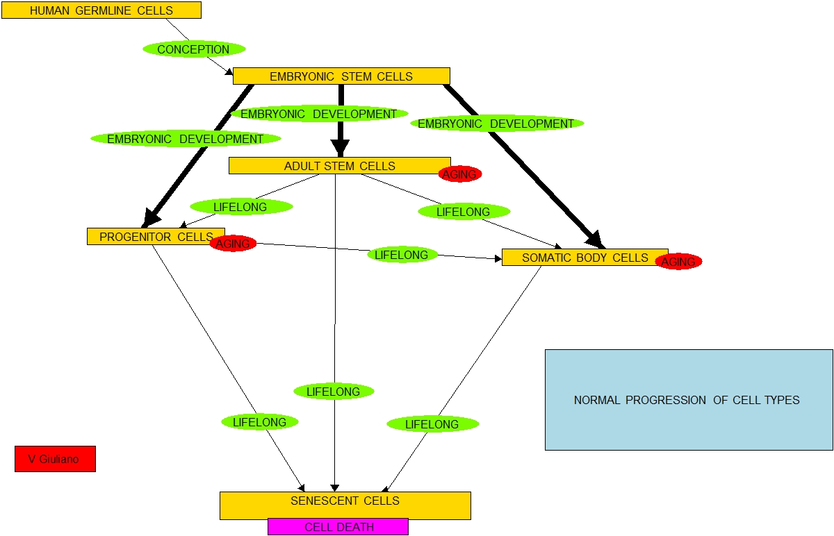
As mentioned, an evolutionary impact in most developing countries resulting from social evolution has been an epigenetically-mediated centuries-old trend to longer lives.  Our genes are the same but we have accommodated to living healthier longer lives.  Fast-food chains thrive, people consume excessive amounts of saturated and trans fats, epigenomes change and there is an inhereted trend towards obesity and diabetes, another trend in evolution.  We build decent cities in which air is clean and people do a lot of walking, and the result is an epigenetically-mediated healthier population.  The same is true for public education campaigns against cigarette smoking where they are carried out.  These biological trends in turn contribute to social trends towards enjoyment of affluence, marrying later in life, having fewer children and declining populations.  Such social-biological evolution exists in Europe, Japan and the US, and is beginning to become manifest in affluent parts of India and China.  So social evolution feeds back into our biological evolution and the other way around.  And the process is extremely complex confounding cause and effect at every stage.

All sciences have always been in a process of evolution.

I comment that the evolution of human evolution does not stand alone but is a component of evolution of the sciences known to govern the universe.  Approximately 13.7 billion years ago, at the initial instant after the big bang, matter, energy, space and time did not exist.  None of the sciences we know of today existed.  Ordinary matter did not exist so there were no laws of physics at work.  Atoms and molecules did not exist so no laws of chemistry were at work, and obviously since there were no living organisms, no laws of biology were in operation.  Each of these sciences emerged in a very crude form at a given time and then evolved rapidly at first and, later, slower.  I believe all are continuing to evolve now.  I am talking about the sciences themselves, the rules of nature and how things work, not about our understanding of them.

Image source

For the first 10–32of a second after the big-bang beginning, none of the current sciences existed.  It was too hot for even the most basic current laws of physics to be operable.  No operable laws of physics, chemistry, biology, sociology, or astronomy existed.  Nor anything that could predict what such laws would eventually look like. 

After the first 10–32of a second after the big-bang, a few of the concepts of very high-energy physics appear to be applicable, the universe being a quark-gluon mix.   Neither ordinary physics or other sciences existed.  Many laws of physics of ordinary cool matter did not fully emerge for another billion years when the universe had cooled.

Image source

The initial laws of chemistry could only emerge after the elements were created within stars and spun out in space leading later to the creation of molecules, starting about a billion years ago.  No laws of biology existed then.  Laws of organic chemistry came much later. 

The initial laws of biology could come into operation only after the origination of life, thought to be about 2 billion years ago on earth.  At any stage of the evolution of life it was impossible to predict what the future would bring.  The same, I believe, holds now.

Image source

Evolution and my motivation

The fact that social evolution can impact on biological evolution impels me to do the work of generating this blog,  I believe increasing awareness as to the possibility of significantly longer lives as well as practical lifestyle interventions to increase health now can and ultimately will assist our evolving to live longer and more productively in a situation of greater democracy and economic equality.  And I believe with longer, healthier and productive lives will ultimately come the wisdom and practical application of that wisdom necessary for societal survival and economic prosperity. 

I am suggesting a positive model for societal evolution.  There is also a negative one that is the opposite. 

We need to move as rapidly as possible in the opposite direction of that prevalent in Yemen, Somalia and other countries in Africa and the Middle East and some of our most distressed inner cities.  In those areas birth rates are very high, most of the population is under 22, lives are short and not worth much, education levels are very low, family structure is threatened, crime and lawlessness and armed gang warfare are the main ways of resolving issues, religious and tribal fanaticism and preoccupation with an enemy population is the norm, social infrastructure no longer functions, the ecology is less and less supportive of food and life, and starvation and hopelessness is the only outlook.

In the US our population’s background and the prevailing ethics are very mixed.  For some of us the rate of social evolution and outlook for health and longevity is very positive.  For others of us, there is real danger of slipping into the declining pattern described in the previous paragraph.  That is our real enemy.  Too much of the debate on these issues has been formulated in terms of polarized right-left politics which has resulted in deadlock.  I am suggesting casting the issue of our future in terms of our health and longevity.  See my blog post Social ethics of longevity.


Phytosubstances – focus on Andrographis, an old medicine with many possible new applications

$
0
0

By Vince Giuliano

This blog entry looks at recent research on Andrographis - a herbal substance that is a staple of traditional Chinese, Indian and South Asian medicine.  It has been used for centuries in countries like China, India, Thailand and Malaysia for the treatment of sore throat, flu and upper respiratory tract infections and snake bites.  Like certain other phytosubstances I have reported on earlier, andrographis has been studied almost only exclusively by Asian scientists and is little known in the Western world except for a few practitioners of herbal medicine and a highly select group of takers of such medicines.  As is the case for dashen root (salvia miltiorrhiza bunge) and gambogenic and gambogic acid, other important traditional Chinese medicines, the documented curative powers of andrographis seem to have escaped the attention of Western medical researchers.  Asian researchers, on the other hand, are seeking to base new therapies for cancer, asthma, stroke and arthritis on compounds in andrographis and their chemical analogs.

Image source

Traditional uses of Andrographis

According to WebMD: “Andrographis is a plant that is native to South Asian countries such as India and Sri Lanka. The leaf and underground stem are used to make medicine. — Andrographis is frequently used for preventing and treating the common cold and flu (influenza). Some people claim andrographis stopped the 1919 flu epidemic in India, although this has not been proven. — Andrographis is also used for a wide assortment of other conditions. It is used for digestive complaints including diarrhea, constipation, intestinal gas, colic, and stomach pain; for liver conditions including an enlarged liver, jaundice, and liver damage due to medications; for infections including leprosy, pneumonia, tuberculosis, gonorrhea, syphilis, malaria, cholera, leptospirosis, rabies, sinusitis, and HIV/AIDS; and for skin conditions including wounds, ulcers and itchiness. — Some people use andrographis for sore throat, coughs, swollen tonsils, bronchitis, and allergies. It is also used for “hardening of the arteries” (atherosclerosis), and prevention of heart disease and diabetes.– Other uses include treatment of snake and insect bites, loss of appetite, kidney problems (pyelonephritis), hemorrhoids, and an inherited condition called familial Mediterranean fever. — Andrographis is also used as an astringent, bacteria killing agent, painkiller, fever reducer, and treatment for worms. — Some Internet vendors offer andrographis products that contain extra amounts of an active ingredient called andrographolide. Some of these products are almost 30% andrographolide. However, be careful; the safety and effectiveness of andrographis preparations with high andrographolide content are unknown.”


Medicine Buddha Dharma  Website for image

Other names for Andrographis

Other names depending on culture and location include: “Andrographis Paniculata, Andrographolide, Bhunimba, Bidara, Carmantina, Carmantine, Chiretta, Chirette Verte, Chirreta, Chuan Xin Lian, Chuanxinlian, Chuan Xin Lin, Creat, Échinacée d’Inde, Fa-Tha-Lai-Jone, Fa-Tha-Lai-Jone, Gubak, Herba Andrographitis, Indian Echinacea, Justicia paniculata, Justicie, Kalamegha, Kalmegh, Kalmegha, Kariyat, King of Bitters, Kirta, Mahalita, Nabin Chanvandi, Poogiphalam, Roi des Amers, Sadilata, Sambilata, Shivaphala, Supari, Takila, Vizra Ufar, Yavatikta.(ref)” 

Andrographolide

Andrographolide is a labdanediterpenoid that is the main bioactive component of the medicinal plant Andrographis paniculata.[1] Andrographolide is an extremely bitter substance extracted from the stem and leaves of the andrographis paniculata, which is grown for medicinal purposes in China and India. — Because of its antiinflammatory, immunosuppressant and neuroprotective effects, andrographolide is used experimentally in different areas of research, including Cell signaling, immunomodulation and stroke.[2] (ref)

Andrographis like most other plant-based bioactive substances is quite chemically complex.  It contains many other bioactive components in addition to andrographoilide.

The 2011 publication [Chemical constituents from roots of Andrographis paniculata] reports “To investigate the chemical constituents of the roots of Andrographis paniculata, 28 compounds were isolated and identified from the 80% ethanol extract. There are 20 flavonoids: 5, 5′-dihydroxy-7, 8, 2′-trimetroxyflavone (1), 5-hydroxy-7, 8, 2′, 6′-tetramethoxyflavone (2), 5, 3′-dihydroxy-7, 8, 4′-trimethoxyflavone (3), 2′-hydroxy-5, 7, 8-trimethoxyflavone (4), 5-hydroxy-7, 8, 2′, 3′, 4′-pentamethoxyflavone (6), wightin (7), 5, 2′, 6′-trihydroxy-7-methoxyflavone 2′-O-beta-D-glucopyranoside (8), 5, 7, 8, 2′-tetramethoxyflavone (10), 5-hydroxy-7, 8-dimethoxyflavanone (11), 5-hydroxy-7, 8-dimethoxyflavone (12), 5, 2′-dihydroxy-7, 8-dimethoxyflavone (13), 5-hydroxy-7, 8, 2′, 5′-tetramethoxyflavone (14), 5-hydroxy-7, 8, 2′, 3′-tetramethoxyflavone (15), 5-hydroxy-7, 8, 2′-trimethoxyflavone (16), 5, 4′-dihydroxy-7, 8, 2′, 3′-tetramethoxyflavone (17), dihydroneobaicalein (18), andrographidine A (19), andrographidine B (20), andrographidine C (21) and 5, 2′-dihydroxy-7, 8-dimethoxyflavone 2′-O-beta-D-glucopyranoside (22); three diterpenoids: andrograpanin (23), neoandrographolide (24) and andrographolide (25); two phenylpropanoids: trans-cinnamic acid (26) and 4-hydroxy-2-methoxycinnamaldehyde (5); and oleanolic acid (9), beta-sitosterol (27) and beta-daucosterol (28). Compound 1 is a new flavone, compound 4 is a new natural product, compounds 2, 3 and 5 were isolated from the Andrographis genus for the first time and compounds 6-9 were isolated from this plant for the first time.”

Andrographis paniculata contains a number of additional bioactive compounds under study, as reported in the May 2012 publication: Rare noriridoids from the roots of Andrographis paniculata: “The rare noriridoids, Andrographidoids A-E (1-5), along with a known iridoid curvifloruside F (6), were isolated from roots of Andrographis paniculata. All noriridoids were aglycones and 1-4 had (semi-) acetal structures located at C-3 but not at C-1. Their structures were established by a series of 1D and 2D NMR analyses. The antibacterial activity of these iridoids was also assessed using the microtitre plate broth dilution method.”

Andrographolide is a powerful inhibitor of inflammation originated by tumornecrosis factor-α (TNF-α), blocks translocation of NF-kappaB into the nucleus and downregulates the P13K/Akt signaling pathways.  These are important themes relevant to many of the actions of andrographis including its anti-cancer activities and therapeutic value for inflammatory diseases..

For example, the November 2011 publicationInhibition of TNF-α-Induced Inflammation by andrographolide via down-regulation of the PI3K/Akt signaling pathwayreports: “Andrographolide (1), an active constituent of Andrographis paniculata, decreased tumor necrosis factor-α (TNF-α)-induced intercellular adhesion molecule-1 (ICAM-1) expression and adhesion of HL-60 cells onto human umbilical vein endothelial cells (HUVEC), which are associated with inflammatory diseases. Moreover, 1 abolished TNF-α-induced Akt phosphorylation. Transfection of an activated Akt1 cDNA vector increased Akt phosphorylation and ICAM-1 expression like TNF-α. In addition, 1 and LY294002 blocked TNF-α-induced IκB-α degradation and nuclear p65 protein accumulation, as well as the DNA-binding activity of NF-κB. Compound 1 exhibits anti-inflammatory properties through the inhibition of TNF-α-induced ICAM-1 expression. The anti-inflammatory activity of 1 may be associated with the inhibition of the PI3K/Akt pathway and downstream target NF-κB activation in HUVEC cells.”

There is significant interest in the anticancer activities of key compounds in andrographis and in basing new anticancer therapies in them.  For example, andrographolide inhibits the proliferation of human gliablastoma cells, at least in vitro.

The June 2012 publication Inactivation of PI3K/Akt signaling mediates proliferation inhibition and G2/M phase arrest induced by andrographolide in human glioblastoma cells relates: “AIMS: Andrographolide, a principal diterpenoid lactone isolated from the traditional herbal medicine Andrographis paniculata, has been reported to show anti-tumor activity. Since the high lipid solubility of andrographolide permits it to penetrate the blood-brain barrier and concentrate in the brain, we hypothesized that andrographolide may be a potential chemotherapeutic agent for the treatment of glioblastomas. To clarify this point, we investigated the growth inhibitory effect and mechanisms of actions of andrographolide on human glioblastoma U251 and U87 cells.  MAIN METHODS: MTT assay and trypan blue exclusion assay were used to investigate the proliferation inhibitory and cytotoxic effects of andrographolide, respectively. Cell cycle distribution was analyzed using flow cytometry. Apoptosis analysis proceeded by detecting the cleavage of caspase-3. The levels of proteins were probed by Western blotting.  KEY FINDINGS: The results showed that non-toxic concentrations of andrographolide inhibited the proliferation of human glioblastoma cells through induction of G2/M arrest, which was accompanied by down-regulating Cdk1 and Cdc25C proteins. Additionally, andrographolide decreased the activity of PI3K/Akt signaling, as demonstrated by down-regulation of the expression of phos-PI3K, phos-Akt, phos-mTOR and phos-p70s6k in U251 and U87 cells. Furthermore, additive effects on the proliferation inhibition, G2/M arrest and down-regulation of G2/M phase-related proteins were observed, when a combined treatment of andrographolide with PI3K inhibitor LY294002 was used in U251 and U87 cells.  SIGNIFICANCE: We prove that andrographolide inhibits the proliferation of human glioblastoma cells via inducing G2/M arrest, which is mediated by inhibiting the activity of PI3K/Akt signaling.”
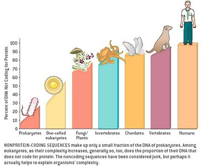
Image source

Figure 2: Effect of andrographolide treatment on cancer cells. Cancer is a multifaceted disease with complex processes and requires a multi-target therapeutic approach to battle it. A similar kind of action is displayed by andrographolide as it modulates various biochemical pathways of cancer cells thereby inhibiting the tumor growth. The compound exerts cytotoxic effect on various cancer cell types in a time and dose dependent manner. Factors required for tumor progression, nourishment and metastasis are down regulated, that is, cyclins A, D, Cdk2, Cdk4, NF-κB, VEGF, E-selectin, VCAM, Akt, TNF, Bcl2, and so forth. On the other hand tumor suppressor elements like p53, caspases, inhibitory proteins p21, p16, p27, and so forth are up regulated as observed in various studies to investigate anti-cancer potential of andrographolide. Up regulation of death receptor 4 to facilitate TRAIL induced apoptosis is of significant interest. The cumulative effect of all these factorial events leads to inhibition of growth in cancer cells.(ref)”

Yet another anti-cancer property of andrographolide is its capability to inhibit angiogenesis in tumors.

The July 2012 publication Andrographolide Inhibits Osteopontin Expression and Breast Tumor Growth Through Down Regulation of PI3 kinase/Akt Signaling Pathwayis one of several recent cell-level and small-animal-level experimental studies on the anticancer capabilities of andrographolide.  “Breast cancer is one of the most common cancers among women in India and around the world. Despite recent advancement in the treatment of breast cancer, the results of chemotherapy to date remain unsatisfactory, prompting a need to identify natural agents that could target cancer efficiently with least side effects. Andrographolide (Andro) is one such molecule which has been shown to possess inhibitory effect on cancer cell growth. In this study, Andro, a natural diterpenoid lactone isolated from Andrographis Paniculata has been shown to inhibit breast cancer cell proliferation, migration and arrest cell cycle at G2/M phase and induces apoptosis through caspase independent pathway. Our experimental evidences suggest that Andro attenuates endothelial cell motility and tumor-endothelial cell interaction. Moreover, Andro suppresses breast tumor growth in orthotopic NOD/SCID mice model. The anti-tumor activity of Andro in both in vitro and in vivo model was correlated with down regulation of PI3 kinase/Akt activation and inhibition of pro-angiogenic molecules such as OPN and VEGF expressions. Collectively, these results demonstrate that Andro may act as an effective anti-tumor and anti-angiogenic agent for the treatment of breast cancer.”
Andrographolide can potentiate the effectiveness of the cancer chemotherapy drug cisplatin in inducing apoptosis in cancer cells via suppression of autophagy.

The March 2012 publication Andrographolide sensitizes cisplatin-induced apoptosis via suppression of autophagosome-lysosome fusion in human cancer cells reports: “Suppression of autophagy has been increasingly recognized as a novel cancer therapeutic approach. Andrographolide (Andro), a diterpenoid lactone isolated from an herbal plant Andrographis paniculata, is known to possess antiinflammatory and anticancer activity. In this study, we sought to examine the effect of Andro on autophagy, and to evaluate whether such effect is relevant to the sensitization effect of Andro on apoptosis induced by DNA damage agents in cancer cells. First, we found that Andro is able to significantly enhance autophagic markers in various cancer cell lines, including GFP-LC3 puncta and LC3-II level. Interestingly, Andro treatment also led to marked increase of p62 protein level and addition of chloroquine (CQ) failed to further enhance either LC3-II or p62 level, indicating that Andro is likely to suppress autophagic flux at the maturation and degradation stage. Next, we provided evidence that Andro inhibits autophagosome maturation not by affecting the lysosomal function, but by impairing autophagosome-lysosome fusion. Lastly, we demonstrated that treatment with cisplatin, a DNA damage agent, induces autophagy in cancer cells. Importantly, Andro is capable of sensitizing cisplatin-induced cell killing determined with both short-term apoptosis assays and long-term clonogenic test, via suppression of autophagy, a process independent of p53. In summary, these observations collectively suggest that Andro could be a promising anti-cancer agent in combination therapy via its potent inhibitory effect on autophagy by disrupting autophagosome-lysosome fusion.”
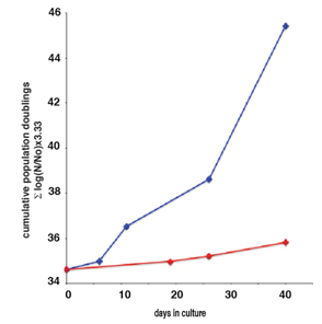
Studies are being conducted relating key compounds in andrographis to the gene-activation pathways they affect in types of cancer cells.  Some compounds works to kill cancer cells by inhibiting the cell cycle or promoting cell cycle arrest.

The February 2012 publicationIdentification of genes involved in the regulation of 14-deoxy-11,12-didehydroandrographolide-induced toxicity in T-47D mammary cells reports: “14-Deoxy-11,12-didehydroandrographolide is one of the principle compounds of the medicinal plant, Andrographis paniculata Nees. This study explored the mechanisms of 14-deoxy-11,12-didehydroandrographolide-induced toxicity and non-apoptotic cell death in T-47D breast carcinoma cells. Gene expression analysis revealed that 14-deoxy-11,12-didehydroandrographolide exerted its cytotoxic effects by regulating genes that inhibit the cell cycle or promote cell cycle arrest. This compound regulated genes that are known to reduce/inhibit cell proliferation, induce growth arrest and suppress cell growth. The growth suppression activities of this compound were demonstrated by a downregulation of several genes normally found to be over-expressed in cancers. Microscopic analysis revealed positive monodansylcadaverine (MDC) staining at 8h, indicating possible autophagosomes. TEM analysis revealed that the treated cells were highly vacuolated, thereby suggesting that 14-deoxy-11,12-didehydroandrographolide may cause autophagic morphology in these cells. This morphology may be correlated with the concurrent expression of genes known to affect lysosomal activity, ion transport, protein degradation and vesicle transport. Interestingly, some apoptotic-like bodies were found, and these bodies contained multiple large vacuoles, suggesting that this compound is capable of eliciting a combination of apoptotic and autophagic-like morphological characteristics.”

Andrographolide inhibits release of NF-kappaB from its binding factor IκB-α and its translocation into the nucleus and activation, and consequentially inhibits release of matrix metalloproteinase-9.  It may thus provide the basis for new therapies for inflammatory processes and certain cancers including leukemia.

The February 2012 e-publication Suppression of matrix metalloproteinase-9 expression by andrographolide in human monocytic THP-1 cells via inhibition of NF-κB activation reports: “There is much evidence indicating that human leukemic cells and monocytes/macrophages synthesize, and secrete, several matrix metalloproteinases (MMPs), and participate in the degradation of extracellular matrix components in tissue lesions. In this study, we investigated the effects and mechanisms of andrographolide, extracted from the herb Andrographis paniculata, on human monocytic MMPs expression and activation. Andrographolide (1-50 μM) exhibited concentration-dependent inhibition of MMP-9 activation, induced by either tumor necrosis factor-α (TNF-α), or lipopolysaccharide (LPS), in THP-1cells. In addition, andrographolide did not present an inhibitory effect on MMP-9 enzymatic activity at a concentration of 50 μM. By contrast, enzyme-linked immunosorbent assay (ELISA) showed that andrographolide partially affect TIMP-1 levels. Western blot analysis showed that both TNF-α, and LPS stimulators attenuated MMP-9 protein expression in a concentration-dependent manner. Using reverse transcription polymerase chain reaction (RT-PCR), we found that andrographolide suppressed expression of MMP-9 messenger RNA. Furthermore, we also found that andrographolide could significantly inhibit the degradation of inhibitor-κB-α (IκB-α) induced by TNF-α. We used electrophoretic mobility shift assay and reporter gene detection to show that andrographolide also markedly inhibited NF-κB signaling, anti-translocation and anti-activation. In conclusion, we demonstrate that andrographolide attenuates MMP-9 expression, and its main mechanism might involve the NF-κB signal pathway. These results provide new opportunities for the development of new anti-inflammatory and leukemic therapies.”

The October 2011 publication Andrographolide inhibits oral squamous cell carcinogenesis through NF-κB inactivation again highlights how inhibition of NF-kappaB is important for andrographolide’s capability to combat cancer.  It reports: “The NF-κB family of transcription factors is essential for promoting cell proliferation and preventing cell apoptosis. We have previously shown that Andrographolide (Andro) isolated from an herbal plant, Andrographis paniculata, covalently modifies reduced cysteine(62) in the oligonucleotide binding pocket of p50 for inhibition of NF-κB activation. Here we report that Andro, but not its inactive structural analog 4H-Andro, potently suppressed squamous cell carcinogenesis induced by 7,12-dimethyl-1,2-benzanthracene (DMBA) in the hamster model of cheek buccal pouch. Compared with 4H-Andro, Andro reduced phosphorylation of p65 (Ser536) and IκBα (Ser32/36) for inhibiting aberrant NF-κB activation, suppressed c-Myc and cyclin D1 expression and attenuated neoplastic cell proliferation, promoted cancerous cell apoptosis, and mitigated tumor-induced angiogenesis. Consistently, Andro retarded growth, decreased proliferation, and promoted apoptosis of Tb cells, a human tongue squamous cell carcinoma cell line, in time- and dose-dependent manners, with concomitant reduction of the expression of NF-κB targeting molecules in vitro. Our results thus demonstrate that NF-κB activation plays important roles in the pathogenesis of chemically induced squamous cell carcinoma. By inhibition of aberrant NF-κB activation, Andro treats chemically induced oral squamous cell carcinogenesis.”

Andrographolide suppresses activation of Nf-kappaB, inhibits the P38/MAPK pathway, activates Nrf2 and upregulates expression of heme oxygenase.  Implications include therapeutic potential for treating coronary heart diseases as well as cancers and multiple other diseases involving runaway inflammatory processes.

The July 2012 publicationSuppression of NF-κB signaling by andrographolide with a novel mechanism in human platelets: regulatory roles of the p38 MAPK-hydroxyl radical-ERK2 cascadereports: “Andrographolide, a novel nuclear factor-κB (NF-κB) inhibitor, is isolated from leaves of Andrographis paniculata. Platelet activation is relevant to a variety of coronary heart diseases. Our recent studies revealed that andrographolide possesses potent antiplatelet activity by activating the endothelial nitric oxide synthase (eNOS)-NO-cyclic GMP pathway. Although platelets are anucleated cells, they also express the transcription factor, NF-κB, that may exert non-genomic functions in platelet activation. Therefore, we further investigated the inhibitory roles of andrographolide in NF-κB-mediated events in platelets. In this study, NF-κB signaling events, including IKKβ phosphorylation, IκBα degradation, and p65 phosphorylation, were time-dependently activated by collagen in human platelets, and these signaling events were attenuated by andrographolide (35 and 75μM). ODQ and KT5823, respective inhibitors of guanylate cyclase and cyclic GMP-dependent kinase (PKG), strongly reversed andrographolide-mediated inhibition of platelet aggregation, relative [Ca(2+)]i mobilization, and IKKβ, and p65 phosphorylation. In addition, SB203580 (an inhibitor of p38 MAPK), but not PD98059 (an inhibitor of ERKs), markedly abolished IKKβ and p65 phosphorylation. SB203580, NAC (a free-radical scavenger), and BAY11-7082 (an inhibitor of NF-κB) all diminished ERK2 phosphorylation, whereas PD98059, BAY11-7082, and NAC had no effects on p38 MAPK phosphorylation. Furthermore, SB203580, but not BAY11-7082 or PD98059, reduced collagen-induced hydroxyl radical (()HO) formation. KT5823 also markedly reversed andrographolide-mediated inhibition of p38 MAPK and ERK2 phosphorylation, and hydroxyl radical formation in platelets. In conclusion, this study demonstrated that andrographolide may involve an increase in cyclic GMP/PKG, followed by inhibition of the p38 MAPK/()HO-NF-κB-ERK2 cascade in activated platelets. Therefore, andrographolide may have a high therapeutic potential to treat thromboembolic disorders and may also be considered for treating various inflammatory diseases.”

Based on its ability to inhibit the impact of tumor necrosis factor α on activation of NF-kappaB and upgrade expression of Nrf2 and heme oxygenase, andrographolide is protective against liver tissue injuries created by cytotoxins such as carbon tetrachloride or paracetamol.

The 2011 publication Protective mechanism of andrographolide against carbon tetrachloride-induced acute liver injury in mice reports: “The aim of this study was to investigate the protective effects of andrographolide (AP), a bioactive component isolated from Andrographis paniculata, on carbon tetrachloride (CCl(4))-induced liver injury as well as the possible mechanisms involved in this protection in mice. Acute liver injury was induced by CCl(4) intoxication in mice. Serum biological analysis, lipid peroxides and antioxidant estimation, histopathological studies, reverse transcription polymerase chain reaction (RT-PCR) and Western blot assay were carried out. CCl(4) treatment resulted in severe hepatic injury, as evidenced by significant elevation of alanine aminotransferase (ALT) and aspartate aminotransferase (AST) levels and typical histopathological changes, such as hepatocyte necrosis. Additionally, CCl(4) administration led to oxidative stress in mice, as indicated by a remarkable increase in the hepatic malondialdehyde (MDA) level, together with a significant decrease in liver reduced glutathione (GSH) content. However, CCl(4)-induced hepatotoxicity was significantly attenuated by pretreatment with AP, as demonstrated by significant reduction of serum ALT, AST levels and hepatic MDA activity, along with a remarkable increase in hepatic GSH content. Histopathological changes induced by CCl(4) were also ameliorated by AP pretreatment. The marked increase of – tumor necrosis factor α (TNF-α) induced by CCl(4) was attenuated by AP, and the dramatic elevation of heme oxygenase-1 (HO-1) at transcriptional and protein levels was augmented following AP pretreatment. AP can effectively prevent liver injury induced by CCl(4), which may be due to inhibition of oxidative stress and inflammatory responses.”

The December 2011 publication Hepatoprotective activity of Andrographis paniculata and Swertia chirayita reported: “ — Oral administration of A. paniculata or S. chirayita extract (100-200mg/kg) offered a significant dose dependent protection against paracetamol induced hepatotoxicity as assessed in terms of biochemical and histopathological parameters.  The paracetamol induced elevated levels of serum marker enzymes such as serum glutamate pyruvate transaminase (GPT), serum glutamate oxaloacetate transaminase (GOT), alkaline phosphatase (ALP), and bilirubin in peripheral blood serum and distorted hepatic tissue architecture along with increased levels of lipid peroxides (LPO) and reduction of superoxide dismutase (SOD), catalase, reduced glutathione (GSH) and glutathione peroxidase (GPx) in liver tissue. Administration of the plant extracts after paracetamol insult restored the levels of these parameters to control (untreated) levels. Thus the present study revealed that the extracts of A. paniculata or S. chirayita offered protection against hepatotoxicity induced by paracetamol.”

Andrographolide appears to be gastroprotective; it is protective against certain duodenal ulcers in rats.

The September 2011 publication Effect of andrographolide on cysteamine-induced duodenal ulcer in rats reports: “The aim of this study was to evaluate the gastroprotective efficacy of andrographolide isolated from Andrographis paniculata in rats induced with duodenal ulcers. Duodenal ulcers were induced by cysteamine administration in rats pretreated with 3mgkg¹ BWday¹ of andrographolide for 30 days. Ulcer score, myeloperoxidase activity, TBARS level, GSH/GSSG ratio and enzyme antioxidants were measured in the duodenal tissue. Brush border and basolateral membranes were isolated to assay sucrase, maltase, alkaline phosphatase and total ATPases. Ulcer score was significantly minimised in rats pretreated with andrographolide. Elevation in myeloperoxidase and TBARS levels were found to be minimised significantly due to andrographolide treatment. Membrane-bound enzyme activities and the thiol redox status of glutathione were significantly maintained in duodenal mucosa of rats that received andrographolide. This study reveals that the major component of A. paniculata, andrographolide, has potent antiulcer properties that are most likely caused by minimising inflammatory changes, counteracting free radical formation and maintaining the thiol redox status in the duodenum.”

The July 2012 publication A biochemical study on the gastroprotective effect of hydroalcoholic extract of Andrographis paniculata in rats speaks to the same point: “Aim: The aim of the present study is to evaluate the gastroprotective effect of hydroalcoholic extract of Andrographis paniculata (HAEAP) in male albino wistar rats.  Materials and Methods: Rats were pretreated with HAEAP (100,200,500mg/kg b. wt for 30 days) and then gastric ulcers were induced by ethanol, aspirin, pylorus ligation and cold restraint stress models. Ulcer score was determined in all the ulcer models. pH, gastric volume, titrable acidity, pepsin, mucin, myeloperoxidase, H+K+ATPase, thiobarbituric acid reacting substances (TBARS) and antioxidant enzyme activities were assayed in ethanol-administered rats. Results: The ulcer score was found to be low in HAEAP-pretreated rats. Among the doses studied, 200 mg/kg b.wt was found to be optimum for significant ulcer reduction. The test drug significantly reduced the acidity, pepsin concentration, myeloperoxidase and H+K+ATPase activities in ethanol-administered rats. The elevated TBARS and decreased glutathione (GSH) and mucin levels observed during ulcerogenesis were found to be altered in HAEAP-received animals. Conclusions: The ulcer preventing effect of HAEAP may partly be due to its regulating effect on H+K+ATPase activity and /or mucin preserving effects. The flavonoids present in the HAEAP might be responsible for the gastroprotective action probably by maintaining the antioxidants and thiol status in the gastrointestinal tract.”

Again relating to the antiulcer capabilities of andrographis is the July 2011 publication Antiulcer activity of Andrographis paniculata (Burm.f.) wall. against cysteamine-induced duodenal ulcer in rats.

Components of andrographis interact with at least one member of the cytochrome P450 family implying that consumption of andrographis may generate a risk of interactions with certain drugs or an enhanced risk of lung cancer in the case of smokers.

The December 2011 publication Different AhR binding sites of diterpenoid ligands from Andrographis paniculata caused differential CYP1A1 induction in primary culture in mouse hepatocytes reports “Andrographis paniculata has been employed as a folklore remedy. Andrographolide (Andro), 14-deoxy-11,12-didehydroandrographolide (DHA), andrographiside (AS), and neoandrographolide (Neo), are major diterpenoids isolated from this plant. In the present study, influence of the four diterpenoids on CYP1A1 mRNA expression was investigated in primary cultured mouse hepatocytes. Additionally, binding of these compounds to aryl hydrocarbon receptor (AhR) was examined using molecular docking analysis to clarify mechanism of CYP1A1 induction. Andro and DHA induced CYP1A1 expression by itself, and co-treatment with a CYP1A1 inducer (BNF, beta-naphthoflavone) showed a synergistic increase of CYP1A1 expression. Andro demonstrated higher enhancing activity than DHA at every similar concentration. On the other hand, Neo suppressed BNF-induced CYP1A1 expression, but AS did not modify the induction. Results from molecular docking analysis of BNF and four diterpenoids on ligand binding domain of AhR were consistent with levels of CYP1A1 mRNA expressions. Furthermore, difference of binding sites of BNF in the presence of diterpenoids might affect the synergism or inhibition of CYP1A1 expression. These results suggest that use of A. paniculata as a health supplement should be concerned in term of herb-drugs interactions or risk of carcinogenesis, according to its ability to influence CYP1A1 expression.”

Regarding CPY1A1: “This gene, “CYP1A1, encodes a member of the cytochrome P450 superfamily of enzymes. The cytochrome P450 proteins are monooxygenases which catalyze many reactions involved in drug metabolism and synthesis of cholesterol, steroids and other lipids. This protein localizes to the endoplasmic reticulum and its expression is induced by some polycyclic aromatic hydrocarbons (PAHs), some of which are found in cigarette smoke. The enzyme’s endogenous substrate is unknown; however, it is able to metabolize some PAHs to carcinogenic intermediates. The gene has been associated with lung cancer risk(ref).”

Effectiveness of andrographolide as a treatment is compromised by its poor oral bioavailability.

The November 2011 publication Poor oral bioavailability of a promising anticancer agent andrographolide is due to extensive metabolism and efflux by P-glycoprotein reports: “Andrographolide (AP), isolated from Andrographis paniculata (Burm. F.) Nees, is an anticancer agent with significant clinical potential. This study determined its oral bioavailability and how intestinal disposition affects its bioavailability. Pharmacokinetics was evaluated in rats. Intestinal disposition was determined using a single-pass rat intestinal perfusion model and the cultured Caco-2 cells and Madin-Darby canine kidney II cells over expressing human P-gp (MDR1-MDCKII). Absolute bioavailability of AP was 2.67%. In the duodenum and jejunum, AP was rapidly metabolized to a sulfonate, identified as 14-deoxy-12-sulfo- andrographolide. AP was also rapidly metabolized by liver S9 fraction and in blank perfusates collected from duodenum and jejunum. The apparent permeability (P(app) ) of AP from basolateral (B) to apical (A) (4.94 × 10 cm/s) in the Caco-2 model was four times higher than the P(app) from A to B (1.14 × 10(-5) cm/s). Moreover, AP was significantly more permeable in the B to A direction than the opposite direction in MDR1-MDCKII cells. In the perfusion model, the effective permeability (P*(eff) ) for AP was highest in the duodenum, followed by jejunum, and then ileum and colon. In the ileum and colon, the P*(eff) for AP was significantly increased by verapamil, a P-glycoprotein (P-gp) inhibitor. AP has poor oral bioavailability because of its rapid biotransformation and efflux by P-gp.”

A less-cytotoxic analog of Andrographolide may provide the basis for a new therapy for asthma.

The June 2011 publication Protective role of 14-deoxy-11,12-didehydroandrographolide, a noncytotoxic analogue of andrographolide, in allergic airway inflammationreports:  “Our group recently reported novel anti-inflammatory effects of andrographolide (2), a bioactive molecule isolated from Andrographis paniculata, in a mouse asthma model. However, 2 has been shown to possess cytotoxic activity. 14-Deoxy-11,12-didehydroandrographolide (1) is an analogue of 2 that can be isolated from A. paniculata. We hypothesized that 1 retains the anti-inflammatory effects for asthma but is devoid of cytotoxicity. In contrast to 2, 1 did not elicit any cytotoxic activity in A549 and BEAS-2B human lung epithelial cells and rat basophilic leukemia (RBL)-2H3 cells using a MTS assay. Compound 1 dose-dependently inhibited ovalbumin (OVA)-induced increases in total and eosinophil counts, IL-4, IL-5, and IL-13 levels in lavage fluid, and serum OVA-specific IgE level in a mouse asthma model. Compound 1 attenuated OVA-induced airway eosinophilia, mucus production, mast cell degranulation, pro-inflammatory biomarker expression in lung tissues, and airway hyper-responsiveness. This substance also blocked p65 nuclear translocation and DNA-binding activity in the OVA-challenged lung and in TNF-α-stimulated human lung epithelial cells. The present findings reveal for the first time that 1 retains the anti-inflammatory activities of 2 for asthma probably through the inhibition of NF-κB. 14-Deoxy-11,12-didehydroandrographolide (1) may be considered as a safer analogue of 2 for the potential treatment of asthma.”

Topical application of andrographis appears to accelerate wound healing, at least in rats.

The June 2012 publication Effect of Andrographis paniculata leaf extract on wound healing in rats reports: “This work was carried out to study the effect of topical application of Andrographis paniculata on the rate of wound enclosure and its histological features. A wound was created in four groups of rat in posterior neck region. Blank placebo was applied topically to the wounds of Group 1. Groups 2 and 3 were dressed with placebo containing 5% and 10% extracts of A. paniculata, respectively. Intrasite gel was applied topically to the wounds of Group 4. Macroscopical examination revealed that the rate of wound healing was significantly accelerated in the wound dressed with A. paniculata extract compared to the blank placebo. The wounds dressed with 10% extract or Intrasite gel healed earlier compared to the wounds dressed with placebo containing 5% A. paniculata extract. Histologically, wounds dressed with A. paniculata extracts showed markedly less scar width and contained large amounts of fibroblast proliferation. More collagen and less angiogenesis with absence of inflammatory cells were seen for wounds dressed with 10% A. paniculata compared to the blank placebo. Conclusion, A. paniculata extracts significantly enhanced rate of wound healing in rats.”

A combined extract of andrographis and danshen together with swimming in a rat model appears to be effective for prevention and treatment of atherosclerosis.

In December 2011 I wrote a comprehensive blog entry on danshen, another important traditional Chinese herbal medicine Focus on phytosubstances – Danshen root – amazing properties of salvia miltiorrhiza Bunge.

The 2011 publicationJoint preventive effects of swimming and Shenlian extract on rat atherosclerosis reports: “This work is to investigate the joint effect of extract from Shenlian (SL, the Chinese abbreviation for Radix Salviae miltiorrhizae and Andrographis paniculata) and swimming on atherosclerosis prevention and treatment. Atherosclerotic rat model was established by combining low shear stress by partial ligation of common carotid artery with afterwards feeding of a hyperlipotic diet. Sprague-Dawley rats after partial ligation of common carotid artery were allotted to a 3 × 3 factorial design with three levels of swimming (0, 1, and 2 hr per day) and three levels of SL extract (0, 2.56, and 5.12 g/kg once daily p.o.) for a total of 9 treatment groups. Then, the feeding of the hyperlipotic diet and the intervention of swimming and SL started at the same time, and lasted for 8 weeks. By the end, blood samples were collected to determine blood viscosity, hematocrit, blood lipids, MCP-1, NF-κB and NO levels. The common carotid arteries of the rats were harvested to investigate pathological changes. The animal model showed early sign of atherosclerosis according to the pathological findings. Joint effects of SL extract and swimming on preventing atherosclerosis appeared significantly: The combination of 1 hour swimming with 2.56 g/kg SL extract showed to be effective for lowering hematocrit, blood viscosity (at 10 s(-1) and 200 s(-1)) and low-density-lipoprotein (p < 0.001). Combined treatment of 2.56 g/kg SL extract with 2 hr swimming led to a synergistic decrease in serum level of MCP-1. As a single factor, SL extract (2.56 g/kg) alone could decrease serum levels of NF-κB significantly (p = 0.003). Swimming alone could decrease cholesterol, triacylglycerols level and increase high-density-lipoprotein. The study demonstrates the combined therapy of oral SL extract with swimming on inhibiting inflammatory factors, improving hemorheological parameters and lipoproteins in rat model of atherosclerosis. It highlights the promising effects of the drugs and shear stress of blood flow, the biomechenopharmacological means, for prevention of atherosclerosis.”

There is considerable activity in developing specialized analogs of Andrographolide that are either more potent in killing cancer cells, less cytotoxic to normal tissues, or have enhanced bioavailability.

The January 2012 publication New substituted C-19-andrographolide analogues with potent cytotoxic activities reports: “Andrographolide, the major diterpenoid lactone from Andrographis paniculata, is toxic against cancer cells. In the present study, we investigated the structure-activity relationships (SARs) of 19 andrographolide analogues which were synthesized by modification at the three hydroxyl groups. A number of the andrographolide analogues showed much higher cytotoxic activities than that of the parent compound on cancer cells including P-388, KB, COL-2, MCF-7, LU-1 and ASK cells. SAR studies of the synthetic analogues indicated that the introduction of silyl ether or triphenylmethyl ether group into C-19 of the parent compound led to increase in toxicity against the cancer cells. The 19-O-triphenylmethyl ether analogue 18 showed higher cytotoxic activity than the potent anticancer drug ellipticine, and this analogue may serve as a potential structure lead for the development of new anticancer drugs.”

The March 2012 publication Andrographolide and its analogues: versatile bioactive molecules for combating inflammation and cancer reports: “1.Andrographis paniculata (Burm. f) Nees, commonly known as ‘king of bitters’, is a herbaceous plant belonging to the Family Acanthaceae. It has been widely used for centuries in Asian countries like China, India, Thailand and Malaysia for the treatment of sore throat, flu and upper respiratory tract infections. 2.Andrographolide, 14-deoxy-11,12-didehydroandrographolide and neoandrographolide are examples of the major labdane diterpenoids isolated from A. paniculata. These bioactive molecules have exhibited varying degrees of anti-inflammatory and anticancer activities in both in vitro and in vivo experimental models of inflammation and cancer. 3.Extensive libraries of andrographolide analogues have been synthesised mainly by modifying the α,β-unsaturated γ-butyrolactone moiety, the two double bonds Δ(8,(17)) and Δ(12,(13)) and the three hydroxyls at C-3 (secondary), C-14 (allylic) and C-19 (primary). Many of these synthetic analogues exhibit superior anticancer activity over the naturally occurring andrographolides. 4.Andrographolide and its derivatives have been shown to have anti-inflammatory effects in experimental models of asthma, stroke and arthritis, as well as in patients with upper respiratory tract infections. Andrographolide reduces the production of cytokines, chemokines, adhesion molecules, nitric oxide and lipid mediators, probably via inhibition of the nuclear factor (NF)-κB signalling pathway. 5.The anticancer mechanisms for andrographolide include inhibition of Janus tyrosine kinases-signal transducers and activators of transcription, phosphatidylinositol 3-kinase and NF-κB signalling pathways, suppression of heat shock protein 90, cyclins and cyclin-dependent kinases, metalloproteinases and growth factors, and the induction of tumour suppressor proteins p53 and p21, leading to inhibition of cancer cell proliferation, survival, metastasis and angiogenesis. 6.Andrographolide drug discovery is a promising strategy for the development of a novel class of anti-inflammatory and anticancer drugs.”

Andrographis can provide significant protection against gastric ulcers.

The November 2011 publication Antioxidant and gastroprotective activities of Andrographis paniculata (Hempedu Bumi) in Sprague Dawley rats reports: “Antioxidant and gastroprotective activities of aqueous and ethanolic extract of Andrographis paniculata leaves in rats have been reported. Sprague Dawley rats, 6 per group were used and rats in groups 1 to 6 were pretreated with (0.25% w/v) carboxymethyl cellulose (negative control, 5 ml/kg), 20 mg/kg omeprazole (positive control), (250 mg/kg and 500 mg/kg) of aqueous leaf extracts (APLAE) and (250 and 500 mg/kg) of ethanol leaf extracts (APLEE) respectively. Animals were orally administered with 95% ethanol (5 ml/kg) 60 min after their pretreatments. Rats were sacrificed 1 h after treatment and gastric contents were collected to measure pH and mucous weight. Stomach was analyzed for gross and histological changes. Ulcer control group showed extensive lesions of gastric mucosal layer, whereas rats pretreated with omeprazole, 250 and 500 mg/kg of APLAE showed significant and dose dependent reduction in gastric lesions with increased pH and mucus content of stomach. Rats pretreated with 250 or 500 mg/kg of APLEE showed significantly better inhibition of gastric mucosal lesions. Further, the in vitro antioxidant studies using 2,2-diphenyl-1-picrylhydrazyl (DPPH) assay showed that ethanol extracts have superior free radical scavenging activity with IC50 value = 10.9 than aqueous extracts with IC50 value = 24.65. Results of this study showed that pretreatment with ethonolic extract of A. paniculata ethanolic provided significant protection against gastric ulcer by regulating of pH, mucous production and antioxidant property.”

Andrographolide may be useful for the treatment of rheumatoid arthritis.

The February 2012 publication Andrographolide induces cell cycle arrest and apoptosis in human rheumatoid arthritis fibroblast-like synoviocytes reports:  “The pseudo-tumoral expansion of fibroblast-like synoviocytes is a hallmark of rheumatoid arthritis (RA), and targeting rheumatoid arthritis fibroblast-like synoviocytes (RAFLSs) may have therapeutic potentials in this disease. Andrographolide, a diterpenoid compound isolated from the herb Andrographis paniculata, has been reported to have potent anti-inflammatory activity. In the present study, we aimed to investigate the effects of andrographolide on human RAFLSs and the underlying molecular mechanism(s). RAFLSs were isolated from patients with RA and treated with or without various concentrations (i.e., 10, 20, and 30 μM) of andrographolide for 48 h. 3-[4,5-Dimethyl-2-yl]-2,5-diphenyl tetrazolium bromide assay revealed that andrographolide treatment decreased the proliferation of RAFLSs in a dose-dependent manner. Cell cycle analysis using propidium iodide (PI) staining showed a G0/G1 cell cycle arrest in andrographolide-treated RAFLSs. Immunoblotting analysis of key cell cycle regulators demonstrated that andrographolide treatment caused a dose-dependent increase in the expression of cell-cycle inhibitors p21 and p27 and a concomitant reduction of cyclin-dependent kinase 4. Exposure to andrographolide-induced apoptosis of RAFLSs measured by annexin V/PI double staining, which was coupled with promotion of cytochrome C release from mitochondria and activation of caspase-3. Moreover, andrographolide-treated RAFLSs displayed a significant decrease in the Bcl-2/Bax ratio compared to untreated cells. In conclusion, our data demonstrate that andrographolide exerts anti-growth and pro-apoptotic effects on RAFLSs, thus may have therapeutic potential for the treatment of RA.”

Andrographolide may be the basis for new generations of mosquito killing and repellant agents, possibly providing an important new defense against malaria.

I know it may seem ridiculous to add this claim to the many others related to andrographis.  However the May 2912 publication Adulticidal and repellent properties of indigenous plant extracts against Culex quinquefasciatus and Aedes aegypti (Diptera: Culicidae)  reports: “Several diseases are associated to the mosquito-human interaction. Mosquitoes are the carriers of severe and well-known illnesses such as malaria, arboviral encephalitis, dengue fever, chikunguniya fever, West Nile virus, and yellow fever. These diseases produce significant morbidity and mortality in humans and livestock around the world. The adulticidal and repellent activities of crude hexane, ethyl acetate, benzene, chloroform, and methanol extracts of leaf of Eclipta alba and Andrographis paniculata were assayed for their toxicity against two important vector mosquitoes, viz., Culex quinquefasciatus and Aedes aegypti (Diptera: Culicidae). The adult mortality was observed after 24 h of exposure. All extracts showed moderate adulticide effects; however, the highest adult mortality was found in methanol extract of A. paniculata against the adults of C. quinquefasciatus and A. aegypti with the LC(50) and LC(90) values were 149.81, 172.37 ppm and 288.12, 321.01 ppm, respectively. The results of the repellent activity of hexane, ethyl acetate, benzene, chloroform, and methanol extract of E. alba and A. paniculata plants at three different concentrations of 1.0, 2.5, and 5.0 mg/cm(2) were applied on skin of forearm in man and exposed against adult female mosquitoes. In this observation, these two plant crude extracts gave protection against mosquito bites without any allergic reaction to the test person, and also, the repellent activity is dependent on the strength of the plant extracts. These results suggest that the leaf solvent plant extracts have the potential to be used as an ideal ecofriendly approach for the control of mosquitoes. This is the first report on the mosquito adulticidal and repellent activities of the reported E. alba and A. paniculata plants.”

The September 2011 report Efficacy of indigenous plant extracts on the malaria vector Anopheles subpictus Grassi (Diptera: Culicidae)concludes similarly: “BACKGROUND &#38; OBJECTIVES: Mosquito control is facing a threat due to the emergence of resistance to synthetic insecticides. Insecticides of plant origin may serve as suitable alternative biocontrol techniques in the future. The purpose of the present study was to assess the ethyl acetate, acetone and methanol extracts of Andrographis paniculata, Eclipta prostrata and Tagetes erecta leaves tested for oviposition-deterrent, ovicidal and repellent activities against malaria vector, Anopheles subpictus Grassi (Diptera: Culicidae). — CONCLUSIONS: The acetone extract of A. paniculata, methanol extract of E. prostrata and T. erecta showed good oviposition-deterrent, ovicidal and repellent activities respectively. These results suggest that the leaf extracts of A. paniculata, E. prostrata and T. erecta may have the potential to be used as an ideal eco-friendly approach for the control of the An. subpictus.”

The March 2011 publication Evaluation of Andrographis paniculata Burm.f. (Family:Acanthaceae) extracts against Culex quinquefasciatus (Say.) and Aedes aegypti (Linn.) (Diptera:Culicidae) speaks again to the same point regarding mosquitos: “CONCLUSIONS: From the results it can be concluded the crude extract of A. paniculata was a potential for controlling Cx. quinquefasciatus and Ae. aegypti mosquitoes.”

Besides possibly providing a powerful agent against malaria-bearing mosquitoes, Andrographolide combined with curcumin might also provide the basis for a powerful anti-malaria therapy for those infected with Plasmodium malaria parasites.

The June 2011 publication Andrographolide: A Novel Antimalarial Diterpene Lactone Compound from Andrographis paniculata and Its Interaction with Curcumin and Artesunate reports: “Andrographolide (AND), the diterpene lactone compound, was purified by HPLC from the methanolic fraction of the plant Andrographis paniculata. The compound was found to have potent antiplasmodial activity when tested in isolation and in combination with curcumin and artesunate against the erythrocytic stages of Plasmodium falciparum in vitro and Plasmodium berghei ANKA in vivo. IC50s for artesunate (AS), andrographolide (AND), and curcumin (CUR) were found to be 0.05, 9.1 and 17.4μM, respectively. The compound (AND) was found synergistic with curcumin (CUR) and addictively interactive with artesunate (AS). In vivo, andrographolide-curcumin exhibited better antimalarial activity, not only by reducing parasitemia (29%), compared to the control (81%), but also by extending the life span by 2-3 folds. Being nontoxic to the in vivo system this agent can be used as template molecule for designing new derivatives with improved antimalarial properties.”

Andrographis can partially neutralize poisonous snake and scorpion venom.

See the 2011 publications Anti-cobra venom activity of plant Andrographis paniculata and its comparison with polyvalent anti-snake venom,Partial in vitro and in vivo red scorpion venom neutralization activity of Andrographis paniculataand Antihaemolytic and snake venom neutralizing effect of some Indian medicinal plants.

Comments

This blog could cite only a representative sample of the current literature coming out of Asia on andrographis and its constituent compounds.  A Pubmed search on andrographis reveals 423 research publications, a significant portion of which written in the last 3-4 years.

Andrographis appears to be a complex substance with a remarkably large number of both traditional and potential new medical and health applications ranging from treatments of cancers to treatments of toxic liver conditions, rheumatoid arthritis, malaria, and gastric ulcers.  It wards off and kills mosquitos and is helpful in cases of venomous snakebites and scorpion stings.

The themes of inhibition of NF-kappaB and activation of Nrf2 emphasized in this blog entry as health-producing strategies are not at all new to my writings.  In my online treatise ANTI-AGING FIREWALLS – THE SCIENCE AND TECHNOLOGY OF LONGEVITYand in previous blog entries I have discussed how inhibition of NF-kappaB and activation of Nrf2 seem to go hand-in-hand in providing a variety of health and possibly anti-aging effects.  Further I have discussed how a variety of phytochemicals that are traditional medicines, like curcumin and ginger, work to do this.  Andrographis seems to be another good example of such a phytochemical.  See the blog entry The pivotal role of Nrf2. Part 2 – foods, phyto-substances and other substances that turn on Nrf2.

I am left with a number of disquieting questions.  With particular reference to andrographis:

  1. Given that:
    1. it appears the Asian scientists who have investigated andrographis and its compounds have drawn on the current research, laboratory tools, theories and thought processes of Western biological and medical sciences and have arrived responsibly at their findings.  It appears they are highly educated in current Western biological sciences and know what they are doing, and given
    2. Asian scientists have repeatedly mentioned the great potential for developing medical therapies based on andrographis and its constituent compounds for cancers and a number of other serious disease conditions of aging,
  2. Why then:
    1. Does it appear that Western scientists and especially those in drug companies have paid no attention to andrographis and its compounds?  These substances just don’t appear to show on the radar screens of Western biological and medical research institutions or in any of the publications emanating from them.
    2. Why have big Western pharma companies not seized on the possibility of developing proprietary analogs of andrographis constituent compounds for particular medical conditions – at this time when traditional drug discovery approaches are running dry.  (Only one clinical trial has been listed for andrographis or andrographolide, and that was completed in 2008.)

The identical questions could be asked about two other important traditional Asian medical substances I have reported on previously  dashen root and gambogenic and gambogic acid.  And the same questions probably can be asked about a number of other traditional folk medicines that have been recently subject to Asian scientific scrutiny. 

Am I missing something or is there a significant gap in East-West medical scientific research?  Information written in English about Asian herbal research is readily available to us in the West.  Why do Western researchers appear to pay no attention to it? What is it that leads us in the West to keep ignoring Asian herbal research while such ignorance does not exist in Asia regarding what we are doing in our labs here in the US?  Does the problem exist because of how our research and how Asian research are funded, because of the research review process where more of the same is the best way to get funded, or because of the nature on academic research institutions?  

Further, why are we here in the West so preoccupied with largely unsuccessful attempts to find magic-bullet kinase inhibitors to cure diseases, simple substances that work on single well-defined pathways [See Victor’s blog entry Kinase Inhibition – A Magic Bullet?], while many Asian researchers are working with impossibly complex natural substances like andrographis long-known to be effective, substances that exercise multiple actions on multiple pathways?  Is the issue simply a philosophical one of Western reductionism vs. Asian holism?


Adaptogens Part 1 – video blog

$
0
0

By Vince Giuliano

This and the Part 2 blog entry are pursuant to one of the key themes of this blog –  the use of phyto substances to promote health and longevity.  Specifically, they are about adaptogens – certain plant substances used in traditional and modern herbal medicine.   The two entries treat adaptogens from two viewpoints.  First, the viewpoint of a practicing herbalist Madelon Hope is conveyed via a video presentation.  Second, the viewpoint of current scientific research is covered in my usual manner: I cite a number of recent research citations relating to adaptogens involving their mechanisms of action and their therapeutic potential.  This blog discusses adaptogens in general.  The following  blog entry Adaptogens Part2 – focus on rhodiola is concerned with one adaptogen of particular interest and power.  Most of my personal commentary is in that blog entry.

Madelon Hope is Director of the Boston School of Herbal Studies and a long-time practicing herbalist.    I asked her for an informal interview which I recorded on a tiny video camera.  The first part of the interview is on adaptogens in general and is included here.  The second part of the interview is included in the Adaptogens Part 2 entry.

MadelonHopePt1 from VinceGiuliano on Vimeo.

From Wikipedia: “An adaptogen is a metabolic regulator which increases the ability of an organism to adapt to environmental factors, and to avoid damage from such factors. Environmental factors can be either physiological (external), such as injury or aging, or psychological (internal), such as anxiety. — An adaptogen must have a normalizing effect, i.e. counteracting or preventing disturbances to homeostasis brought about by stressors. Moreover, it must be innocuous with a broad range of therapeutic effects without causing any major side effects. The adaptogen concept does not fit easily into the Western model of medicine.  — According to the original definition adaptogens are non-specific remedies that increase resistance to a broad spectrum of harmful factors – “stressors” – of different physical, chemical and biological natures.[1][2][4]  – This definition has been updated and today adaptogens are defined as a “new class of metabolic regulators which increase the ability of an organism to adapt to environmental factors and to avoid damage from such factors.”[2][4] — Despite an extensive amount of research in the USSR, (by 1984, more than 1,500 pharmacological and clinical published studies),[4] the concept is not generally recognized in Western countries as it seemed to be in contrast to some of the key concepts of modern pharmacology: potency, selectivity and with efficacy balanced by an accepted level of toxicity.[2][3] In 1998, however, the term adaptogen was allowed as a functional claim for certain products by US Food and Drug Administration and it is now a generally accepted concept,[2] also by the European Medicines Agency and EFSA.[5][6][7(ref)”

Most of the research literature relating to adaptogens originates in Asia or Europe; relatively little comes from the US or Canada.  Among the topics treated in the literature are actions of adaptogens in the cases of immune system response, energy metabolism, endurance exercise performance, acute lung injury, ischemia-reperfusion injury, early left ventricular regional systolic dysfunction, asthma, metabolic syndrome, type 2 diabetes, Parkinson’s disease, mental and physical fatigue, hypoxia and acute mountain sickness, hypoglycemia, toxicity-induced apoptosis, inflammatory cytokinesm DNA repair, heat shock protein interactions, particular bacteria including propionibacterium, and multiple types of cells including those of the liver and heart, cortical neurons, neuroglia and certain cancer cells.  Most of the studies reported recently are on the molecular biology, cell, and small-animal levels.  Although I have seen many mentions of clinical trials these appear to be mostlyolder, specialized and conducted on a small scale.  The concept of adaptogens appears to be unique to herbal medicine and mainly not recognized from the viewpoint of Western allopathic medicine.

I cite a few citations applicable to adaptogens in general here.  Since a great deal of the research on adaptogens is focused on rhodiola, the following blog entry cites a more interesting and extensive collection of publications.

Chinese Medicine and Ayurvedic medicine, the two most comprehensive schools of traditional medicine going back thousands of years, each recognizes a number of what are now called adaptogens. 

The 2010 review  publication Scientific basis for the use of Indian ayurvedic medicinal plants in the treatment of neurodegenerative disorders: ashwagandha is interesting in that  it discusses an important adaptogenic herb in the historical context of Ayurvedic medicine.  “Scientific basis for the use of Indian ayurvedic medicinal plants in the treatment of neurodegenerative disorders: ashwagandha.  “Ayurveda is a Sanskrit word, which means "the scripture for longevity". It represents an ancient system of traditional medicine prevalent in India and in several other south Asian countries. It is based on a holistic view of treatment which is believed to cure human diseases through establishment of equilibrium in the different elements of human life, the body, the mind, the intellect and the soul [1]. Ayurveda dates back to the period of the Indus Valley civilization (about 3000 B.C) and has been passed on through generations of oral tradition, like the other four sacred texts (Rigveda, Yajurveda, Samaveda and Atharvanaveda) which were composed between 12(th) and 7(th) century B.C [2, 3]. References to the herbal medicines of Ayurveda are found in all of the other four Vedas, suggesting that Ayurveda predates the other Vedas by at least several centuries. It was already in full practice at the time of Buddha (6(th) century B.C) and had produced two of the greatest physicians of ancient India, Charaka and Shushrutha who composed the basic texts of their trade, the Samhitas. By this time, ayurveda had already developed eight different subspecialties of medical treatment, named Ashtanga, which included surgery, internal medicine, ENT, pediatrics, toxicology, health and longevity, and spiritual healing [4]. Ayurvedic medicine was mainly composed of herbal preparations which were occasionally combined with different levels of other compounds, as supplements [5]. In the Ayurvedic system, the herbs used for medicinal purposes are classed as brain tonics or rejuvenators. Among the plants most often used in Ayurveda are, in the descending order of importance: (a) Ashwagandha, (b) Brahmi, (c) Jatamansi, (d) Jyotishmati, (e) Mandukparni, (f) Shankhapushpi, and (g) Vacha. The general appearance of these seven plants is shown in Fig.1. Their corresponding Latin names, as employed in current scientific literature, the botanical families that each of them belongs to, their normal habitats in different areas of the world, as well as the common synonyms by which they are known, are shown in the Table 1. The scientific investigations concerning the best known and most scientifically investigated of these herbs, Ashwagandha will be discussed in detail in this review. Ashwagandha (Withania somnifera, WS), also commonly known, in different parts of the world, as Indian ginseng, Winter cherry, Ajagandha, Kanaje Hindi and Samm Al Ferakh, is a plant belonging to the Solanaceae family. It is also known in different linguistic areas in India by its local vernacular names [6]. It grows prolifically in dry regions of South Asia, Central Asia and Africa, particularly in India, Pakistan, Bangladesh, Sri Lanka, Afghanistan, South Africa, Egypt, Morocco, Congo and Jordon [7]. In India, it is cultivated, on a commercial scale, in the states of Madhya Pradesh, Uttar Pradesh, Punjab, Gujarat and Rajasthan [6]. In Sanskrit, ashwagandha, the Indian name for WS, means “odor of the horse”, probably originating from the odor of its root which resembles that of a sweaty horse. The name”somnifera” in Latin means “sleep-inducer” which probably refers to its extensive use as a remedy against stress from a variety of daily chores. Some herbalists refer to ashwagandha as Indian ginseng, since it is used in India, in a way similar to how ginseng is used in traditional Chinese medicine to treat a large variety of human diseases [8]. Ashwagandha is a shrub whose various parts (berries, leaves and roots) have been used by Ayurvedic practitioners as folk remedies, or as aphrodisiacs and diuretics. The fresh roots are sometimes boiled in milk, in order to leach out undesirable constituents. The berries are sometimes used as a substitute to coagulate milk in cheese making. In Ayurveda, the herbal preparation is referred to as a “rasayana”, an elixir that works, in a nonspecific, global fashion, to increase human health and longevity. It is also considered an adaptogen, a nontoxic medication that normalizes physiological functions, disturbed by chronic stress, through correction of imbalances in the neuroendocrine and immune systems [9, 10]. The scientific research that has been carried out on Ashwagandha and other ayurvedic herbal medicines may be classified into three major categories, taking into consideration the endogenous or exogenous phenomena that are known to cause physiological disequilibrium leading to the pathological state; (A) pharmacological and therapeutic effects of extracts, purified compounds or multi-herbal mixtures on specific non-neurological diseases; (B) pharmacological and therapeutic effects of extracts, purified compounds or multi-herbal mixtures on neurodegenerative disorders; and (C) biochemical, physiological and genetic studies on the herbal plants themselves, in order to distinguish between those originating from different habitats, or to improve the known medicinal quality of the indigenous plant. Some of the major points on its use in the treatment of neurodegenerative disorders are described below.”  Followers of this blog may know that I have long incorporated Ashwagandha as part of the suggested anti-aging supplement firewall in my online treatise ANTI-AGING FIREWALLS – THE SCIENCE AND TECHNOLOGY OF LONGEVITY.

From the Chinese/Japanese point of view related to the adaptogens ginseng for example, we have the publication [Medicinal history and ginsenosides composition of Panax ginseng rhizome, "Rozu"] .  “Ginseng is prepared from Panax ginseng C.A. Meyer root. The root of wild P. ginseng has long tortuous rhizome called traditionally “Rozu” in Japanese. In the present historical studies on ginseng, it has been proven that ginseng has sometimes been used after removing “Rozu” due to its emetic effects. However, ginseng with “Rozu” is prescribed in almost all the present Kampo formulations used clinically in China and Japan. Possible reasons for this are (1) some formulations including “Rozu” have been used for vomiting resulting from the retention of fluid in the intestine and stomach, “tan-in” in Japanese, and (2) the present cultivated ginseng has shorter “Rozu” than wild ginseng. Furthermore, it is proved that “Rozu”, rich in ginsenoside Ro with oleanane-type aglycone, is distinguished from ginseng roots rich in ginsenosides Rb1 and Rg1 with dammarane-type aglycone. This is the first report to declare the distribution of ginsenosides in underground parts of wild P. ginseng. Ginsenoside Ro is a minor ginsenoside in ginseng whereas it is the major ginsenoside in P. japonicus rhizome (chikusetsu-ninjin in Japanese). Ginsenoside Ro is characterized by antiinflammatory effects which differ from ginsenosides Rb1 and Rg1 responsible for adaptogenic effects of ginseng. These results suggest that “Rozu” containing both oleanane- and dammarane-type ginsenosides might be a promising raw material distinct from ginseng root or P. japonicus rhizome.”

Upstream targets of adaptogens in response to stress appear to include the neuropeptide NPY and the heat shock protein HSP72.

The June 2012 publication Adaptogens stimulate neuropeptide y and hsp72 expression and release in neuroglia cells reports: “The beneficial stress-protective effect of adaptogens is related to the regulation of homeostasis via mechanisms of action associated with the hypothalamic-pituitary-adrenal axis and the regulation of key mediators of the stress response, such as molecular chaperones, stress-activated c-Jun N-terminal protein kinase, forkhead box O transcription factor, cortisol, and nitric oxide (NO). However, it still remains unclear what the primary upstream targets are in response to stimulation by adaptogens. The present study addresses this gap in our knowledge and suggests that an important target for adaptogen mediated stress-protective effector functions is the stress hormone neuropeptide Y (NPY). We demonstrated that ADAPT-232, a fixed combination of adaptogens Eleutherococcus senticosus root extract, Schisandra chinensis berry extract, Rhodiola rosea root extract SHR-5, and its active constituent salidroside, stimulated the expression of NPY and 72 kDa heat shock protein (Hsp72) in isolated human neuroglia cells. The central role of NPY was validated in experiments in which pre-treatment of human neuroglia cells with NPY-siRNA and HSF1-siRNA resulted in the significant suppression of ADAPT-232-induced NPY and Hsp72 release. Taken together our studies suggest that the stimulation and release of the stress hormones, NPY and Hsp72, into systemic circulation is an innate defense response against mild stressors (ADAPT-232), which increase tolerance and adaptation to stress.”

Note that “Enhanced expression of Hsp72 has been shown to reduce tissue injury in response to stress stimuli and improve cell survival in experimental models of stroke, sepsis, renal failure, and myocardial ischemia. Hsp72 inhibits several features of the intrinsic apoptotic pathway(ref).” “Neuropeptide Y (NPY) is a 36-amino acid peptide neurotransmitter found in the brain and autonomic nervous system. It regulates energy usage, and is involved in learning, memory processing, and epilepsy.[1] The main effect of its level/activity elevation is increased food intake and decreased physical activity. NPY is secreted by the hypothalamus, and, in addition to increasing food intake, it increases the proportion of energy stored as fat and blocks nociceptive signals to the brain.[2] NPY also augments the vasoconstrictor effects of noradrenergic neurons(ref).”

Multiple studies of adaptogens on humans appear consistently to show beneficial effects.  However the studies have generally been small and administered under differing conditions making it difficult to draw rigorous general conclusions.  So, the evidence favoring their use appears to be qualitatively different than that for Western pharmacological substances which have been subject to  large-scale clinical trials.

An example is described in the 2010 publication Double-blind, placebo-controlled, randomised study of single dose effects of ADAPT-232 on cognitive functions.  “The aim of this study was to assess the effect of a single dose of ADAPT-232 (a standardised fixed combination of Rhodiola rosea L., Schisandra chinensis (Turcz.) Baill., and Eleutherococcus senticosus Maxim) extracts on mental performance, such as attention, speed and accuracy, in tired individuals performing stressful cognitive tasks. The pilot study (phase IIa) clinical trial took the form of a double-blind, placebo-controlled, randomised, with two parallel groups. Forty healthy females aged between 20-68 years, who claimed to have felt stressed over a long period of time due to living under psychologically stressful conditions were selected to participate in the pilot study. In addition, a Stroop Colour-Word test (Stroop CW) was used to exhaust/prepare the volunteers prior to the d2 test used for assessment of cognitive function of patients. The participants were randomised into two groups, one (n=20) of which received a single tablet of ADAPT-232 (270mg), while a second (n=20) received a single tablet of placebo. The effects of the extract were measured prior to treatment and two hours after treatment using the d2 Test of Attention (d2). The results of the d2 test showed a significant difference (p<0.05) in attention, speed, and accuracy (TN-E scores) between the two treatment groups. The subjects in the ADAPT-232 group quickly (two hours after verum was taken) gained improved attention and increased speed and accuracy during stressful cognitive tasks, in comparison to placebo. There was also a tendency of ADAPT-232 to reduce percentage of errors, which means better accuracy, quality of the work, and degree of care in the volunteers under stressful conditions. No serious side effects were reported, although a few minor adverse events, such as sleepiness and cold extremities, were observed in both treatment groups.”Although the study was controlled in several important respects, it only measured a single outcome: attention, speed and accuracy of pursuing cognitive tasks two hours after administration of a single dose of an adaptogens cocktail.

Another example human study related to stress is the study reported in the 2010 publication Adaptogenic potential of a polyherbal natural health product: report on a longitudinal clinical trial.  “Stress is a risk factor for a number of diseases and is an important predictor of health in general. Herbal medicines have been used as adaptogens to regulate and improve the stress response and there is evidence to support the use of herbal medicines for this purpose. We conducted an open-label longitudinal study on the natural health product, OCTA(c), a compound mixture of eight herbs, to determine its effects on perceptions of stress. Eighteen participants were enrolled in the study and were followed over a period of 3 months. Primary endpoints included scores from four validated questionnaires (SF-36v2, PSS, STAI and BDI-II), serum DHEA, ALT, AST and creatinine all measured at 12 weeks. Seventeen patients completed the study. Except for the physical summary score of the SF36 questionnaire, all the subjective scores indicated a highly significant (P < 0.0001) improvement in the participants’ ability to cope with stress. No adverse effects were reported and there was no evidence of damage to the liver or kidney based on serum markers. Initial evidence for this polyherbal compound supports its potential as an effective ‘adaptogenic’ aid in dealing with stress. Further research using a randomized controlled design is necessary to confirm the findings from this pilot study.”

Yet another controlled human study is reported for the adaptogens Bacopa monnieri in the 2008 publication Effects of a standardized Bacopa monnieri extract on cognitive performance, anxiety, and depression in the elderly: a randomized, double-blind, placebo-controlled trial.  “OBJECTIVES: Study aims were to evaluate effects of Bacopa monnieri whole plant standardized dry extract on cognitive function and affect and its safety and tolerability in healthy elderly study participants.  DESIGN: The study was a randomized, double-blind, placebo-controlled clinical trial with a placebo run-in of 6 weeks and a treatment period of 12 weeks. Setting/location: Volunteers were recruited from the community to a clinic in Portland, Oregon by public notification.  SUBJECTS: Fifty-four (54) participants, 65 or older (mean 73.5 years), without clinical signs of dementia, were recruited and randomized to Bacopa or placebo. Forty-eight (48) completed the study with 24 in each group.  INTERVENTIONS: Standardized B. monnieri extract 300 mg/day or a similar placebo tablet orally for 12 weeks. OUTCOME MEASURES: The primary outcome variable was the delayed recall score from the Rey Auditory Verbal Learning Test (AVLT). Other cognitive measures were the Stroop Task assessing the ability to ignore irrelevant information, the Divided Attention Task (DAT), and the Wechsler Adult Intelligence Scale (WAIS) letter-digit test of immediate working memory. Affective measures were the State-Trait Anxiety Inventory, Center for Epidemiologic Studies Depression scale (CESD)-10 depression scale, and the Profile of Mood States. Vital signs were also monitored.  RESULTS: Controlling for baseline cognitive deficit using the Blessed Orientation-Memory-Concentration test, Bacopa participants had enhanced AVLT delayed word recall memory scores relative to placebo. Stroop results were similarly significant, with the Bacopa group improving and the placebo group unchanged. CESD-10 depression scores, combined state plus trait anxiety scores, and heart rate decreased over time for the Bacopa group but increased for the placebo group. No effects were found on the DAT, WAIS digit task, mood, or blood pressure. The dose was well tolerated with few adverse events (Bacopa n = 9, placebo n = 10), primarily stomach upset.  CONCLUSIONS: This study provides further evidence that B. monnieri has potential for safely enhancing cognitive performance in the aging.”

Numerous small-animal studies of various adaptogens over the years appear to indicate that they are generally safe and efficacious, particularly for relieving stress.

An example is discussed in the 2011 publication related to curcumin Adaptogenic potential of curcumin in experimental chronic stress and chronic unpredictable stress-induced memory deficits and alterations in functional homeostasis.  [J Nat Med. 2011] “The present study was designed to investigate the role of curcumin in chronic stress and chronic unpredictable stress-induced memory deficits and alteration of functional homeostasis in mice. Chronic stress was induced by immobilizing the animal for 2 h daily for 10 days, whereas chronic unpredictable stress was induced by employing a battery of stressors of variable magnitude and time for 10 days. Curcumin was administered to drug-treated mice prior to induction of stress. Body weight, adrenal gland weight, ulcer index and biochemical levels of glucose, creatine kinase, cholesterol, corticosterone, thiobarbituric acid reactive substances (TBARS) and reduced glutathione (GSH) were evaluated to assess stress-induced functional changes. Memory deficits were evaluated using the elevated plus maze (EPM) model. Chronic stress and chronic unpredictable stress significantly increased the levels of corticosterone, glucose and creatine kinase and decreased cholesterol levels. Moreover, chronic stress and chronic unpredictable stress resulted in severe memory deficits along with adrenal hypertrophy, weight loss and gastric ulceration. Chronic stress and chronic unpredictable stress also increased oxidative stress assessed in terms of increase in TBARS and decrease in GSH levels. Pretreatment with curcumin (25 and 50 mg/kg p.o.) attenuated chronic stress and chronic unpredictable stress-associated memory deficits, biochemical alterations, pathological outcomes and oxidative stress. It may be concluded that curcumin-mediated antioxidant actions and decrease in corticosterone secretion are responsible for its adaptogenic and memory restorative actions in chronic and chronic unpredictable stress.”

There is a significant collection of earlier studies related to adaptogens, many based on mouse and rat models, including:

Adaptogenic activity of Siotone, a polyherbal formulation of Ayurvedic rasayanas.

Bhattacharya SK, Bhattacharya A, Chakrabarti A. Indian J Exp Biol. 2000 Feb; 38(2):119-28.

Effect of poly herbal formulation, EuMil, on neurochemical perturbations induced by chronic stress.

Bhattacharya A, Muruganandam AV, Kumar V, Bhattacharya SK. Indian J Exp Biol. 2002 Oct; 40(10):1161-3.

Adaptogenic activity of Withania somnifera: an experimental study using a rat model of chronic stress.

Bhattacharya SK, Muruganandam AV. Pharmacol Biochem Behav. 2003 Jun; 75(3):547-55.

Review Adaptogenic properties of six rasayana herbs used in Ayurvedic medicine.

Rege NN, Thatte UM, Dahanukar SA. Phytother Res. 1999 Jun; 13(4):275-91.

Again, these publications all suggest that the adaptogens discussed indeed have health-producing properties.

Except for one important comment which I don’t want to hold back, my impression of adaptogens is discussed at the end of the following blog entry. Adaptogens Part 2 – focus on rhodiola.  The comment is that virtually all of the research literature on adaptogens misses a crucial new point of view – that the stress-relieving, antioxidant and other important health-producing properties of adaptogens are almost certainly due to their being activators of Nrf2, and that they act via hormesis.  See the blog entries The pivotal role of Nrf2. Part 1 – a new view on the control of oxidative damage and generation of hormetic effects, The pivotal role of Nrf2. Part 2 – foods, phyto-substances and other substances that turn on Nrf2, and The pivotal role of Nrf2. Part 3 – Part 3 – Is promotion of Nrf2 expression a viable strategy for human human healthspan and lifespan extension?  The Nrf2 viewpoint is a new way for looking at the actions of many phytosubstances that did not exist when much of the research literature on adaptogens was generated.  But even the current publications on adaptogens I have seen do not mention Nrf2.

Viewing all 149 articles
Browse latest View live