Quantcast
Viewing all 149 articles
Browse latest View live

Adaptogens Part 2: – focus on rhodiola – video blog

By Vince Giuliano

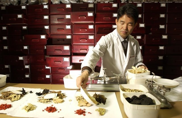
This and the Part 1 blog entry Adaptogens Part 1 are pursuant to one of the key themes of this blog -  the use of phyto-substances to promote health and longevity.  The Adaptogens Part 1 blog entry discusses adaptogen herbs in general. This Part 2 blog entry is about a particular well studied adaptogen herb – rhodiola.   Rhodiola is treateds from two viewpoints.  First, the viewpoint of a practicing herbalist, Madelon Hope, is conveyed via a video presentation.  Second, the viewpoint of current scientific research is covered in my usual manner: I cite a number of recent research citations relating to rhodiola involving its mechanisms of action and therapeutic potential.      Most of my personal commentary regarding both adaptogens and rhodiola is in the concluding part of this blog entry.

The video features Madelon Hope, Director of the Boston School of Herbal Studies and a long-time practicing herbalist. I asked her for an informal interview which I recorded on a tiny video camera. The first part of the interview, on adaptogens in general, is included in theAdaptogens Part 1 blog entry. The segment of the video interview specific to rhodiola is included here.


MadelonHopePt2 from VinceGiuliano on Vimeo.

Some basics on rhodiola

There are many important subspecies of rhodiola plants, but the best known for health and medicinal purposes is Rhodiola roseaOther subspecies mentioned here include Rhodiola Sachalinensis, Rhodiola Crenulataand Rhodiola integrifolia.  Some scholars lump several subspecies under rhodiola rosea, others may see subspecies such as Rhodiola integrifolia as quite separate(ref). To further complexify the matter, the chemical constituents in the plant may vary in different subspecies and vary further due to soil and climate conditions.

According to Wikipedia: “Rhodiola rosea (also Golden Root, Rose Root, Roseroot, Aaron’s Rod, Arctic root, King’s Crown, Lignum Rhodium, Orpin Rose) is a plant in the Crassulaceae family that grows in cold regions of the world. These include much of the Arctic, the mountains of Central Asia, the Rocky Mountains, and mountainous parts of Europe, such as the Alps, Pyrenees, Carpathian Mountains, Scandinavia, Iceland, Great Britain and Ireland. — Rhodiola rosea may be effective for improving mood and alleviating depression. Pilot studies on human subjects[2][3][4] showed that it improves physical and mental performance, and may reduce fatigue. — Rhodiola rosea’s effects are potentially mediated by changes in serotonin and dopamine levels due to monoamine oxidase inhibition and its influence on opioidpeptides such as beta-endorphin,[5] although these specific neurochemical mechanisms have not been clearly documented with scientific studies. — Rhodiola is included among a class of plant derivatives called adaptogens which differ from chemical stimulants, such as nicotine, and do not have the same physiological effects. — In Russia and Scandinavia, Rhodiola rosea has been used for centuries to cope with the cold Siberian climate and stressful life.[6][citation needed] Such effects were provided with evidence in laboratory models of stress using the nematodeC. elegans,[7] and in rats in which Rhodiola effectively prevented stress-induced changes in appetite, physical activity, weight gain and the estrus cycle.[8] — Rhodiola has been used in traditional Chinese medicine, where it is called hóng jǐng tiān (). — Rhodiola rosea contains a variety of compounds that may contribute to its effects,[10] including the class of rosavins which include rosavin, rosarin, and rosin. Several studies have suggested that the most active components are likely to be rhodioloside and tyrosol,[11] with other components being inactive when administered alone, but showing synergistic effects when a fixed combination of rhodioloside, rosavin, rosarin and rosin was used.[12]” 

Salidroside (Rhodioloside) is a glucoside of tyrosol found in the plant Rhodiola rosea. It is thought to be one of the compounds responsible for the antidepressant and anxiolytic actions of this plant, along with rosavin.[1][2] Salidroside may be more active than rosavin,[3] even though many commercially marketed Rhodiola rosea extracts are standardised for rosavin content rather than salidroside(ref).”

Until relatively recently it was thought that salidroside was a relatively inactive ingredient in rhodiola rosea and other key subspecies, probably not responsible for their major health benefits. “Although rosavin, rosarin, rosin and salidroside (and sometimes p-tyrosol, rhodioniside, rhodiolin and rosiridin) are among suspected active ingredients of Rhodiola rosea, these compounds are mostly polyphenols for which no physiological effect in humans is proved to prevent or reduce risk of disease.[13] (ref).”  Now that view appears to be reversed and much of the recent literature reviewed below is concerned with health benefits of salidroside.

 Image may be NSFW.
Clik here to view.
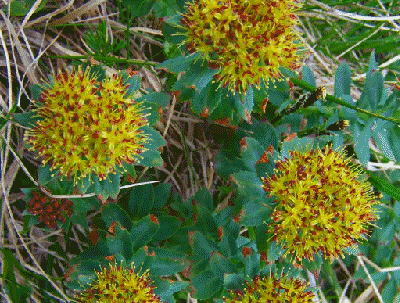
Rhodiola rosea     Image source

A number of research publications deal with the capabilities of rhodiola to ameliorate conditions of stress and fatigue.  Almost all the publications are positive with respect to the capabilities of the substance to exercise positive benefits.  From the viewpoint of Western pharmaceutical science, however, the studies may be biased and collectively are probably inconclusive.  More careful studies are needed.

The May 2012 review publication Rhodiola rosea for physical and mental fatigue: a systematic review represents this conservative point of view.  “BACKGROUND: Rhodiola rosea (R. rosea) is grown at high altitudes and northern latitudes. Due to its purported adaptogenic properties, it has been studied for its performance-enhancing capabilities in healthy populations and its therapeutic properties in a number of clinical populations.  OBJECTIVE: To systematically review evidence of efficacy and safety of R.rosea for physical and mental fatigue.  METHODS: Six electronic databases were searched to identify randomized controlled trials (RCTs) and controlled clinical trials (CCTs), evaluating efficacy and safety of R. rosea for physical and mental fatigue. Two reviewers independently screened the identified literature, extracted data and assessed risk of bias for included studies.  RESULTS: Of 206 articles identified in the search, 11 met inclusion criteria for this review. Ten were described as RCTs and one as a CCT. Two of six trials examining physical fatigue in healthy populations report R.rosea to be effective as did three of five RCTs evaluating R. rosea for mental fatigue. All of the included studies exhibit either a high risk of bias or have reporting flaws that hinder assessment of their true validity (unclear risk of bias).  CONCLUSION: Research regarding R.rosea efficacy is contradictory. While some evidence suggests that the herb may be helpful for enhancing physical performance and alleviating mental fatigue, methodological flaws limit accurate assessment of efficacy. A rigorously-designed well reported RCT that minimizes bias is needed to determine true efficacy of R.rosea for fatigue.” 

Personally, looking at many of the specific research publications, I find this conclusion rather harsh.  Using the same logic, one could conclude that there are no clearly documented health benefits of drinking water, and a large scale clinical trial would be needed to clearly establish that there are such health benefits.  My readers are invited to read on below and draw their own conclusions.

Some more-recent rat model studies have looked at actions of rhodiola components, particularly salidroside, on basic biological processes which happen in aging such as as decline in immune function, decline in DNA repair effectiveness, and increase in senescent cells.  The reported results appear to be interesting if not dramatic.

The February 2012 e-publication Rejuvenating activity of salidroside (SDS): dietary intake of SDS enhances the immune response of aged rats reports: “It is well known that immune response decreases with aging. Salidroside (SDS), an antioxidant component isolated from the traditional Chinese medicine roseroot Rhodiola rosea, has been demonstrated to possess potent anti-aging and health-promoting activities. However, the mechanism underlying these activities is poorly understood. In this study, we clearly demonstrated that (1) dietary intake of SDS induced a considerable increase in total T cells (CD3(+)) and T helper cells (CD4(+)) in aged (21 months old) Wistar male rats; (2) SDS supplementation significantly increased the DTH response, a T cell-mediated immune response, in aged rats; and (3) SDS supplementation remarkably promoted the production of total anti-KLH IgG, anti-KLH IgG(1), and anti-KLH IgG(2α) in aged rats without disturbing immune homeostasis. These indicate that SDS is able to counteract immunosenescence, thereby resulting in rejuvenation. Practically, SDS may be used to help the elderly to generate an improved response to vaccine with stronger humoral and cell-mediated immune responses.”  If this finding could be shown to carry over to humans, I believe that would be very exciting.

The May 2012 publication Salidroside stimulates DNA repair enzyme Parp-1 activity in mouse HSC maintenance also suggests a rejuvenating role of salidroside related to DNA repair and maintenance of hematopoietic stem cell pools.    “Salidroside is a phenylpropanoid glycoside isolated from the medicinal plant Rhodiola rosea, which has potent antioxidant properties. Here we show that salidroside prevented the loss of hematopoietic stem cells (HSCs) in mice under oxidative stress. Quiescent HSCs were recruited into cell cycling on in vivo challenge with oxidative stress, which was blocked by salidroside. Surprisingly, salidroside does not prevent the production of reactive oxygen species but reduces hydrogen peroxide-induced DNA-strand breaks in bone marrow cells enriched for HSCs. We tested whether salidroside enhances oxidative DNA damage repair in mice deficient for 5 DNA repair pathways known to be involved in oxidative DNA damage repair; we found that salidroside activated poly(ADP-ribose)polymerase-1 (PARP-1), a component of the base excision repair pathway, in mouse bone marrow HSCs as well as primary fibroblasts and human lymphoblasts. PARP-1 activation by salidroside protects quiescent HSCs from oxidative stress-induced cycling in native animals and self-renewal defect in transplanted recipients, which was abrogated by genetic ablation or pharmacologic inhibition of PARP-1. Together, these findings suggest that activation of PARP-1 by salidroside could affect the homeostasis and function of HSCs and contribute to the antioxidant effects of salidroside.”  As suggested in the previous Part 1 blog entry, I very strongly suspect that salidroside in part works by upregulating the expression of Nrf2 thus activating the body’s own natural antioxidant and repair defense systems.

The November 2010 publication Salidroside protects human fibroblast cells from premature senescence induced by H(2)O(2) partly through modulating oxidative status reports “Although salidroside and salidroside-like compounds are considered as most critical constitutes needed and responsible for multiple therapeutic benefits of Rhodiola rosea L., including anti-aging, direct demonstration regarding the role of salidroside in anti-aging process is still deficient. In this study, we selected the H(2)O(2)-induced premature senescence model in human fetal lung diploid fibroblasts to investigate the protection of salidroside against aging in vitro and associated molecular mechanisms. We found that salidroside considerably reversed senescence-like phenotypes in the oxidant challenged model, including alterations of morphology, cell cycle, SA-β-gal staining, DNA damage, as well as related molecules expression such as p53, p21 and p16. The protection occurred in a dose-dependent manner, with 5μM offering best efficacy. The proposed antioxidant property of the compound was confirmed in this cellular system, and thus at least partially accounted for the protection of the compound against premature senescence. Similar protection of salidroside against replicative senescence was observed as well. Interestingly, the regulation of senescence-related molecules by salidroside involved ROS-irrelevant mechanisms in both models. This finding presents salidroside as an attractive agent with potential to retard aging and attenuate age-related diseases in humans”

Multiple recent cell-level studies establish that salidroside definitely exercises cytoprotective effects, in part by inhibiting apoptosis in the presence of oxidative stress.  Again. I suspect the effect is due to promotion of expression of Nrf2.

I cite only a few examples.  For example, the April 2012 publication Salidroside protects human erythrocytes against hydrogen peroxide-induced apoptosis reports “Rhodiola rosea is a commonly used folk medicine for the treatment of high altitude sickness, mountain malhypoxia, and anoxia. Its active ingredient, salidroside [2-(4-hydroxyphenyl)ethyl β-D-glucopyranoside (1)], has been reported to have a broad spectrum of biological effects. However, the protective role of 1 in human erythrocytes remains unclear. This study therefore has investigated the effects of 1 on oxidative stress-induced apoptosis in human erythrocytes (also known as eryptosis or erythroptosis). Compound 1 increased cell survival significantly and prevented human erythrocytes from undergoing eryptosis/erythroptosis mediated by H(2)O(2), as confirmed by the decreased expression of phosphatidylserine on the cell surface and reduced leakage of calcein through the damaged membrane. Mechanistically, 1 was found to exert its protective effects through its antioxidative activity and the inhibition of caspase-3 activation and stress-induced intracellular Ca(2+) rise in a dose-dependent manner. Compound 1 is a protective agent in human erythrocytes against oxidative stress and may be a good adaptogen to enhance the body’s resistance to stress and fatigue.”

The July 2012 publication Salidroside and tyrosol from Rhodiola protect H9c2 cells from ischemia/reperfusion-induced apoptosis reports: “AIMS: Heart disease is the leading cause of death worldwide. Ischemia-reperfusion injury can lead to apoptotic death of heart cells and subsequently heart failure. Rhodiola is an herbal medicine with two main bioactive compounds – salidroside (SAL) and tyrosol (TYR). This study aimed to investigate whether these two compounds can prevent ischemia/reperfusion-induced apoptosis in H9c2 cells.  MAIN METHODS: Assays for total phenolics assay and Oxygen Radical Absorbance Capacity showed high antioxidant capacity of SAL and TYR. H9c2 cells were subjected to simulated ischemia/reperfusion (IR) in the presence and absence of SAL and/or TYR, and nuclei condensation, caspase-3 activity, cytochrome c release and JNK phosphorylation were determined.  KEY FINDINGS: In H9c2 cells, IR can lead to a 5-fold increase in p-JNK level. Apoptotic nuclei condensation, caspase-3 activity and cytochrome c release were markedly elevated, indicating the occurrence of apoptosis. SAL and TYR caused a dose-dependent inhibition of nuclear condensation. Furthermore, SAL and TYR, separately and in combination, significantly reduced caspase-3 activity, cytochrome c release and JNK activation. The anti-apoptotic effect of the combination was markedly higher than that of SAL or TYR alone.  SIGNIFICANCE: The inhibition of the JNK signaling pathway is the key mechanism for the cytoprotective effect of SAL and TYR in IR-induced apoptosis.”

The August 2012 publication Protective effects of salidroside from Rhodiola rosea on LPS-induced acute lung injury in mice reports: “Salidroside is a major component extracted from Rhodiola rosea. In this study, we investigated protective effects of salidroside on lipopolysaccharide (LPS)-induced acute lung injury (ALI) in mice. In the mouse model, we found that pretreatment with a single 120 mg/kg dose of salidroside prior to the administration of intratracheal LPS induced a significant decrease in the W/D ratio and mouse myeloperoxidase activity of lung, reduction protein concentration, the number of total cells, neutrophils and macrophages in the bronchoalveolar lavage fluid. In addition, salidroside also inhibited the production of several inflammatory cytokines, including tumor necrosis factor-α, interleukin-6 (IL-6) and IL-1β, and the NF-κB DNA-binding activation after LPS challenge. These results indicated that salidroside possess a protective effect on LPS-induced ALI in mice.”

The 2011 publication Salidroside promotes erythropoiesis and protects erythroblasts against oxidative stress by up-regulating glutathione peroxidase and thioredoxin relates: “ETHNOPHARMACOLOGICAL RELEVANCE: Rhodiola rosea is commonly used in China and Tibet folk medicine for the treatment of high altitude sickness, anoxia and mountain malhypoxia.  AIM OF STUDY: Salidroside (SDS) is an active ingredient of Rhodiola rosea. This study attempted to examine the potential erythropoiesis-stimulating and anti-oxidative effect of SDS in TF-1 erythroblasts.  MATERIALS AND METHODS: The erythropoiesis-promoting effect was determined by treating human TF-1 cells, one of the popular in vitro models for studying erythropoiesis, with SDS in the presence and absence of erythropoietin (EPO) through the measurement of the expression of a series of erythroid markers such as glycophorin A (GPA), transferrin receptor (CD71) and hemoglobin (Hb). The potential protective effect of SDS against H(2)O(2)-induced apoptosis and its underlying mechanism in TF-1 erythroblasts were examined by flow cytometry and Western blot analysis.  RESULTS: SDS promotes erythropoiesis in the EPO-treated cells and it also reduces the number of apoptotic cells in TF-1 erythroblasts after H(2)O(2) treatment probably through the up-regulation of protective proteins thioredoxin-1 (Trx1) and glutathione peroxidase-1 (GPx1). CONCLUSION: Our study provides evidence to explain the ethnopharmacological role of SDS and Rhodiola rosea in Chinese medicine. Our findings also support the use of SDS as an erythropoiesis-adjuvant agent to correct anemia and malhypoxia.”  Again, we have here the familiar themes of protection against oxidative stress, prevention of apoptosis and stimulating stem cell differentiation.

The October 2011 publication Salidroside protects against hydrogen peroxide-induced injury in cardiac H9c2 cells via PI3K-Akt dependent pathway reports: “Oxidative stress induces serious tissue injury in cardiovascular diseases. Salidroside, with its strong antioxidative and cytoprotective actions, is of particular interest in the development of antioxidative therapies for oxidative injury in cardiac diseases. We examined the pharmacological effects of salidroside on H9c2 rat cardiomyoblast cells under conditions of oxidative stress induced by hydrogen peroxide (H2O2) challenge. Salidroside attenuated H2O2-impaired cell viability in a concentration-dependent manner, and effectively inhibited cellular malondialdehyde production, lethal sarcolemmal disruption, cell necrosis, and apoptosis induced by H2O2 insult. Salidroside significantly augmented Akt phosphorylation at Serine 473 in the absence or presence of H2O2 stimulation; wortmannin, a specific inhibitor of PI3K, abrogated salidroside protection. Salidroside increased the intracellular mRNA expression and activities of catalase and Mn-superoxide dismutases in a PI3K-dependent manner. Our results indicated that salidroside protected cardiomyocytes against oxidative injury through activating the PI3K/Akt pathway and increasing the expression and activities of endogenous PI3K dependent antioxidant enzymes.”

Another study relating salidroside to cardiac cells is reported in the 2009 publication Salidroside protects cardiomyocyte against hypoxia-induced death: a HIF-1alpha-activated and VEGF-mediated pathway.  Cardiomyocyte death (necrosis and apoptosis) plays a critical role in the progress of heart diseases. Salidroside, a phenylpropanoid glycoside isolated from Rhodiola rosea L, has shown cardioprotective effects in vivo. However, whether salidroside has a protective effect against cardiomyocyte death is poorly understood. The present study was aimed to investigate the cardioprotective role of salidroside and the underlying mechanisms in hypoxia-induced cardiomyocyte death. Cardiomyocytes pretreated with or without salidroside for 24 h were exposed to hypoxic condition for 6 h and then cell viability, necrosis, apoptosis, the expressions of HIF-1alpha and VEGF were investigated. Pretreatment with salidroside markedly attenuated hypoxia-induced cell viability loss, cell necrosis and apoptosis in a dose-dependent manner. Mechanistically, pretreatment with salidroside up-regulated the HIF-1alpha protein expression and induced its translocation. Moreover, the level of VEGF, a downstream target of HIF, was significantly increased in parallel with the level of HIF-1alpha following pretreatment with salidroside. However, 2-methoxyestradiol (2-ME2), a HIF-1alpha inhibitor, attenuated the protection of salidroside and blocked the increase of HIF-1alpha and VEGF. These data indicated that salidroside has protective effect against hypoxia-induced cardiomyocytes necrosis and apoptosis by increasing HIF-1alpha expression and subsequently up-regulating VEGF levels.”

A number of cell-level studies look at the protective effects of salidroside of neural cells and the ability of salidroside to promote neurogenesis under various in-vitro conditions and speculate whether it might become the basis for treatment of Parkinson’s disease, Alzheimer’s disease and other neurodegenerative disorders.

The January 2012 publication Protective effects of a Rhodiola crenulata extract and salidroside on hippocampal neurogenesis against streptozotocin-induced neural injury in the rat is a case in point. “Previously we have demonstrated that a Rhodiola crenulata extract (RCE), containing a potent antioxidant salidroside, promotes neurogenesis in the hippocampus of depressive rats. The current study was designed to further investigate the protective effect of the RCE on neurogenesis in a rat model of Alzheimer’s disease (AD) induced by an intracerebroventricular injection of streptozotocin (STZ), and to determine whether this neuroprotective effect is induced by the antioxidative activity of salidroside. Our results showed that pretreatment with the RCE significantly improved the impaired neurogenesis and simultaneously reduced the oxidative stress in the hippocampus of AD rats. In vitro studies revealed that (1) exposure of neural stem cells (NSCs) from the hippocampus to STZ strikingly increased intracellular reactive oxygen species (ROS) levels, induced cell death and perturbed cell proliferation and differentiation, (2) hydrogen peroxide induced similar cellular activities as STZ, (3) pre-incubation of STZ-treated NSCs with catalase, an antioxidant, suppressed all these cellular activities induced by STZ, and (4) likewise, pre-incubation of STZ-treated NSCs with salidroside, also an antioxidant, suppressed all these activities as catalase: reduction of ROS levels and NSC death with simultaneous increases in proliferation and differentiation. Our findings indicated that the RCE improved the impaired hippocampal neurogenesis in the rat model of AD through protecting NSCs by its main ingredient salidroside which scavenged intracellular ROS.”

The 2009 publication Protective effect of salidroside against H2O2-induced cell apoptosis in primary culture of rat hippocampal neurons relates : “Salidroside, a phenylpropanoid glycoside separated from a medicinal plant Rhodiola rosea, has been documented to have protective effects on neuronal cells in vitro. This study investigated whether salidroside was able to extend its unique neuroprotection to primary cultured rat hippocampal neurons against hydrogen peroxide (H(2)O(2))-induced cell damage. Cell viability tests and cell apoptosis assays confirmed that salidroside pretreatment attenuated H(2)O(2)-stimulated apoptotic cell death in primary culture of hippocampal neurons in a concentration-dependent manner. The measurements of caspase-3 activity, nitric oxide (NO) production, and NO synthase (NOS) activity suggest that the protection of salidroside, shown in this study, might be mediated by inhibiting caspase-3 activity, and antagonizing NO production and NOS activity during H(2)O(2) stimulation. Perhaps, this study might contribute to the development of salidroside as a broad-spectrum agent for preventing and/or treating neuronal damage in neurodegenerative disorders.”

The 2010 publication Neuroprotective effects of salidroside against beta-amyloid-induced oxidative stress in SH-SY5Y human neuroblastoma cells reported: “Beta-amyloid (Abeta) peptide, the hallmark of Alzheimer’s disease (AD), invokes a cascade of oxidative damages to neurons and eventually leads to neuronal death. In this study, salidroside (Sald), an active compound isolated from a traditional Chinese medicinal plant, Rhodiola rosea L., was investigated to assess its protective effects and the underlying mechanisms against Abeta-induced oxidative stress in SH-SY5Y human neuroblastoma cells. Abeta(25-35)-induced neuronal toxicity was characterized by the decrease of cell viability, the release of lactate dehydrogenase (LDH), morphological alterations, neuronal DNA condensation, and the cleavage of poly(ADP-ribose) polymerase (PARP) by activated caspase-3. Pretreatment with salidroside markedly attenuated Abeta(25-35)-induced loss of cell viability and apoptosis in a dose-dependent manner. The mechanisms of salidroside protected neurons from oxidative stress included the induction of antioxidant enzymes, thioredoxin (Trx), heme oxygenase-1 (HO-1), and peroxiredoxin-I (PrxI); the downregulation of pro-apoptotic protein Bax and the upregulation of anti-apoptotic protein Bcl-X(L). Furthermore, salidroside dose-dependently restored Abeta(25-35)-induced loss of mitochondrial membrane potential (MMP) as well as suppressed the elevation of intracellular reactive oxygen species (ROS) level. It was also observed that Abeta(25-35) stimulated the phosphorylation of mitogen-activated protein (MAP) kinases, including c-Jun NH(2)-terminal kinase (JNK) and p38 MAP kinase, but not extracellular signal-regulated kinase1/2 (ERK1/2). Salidroside inhibited Abeta(25-35)-induced phosphorylation of JNK and p38 MAP kinase, but not ERK1/2. These results suggest that salidroside has protective effects against Abeta(25-35)-induced oxidative stress, which might be a potential therapeutic agent for treating or preventing neurodegenerative diseases.”

Earlier publications on the effects of salidroside on neuroblastoma cells include the 2007 report Protective effects of salidroside on hydrogen peroxide-induced apoptosis in SH-SY5Y human neuroblastoma cells.

The neuroprotective effects of salidroside and the possibility of creating therapies for neurodegenerative diseases based on rhodiola compounds is far from new.  For example there is a 2003 publication Salidroside from Rhodiola sachalinensis protects neuronal PC12 cells against cytotoxicity induced by amyloid-beta“The amyloid beta-peptide (Abeta)-induced oxidative stress is a well-established pathway of neuronal cell death in Alzheimer’s disease (AD). Salidroside, one of the major compounds from the roots of Rhodiola species (Crassulaceae), was investigated in vitro for its cytoprotection against Abeta-induced toxicity on rat neuronal PCl2 cells. Salidroside significantly reduced Abeta-induced cytotoxicity in a dose-dependent manner. Salidroside also reduced Abeta-mediated intracellular accumulation of reactive oxygen species and malondialdehyde (MDA), a product of lipid peroxides, by preventing Abeta-induced decline of antioxidant enzyme activities. These results suggest that salidroside protects neuronal PC12 cells from Abeta-induced cytotoxicity via its antioxidant pathway.”

Fast-forwarding eight years to 2011, another report related to PC12 cells is Salidroside protects against MPP(+)-induced apoptosis in PC12 cells by inhibiting the NO pathway. “Oxidative stress plays an important role in Parkinson’s disease and other neurodegenerative disorders. Salidroside, a phenylpropanoid glycoside isolated from Rhodiola rosea L., has potent antioxidant properties. In the present study, we investigated the protective activity of salidroside against 1-methyl-4-phenylpyridinium (MPP(+))-induced apoptosis in PC12 cells. We found that incubation of PC12 cells with salidroside prior to MPP(+) exposure significantly reduced cell apoptosis and attenuated collapse of the mitochondrial membrane potential (MMP). Furthermore, salidroside inhibited the MPP(+)-induced nitric oxide (NO) increase and overexpression of nNOS and iNOS and suppressed accumulation of reactive oxygen species (ROS) and intracellular free Ca(2+). Our results show that the protective effects of salidroside on PC12 cells are mediated, at least in part, by inhibition of the NO pathway.”

Moving to right now, August 2012, we have the publication Salidroside protects PC12 cells from MPP(+)-induced apoptosis via activation of the PI3K/Akt pathway“Oxidative stress plays an important role in the pathogenesis of Parkinson’s disease (PD). Salidroside (SAL), a phenylpropanoid glycoside isolated from Rhodiola rosea L., can exert potent antioxidant properties. In this study, we investigated the protective effects, and the possible mechanism of action, of SAL against 1-methyl-4-phenylpyridinium (MPP(+))-induced cell damage in rat adrenal pheochromocytoma PC12 cells. Pretreatment of PC12 cells with SAL significantly reduced the ability of MPP(+) to induce apoptosis in a dose and time-dependent manner. SAL significantly and dose-dependently inhibited MPP(+)-induced chromatin condensation and MPP(+)-induced release of lactate dehydrogenase by PC12 cells. SAL enhanced Akt phosphorylation in PC12 cells, and the protective effects of SAL against MPP(+)-induced apoptosis were abolished by LY294002, a specific inhibitor of phosphatidylinositol 3-kinase (PI3K) phosphorylation. These findings suggest that SAL prevents MPP(+)-induced apoptosis in PC12 cells, at least in part through activation of the PI3K/Akt pathway.”

Different species of rhodiola may induce differing biological effects.

The 2006 publication Evaluation of Rhodiola crenulata and Rhodiola rosea for management of type II diabetes and hypertension reported: “In the current study, we investigated 2 species of the genus Rhodiola for the inhibition of alpha-amylase,alpha-glucosidase and angiotensin converting enzyme (ACE) inhibitory activity. Water extracts of Rhodiola crenulata had the highest alpha-amylase inhibitory activity (IC50,98.1 microg total phenolic /ml) followed by ethanol extract of R.crenulata (IC50, 120.9 microg total phenolic/ml) and ethanol extract of R.rosea (IC50, 173.4 microg total phenolic /ml). Ethanol R.rosea (IC50, 44.7 microg total phenolic/ml), water extract of R.rosea (IC50, 52.3 microg total phenolic/ml), water extract of R.crenulata (IC50, 60.3 microg total phenolic /ml) and ethanol extract of R.crenulata (IC50, 60.2 microg total phenolic/ml) also showed significant alpha-glucosidase inhibitory activity. The alpha-glucosidase inhibitory activity of the extracts was compared to standard tyrosol, which was significantly detected in the extracts using HPLC. Tyrosol had strong alpha-glucosidase inhibitory activity (IC50, 70.8 microg total phenolic/ml) but did not have any inhibitory effect on the alpha-amylase activity. Results suggested that alpha-glucosidase inhibitory activities of both Rhodiola extracts correlated to the phenolic content, antioxidant activity and phenolic profile of the extracts. The ability of the above Rhodiola extracts to inhibit rabbit lung angiotensin I-converting enzyme (ACE) was investigated. The ethanol extracts of R.rosea had the highest ACE inhibitory activity (38.5 %) followed by water extract of R.rosea (36.2 %) and R.crenulata (15.4 %).”

Rhodiola may be useful for treatment of metabolic syndrome or Type II diabetes.

Pursuing a theme of the 2006 publication just mentioned , a new (August 2012) publication Rhodiola crenulata root ameliorates derangements of glucose and lipid metabolism in a rat model of the metabolic syndrome and type 2 diabetes aeports: “AIM OF THE STUDY: To examine the effects of Rhodiola on glucose and lipid metabolism in the metabolic syndrome and type 2 diabetes.  MATERIALS AND METHODS: Zucker diabetic fatty (ZDF) rats were treated with Rhodiola crenulata root (RCR) powder (100 and 500mg/kg, by gavage, once daily for 4 weeks). In addition, the effects of RCR on sucrose-induced acute hyperglycemia in mice and olive oil-induced hypertriglyceridemia in rats were also examined. Biochemical variables were determined enzymatically or by ELISA.  RESULTS: In ZDF rats, RCR treatment decreased the increased plasma insulin and triglyceride concentrations at baseline, the index of the homeostasis model assessment of insulin resistance (HOMA-IR) and excessive hepatic triglyceride accumulation. This treatment also inhibited abnormal increases in plasma glucose and insulin concentrations during oral glucose tolerance test. Furthermore, RCR reversed the increased adipose insulin resistance index, and accelerated the decline of plasma concentrations of non-esterified fatty acids after exogenous glucose stimulation. However, RCR minimally affected sucrose-induced acute hyperglycemia in mice and olive oil-induced acute hypertriglyceridemia in rats.  CONCLUSIONS: The present results demonstrate that RCR treatment improves metabolic derangements in animal model of the metabolic syndrome and type 2 diabetes. Our findings may provide new pharmacological basis of therapeutics for the adaptogenic plants to treat metabolic derangements-associated disorders, such as asthenia.”

Some small clinical trials point to the safety and effectiveness of salidroside as a therapy or adjunct therapy for special disease conditions.

An example is discussed in the June 2012 publication Protective effects of salidroside on epirubicin-induced early left ventricular regional systolic dysfunction in patients with breast cancer“Background: Salidroside [2-(4-hydroxyphenyl)ethyl-β-D-glucopyranoside], one of the most potent ingredients extracted from the plant Rhodiola rosea L., has been shown to have a cardiovascular protective effect as an antioxidant, and early treatment of epirubicin-induced cardiotoxicity has been the focus of clinical chemotherapy in patients with breast cancer. However, the cardioprotective effects of salidroside on epirubicin-induced cardiotoxicity, especially early left ventricular regional systolic dysfunction, have to date been sparsely investigated. Objective: The aim of this study was to investigate the protective effects of salidroside in preventing early left ventricular regional systolic dysfunction induced by epirubicin. Methods: Sixty patients with histologically confirmed breast cancer were enrolled. Eligible patients were randomized to receive salidroside (600 mg/day; n = 30) or placebo (n = 30) starting 1 week before chemotherapy. Patients were investigated by means of echocardiography and strain rate (SR) imaging. We also measured plasma concentrations of reactive oxygen species (ROS). All parameters were assessed at baseline and 7 days after each new epirubicin dose of 100 mg/m2. Results: A decline of the SR peak was observed at an epirubicin dose of 200 mg/m2, with no significant differences between salidroside and placebo (1.35 ± 0.36 vs 1.42 ± 0.49/second). At growing cumulative doses of epirubicin, the SR normalized only with salidroside, showing a significant difference in comparison with placebo at epirubicin doses of 300 mg/m2 (1.67 ± 0.43 vs 1.32 ± 0.53/second, p < 0.05) and 400 mg/m2 (1.68 ± 0.29 vs 1.40 ± 0.23/second, p < 0.05). Moreover, a significant increase in plasma concentrations of ROS was found with placebo, but they remained unchanged with salidroside. Conclusion: Salidroside can provide a protective effect on epirubicin-induced early left ventricular regional systolic dysfunction in patients with breast cancer.”

Small-scale human trials as well as animal studies suggests that salidroside can enhance endurance exercise performance.

The May 2012 publication The Effects of an Acute Dose of Rhodiola Rosea on Endurance Exercise Performance reports: “The purpose of this study was to determine the effects of an acute oral dose of 3mg/kg of Rhodiola Rosea (Rr) on endurance exercise performance, perceived exertion, mood, and cognitive function. Subjects (n=18) ingested either Rr or a carbohydrate placebo 1 h prior to testing in a double blind, random crossover manner. Exercise testing consisted of a standardized 10 min warm-up, followed by a 6-mile time trial (TT) on a bicycle ergometer. Perceived exertion (RPE) was measured every 5 min during the TT using a BORG 10 pt scale. Blood lactate concentration, salivary cortisol and salivary alpha amylase were measured pre warm-up, 2 min post warm-up, and 2 min post TT (n=15). A Profile of Mood States (POMS) questionnaire and a Stroop’s Color Test were completed pre-warm up and post TT. Testing was repeated 2-7d later with the other condition. Rr ingestion significantly decreased heart rate during the standardized warm up (Rr=136+17bpm; Placebo=140+17bpm; mean+SD; p=0.001). Subjects completed the TT significantly faster following Rr ingestion (Rr=25.4+2.7 min; placebo=25.8+3.0 min; p=0.037). The mean RPE was lower in the Rr trial (Rr= 6.0+0.9; placebo= 6.6+1.0; p=0.04). This difference was even more pronounced when a ratio of the RPE relative to the workload was calculated (Rr= 0.048+0.01; placebo= 0.057+0.02; p=0.007). No other statistically significant differences were observed. Acute Rr ingestion decreases heart rate response to sub-maximal exercise, and appears to improve endurance exercise performance by decreasing the perception of effort.”

Biological impacts of salidroside under exercise conditions are examined in a mouse model in the January 2012 publication [Influence of salidroside from Rhodiola Sachalinensis A. Bor on some related indexes of free radical and energy metabolism after exercise in mice]“OBJECTIVE: To study the anti-fatigue mechanism of salidroside from Rhodiola SachalinensisA.Bor (SRS)in anti-oxidation and energy metabolic systems in mice , some related indexes of free radical and energy metabolism after exercise were measured.  METHODS: Forty male mice were divided into four groups (n = 10): SRS sport group(SS), SRS quiet group(SQ),sport control group(SC), quiet control group (QC). The mice of SS and SQ groups received SRS solution of 150 ml/kg body weight per day for two weeks, while the mice of SC and QC groups received the same volume of distilled water. 30 min after the last treatment, the mice of SS and SC groups were forced to swim for 120 min without loads. then the biochemical parameters related to fatigue were determined.  RESULT: SRS could increase liver superoxide dismutase (SOD) , glutathione peroxidase (GSH-Px) activity of antioxidant enzymes and reduce the malondialdehyde (MDA) content, which might increase the body activity of antioxidant enzymes to play the role of anti-oxidation; SRS had some effect of stabilizing blood sugar, increasing liver glycogen and muscle glycogen reserves, preventing blood sugar, liver glycogen and muscle glycogen levels from reducing in long time exercise on mice; SRS could increase plasma total cholesterol (TC), triglyceride (TG) and free fatty acid (FFA) levels in exercise mice, and it had some effect on metabolism of fat under different conditions, and promoted the use of the role of fat.  CONCLUSION: Influence of SRS on some related indexes of free radical and energy metabolism is one of the mechanisms of anti-exercise-induced fatigue of SRS.”

A 2010 publication Effects of chronic Rhodiola Rosea supplementation on sport performance and antioxidant capacity in trained male: preliminary results relates: “AIM: Rhodiola Rosea, is an adaptogen plant which has been reported to promote fatty acids utilisation, to ameliorate antioxidant function, and to improve body resistance to physical strenuous efforts. The purpose of the present study was to investigate the effects on physical performance as well as on the redox status of a chronic Rhodiola Rosea supplementation in a group of competitive athletes during endurance exercise.  METHODS: Following a chronic supplementation with Rhodiola Rosea for 4 weeks, 14 trained male athletes underwent a cardio-pulmonary exhaustion test and blood samples to evaluate their antioxidant status and other biochemical parameters. These data were compared with those coming from the same athletes after an intake of placebo. RESULTS: The evaluation of physical performance parameters showed that HR Max, Borg Scale level, VO(2) max and duration of the test were essentially unaffected by Rhodiola Rosea assumption. On the contrary, Rhodiola Rosea intake reduced, in a statistically significative manner, plasma free fatty acids levels. No effect on blood glucose was found. Blood antioxidant status and inflammatory parameters resulted unaffected by Rhodiola Rosea supplementation. Blood lactate and plasma creatine kinase levels were found significantly lower (P<0.05) in Rhodiola Rosea treated subjects when compared to the placebo treated group. CONCLUSION: Chronic Rhodiola Rosea supplementation is able to reduce both lactate levels and parameters of skeletal muscle damage after an exhaustive exercise session. Moreover this supplementation seems to ameliorate fatty acid consumption. Taken together those observation confirm that Rhodiola Rosea may increase the adaptogen ability to physical exercise.”

Also relevant to the exercise issue is the 2009 study Attenuation of long-term Rhodiola rosea supplementation on exhaustive swimming-evoked oxidative stress in the rat.   “Rhodiola rosea improves exercise endurance and fatigue. We hypothesized that ingredients in Rhodiola rosea may increase antioxidant capability against swimming induced oxidative stress. In this study, we have identified the Rhodiola rosea ingredients, p-tyrosol, salidroside, rosin, rosavin and rosarin by high performance liquid chromatography-mass spectrometer and evaluated their O2(-)*, H2O2, and HOCl scavenging activities by a chemiluminescence analyzer. We next explored the effect and mechanism of Rhodiola rosea on 90-min swimming-induced oxidative stress in male Wistar rats fed with three doses of Rhodiola rosea extracts in drinking water (5, 25, 125 mg/day/rat) for 4 weeks. Our results showed that the 4 major ingredients (salidroside, rosin, rosavin and rosarin) from Rhodiola rosea extracts scavenged O2(-)*, H2O2, and HOCl activity in a dose-dependent manner. The ninety-min swimming exercise increased the O2(-)* production in the order: liver > skeletal muscle > blood, indicating that liver is the most sensitive target organ. The level of plasma malonedialdehyde, a lipid peroxidation product, was also increased after exercise. Treatment of 4 weeks of Rhodiola rosea extracts significantly inhibited swimming exercise-enhanced O2(-)* production in the blood, liver and skeletal muscle and plasma malonedialdehyde concentration. The expression in Mn-superoxide dismutase Cu/Zn-superoxide dismutase, and catalase in livers were all enhanced after 4 weeks of Rhodiola rosea supplementation especially at the dose of 125 mg/day/rat. Treatment of Rhodiola rosea extracts for 4 weeks significantly increased swimming performance. In conclusion, treatment of Rhodiola rosea extracts for 4 weeks could reduce swimming-enhanced oxidative stress possibly via the reactive oxygen species scavenging capability and the enhancement of the antioxidant defense mechanisms.”

Rhodiola might be useful for treating opioid addiction.

A May 2012 publication Effects of a Rhodiola rosea L. extract on the acquisition, expression, extinction, and reinstatement of morphine-induced conditioned place preference in mice reports: “RATIONALE: Opioid addiction is a chronic, recurrent brain disease that is characterised by compulsive drug seeking and a high rate of relapse even after long periods of abstinence. Prevention of relapse is the primary goal of addiction treatment and is still the major limitation in drug therapy.  OBJECTIVES: The present study investigated the effects of a Rhodiola rosea L. hydroalcoholic extract (RHO), a well-known traditional oriental medicine, on establishment and reinstatement of morphine-induced conditioned place preference (CPP) in mice.  METHODS: CPP was induced by intraperitoneal injection of morphine (10 mg/kg) as an 8-day conditioning schedule. The effects of RHO on the rewarding properties of morphine were tested in mice receiving oral administration of RHO (10, 15, and 20 mg/kg) 60 min prior to each morphine injection (acquisition) or prior to the CPP test on day 9 (expression). Once established, CPP was extinguished by repeated testing, during which conditioned mice were injected daily with different doses of RHO. Finally, the efficacy of RHO in blocking reinstatement of CPP provoked by priming injections and physical stress was also evaluated.  RESULTS: RHO administration showed dose dependency for prevention of establishment of CPP and was effective in facilitating extinction of morphine-induced CPP. RHO suppressed both priming- and stress-induced reinstatement of CPP in a dose-dependent manner.  CONCLUSIONS: In conclusion, as RHO was effective for reducing craving and vulnerability to relapse, it might be a very effective natural remedy for the treatment of opioid addiction.”

One of the traditional uses of rhodiola is for treatment of high-altitude mountain sickness.  The benefits are probably due to the presence of salidroside

The March 2012 publication Salidroside stimulates the accumulation of HIF-1α protein resulted in the induction of EPO expression: a signaling via blocking the degradation pathway in kidney and liver cells reports: “Rhodiolae Crenulatae Radix et Rhizoma (Rhodiola), the root and rhizome of Rhodiola crenulata (Hook. f. et Thoms.) H. Ohba, has been used as a traditional Chinese medicine (TCM) to increase the body resistance to mountain sickness in preventing hypoxia; however, the functional ingredient responsible for this adaptogenic effect has not been revealed. Here, we have identified salidroside, a glycoside predominantly found in Rhodiola, is the chemical in providing such anti-hypoxia effect. Cultured human embryonic kidney fibroblast (HEK293T) and human hepatocellular carcinoma (HepG2) were used to reveal the mechanism of this hematopoietic function mediated by salidroside. The application of salidroside in cultures induced the expression of erythropoietin (EPO) mRNA from its transcription regulatory element hypoxia response element (HRE), located on EPO gene. The application of salidroside stimulated the accumulation of hypoxia-inducible factor-1α (HIF-1α) protein, but not HIF-2α protein: the salidroside-induced HIF-1α protein was via the reduction of HIF-1α degradation but not the mRNA induction. The increased HIF-1α could account for the activation of EPO gene. These results supported the notion that hematopoietic function of Rhodiola was triggered, at least partially, by salidroside.”

Comments

I could go on with many more citations and observations but think what is included above is sufficient to convey a good idea of what rhodiola’s capabilities are.

Collectively, these citations lead me to general observations applicable not only to salidroside and rhodiola but to most other traditional folk remedies:  There have been more and more cell-level studies over the years establishing safety and efficacy of rhodiola and salidroside as an anti-oxidant and anti-apoptotic on the cell level. These cell-level studies are relatively easy and cheap to do and appear to me to be somewhat redundant over the years.  Many of the newer studies are also unveiling the molecular pathways through which rhodiola and its important constituent compounds such as salidroside work.  A smaller number of small-animal studies also establishing safety and efficacy for a number of health and medical conditions, and a yet-smaller collection of small-scale human trials have been held under various specialized conditions. 

But, in the West at least, mainline salidroside-based clinical therapies that have been subject to pharma-scale clinical trials are yet to emerge.  So salidroside and other traditional medical substances like andrographis, gambogic and gambogenic acids and icarin remain in the shadows.  They are unknown to most physicians and unused in Western clinical practice.  They may even be viewed as dangerous because of the possibility of their interactions with pharmaceutical drugs. 

It is difficult to establish whether this is due to the weaknesses of the substances as drug candidates or due to the economics of clinical drug development in the Western model of medical practice involving pharmaceutical companies with a strong profit motive and the FDA as a watchdog.  What drug company would want to spend a billion dollars and ten years to bring to market a natural substance that is not patentable, or an analog of such a natural substance that may be little or no better than the substance itself?  And how many doctors are willing to risk administering substance therapies that are not FDA sanctioned?

So, in the US and other advanced Western countries, herbal healing remains a splinter activity unconnected with mainline medical practice.  This may be slowly changing as 1.  More medical schools and health institutions are starting programs in complementary, alternative or integrative medicine, 2.  Big pharma and biotech companies turn more to traditional folk medicines in their searches for new drug candidates, and 3. There is increased public awareness among people that individuals need to take responsibility for their own health and longevity.


MEDICAL DISCLAIMER

FROM TIME TO TIME, THIS BLOG DISCUSSES DISEASE PROCESSES. THE INTENTION OF THOSE DISCUSSIONS IS TO CONVEY CURRENT RESEARCH FINDINGS AND OPINIONS, NOT TO GIVE MEDICAL ADVICE. THE INFORMATION IN POSTS IN THIS BLOG IS NOT A SUBSTITUTE FOR A LICENSED PHYSICIAN’S MEDICAL ADVICE. IF ANY ADVICE, OPINIONS, OR INSTRUCTIONS HEREIN CONFLICT WITH THAT OF A TREATING LICENSED PHYSICIAN, DEFER TO THE OPINION OF THE PHYSICIAN. THIS INFORMATION IS INTENDED FOR PEOPLE IN GOOD HEALTH. IT IS THE READER’S RESPONSIBILITY TO KNOW HIS OR HER MEDICAL HISTORY AND ENSURE THAT ACTIONS OR SUPPLEMENTS HE OR SHE TAKES DO NOT CREATE AN ADVERSE REACTION.


Radiation hormesis

By Vince Giuliano

Small doses of radiation, such as from occasional X-rays or living at a high altitude, may actually be good for you according to the radiation hormesis hypothesis discussed here.  Although this hypothesis is about 30 years old now, its history has been controversial and related to political issues such as the safety of nuclear energy.  The hypothesis has not been embraced by those concerned with radiation safety in X-ray applications, nuclear plants and space travel.  Recent research lends strong support to the hypothesis however, and characterizes the biological pathways through which it works.  This blog entry briefly reviews the concept of hormesis and looks at a representative sample of hundreds of research articles related to radiation hormesis.

About hormesis

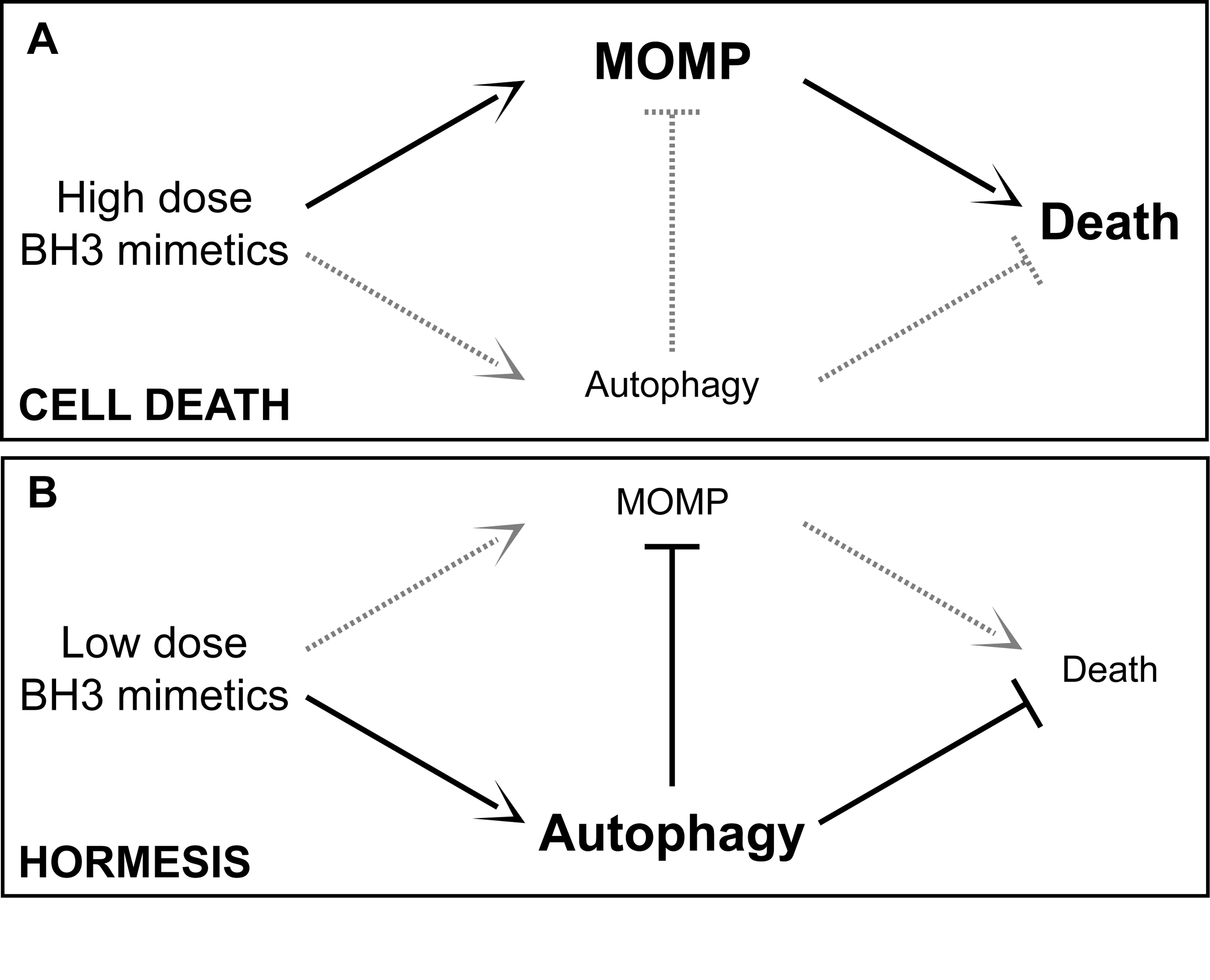
I introduced the concept of hormesis in this blog in November 2009.  My blog posting Hormesis and age retardation describes hormesis as a process of “challenging cells and body systems by mild stress resulting in them becoming stronger and resistant to aging(ref). The stress can be physical, chemical and even possibly psychological.” Exercise is an example. That blog entry reviews the science behind hormesis and some of its demonstrable anti-aging effects. Also, see my blog entry Stress and longevity for further discussion of how moderate stresses confer longevity.

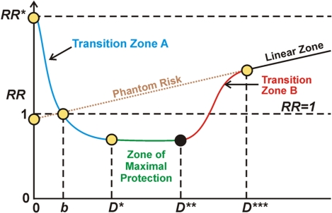
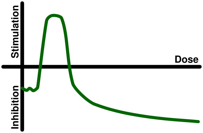
From Wikipedia: “Hormesis (from Greek hórmēsis “rapid motion, eagerness,” from ancient Greek hormáein “to set in motion, impel, urge on”) is the term for generally favorable biological responses to low exposures to toxins and other stressors. A pollutant or toxin showing hormesis thus has the opposite effect in small doses as in large doses. — In toxicology, hormesis is a dose response phenomenon characterized by a low dose stimulation, high dose inhibition, resulting in either a J-shaped or an inverted U-shaped dose response.  The hormesis model of dose response is vigorously debated.[1] The notion that hormesis is a widespread or important phenomenon in biological systems is not widely accepted.[2]The biochemical mechanisms by which hormesis works are not well understood. It is conjectured that low doses of toxins or other stressors might activate the repair mechanisms of the body. The repair process fixes not only the damage caused by the toxin, but also other low-level damage that might have accumulated before without triggering the repair mechanism. Image may be NSFW.
Clik here to view.


Image source

A good review of the current status of hormesis is provided in the 2011 e-publication Hormesis pervasiveness and its potential implications for pharmaceutical research and development. “ The volume of literature published in this area each year has grown 10-fold in large part due to single, original papers in a variety of high impact factor, scientific, peer-reviewed international journals, but also due to entire volumes of journals which have been devoted to hormesis. Some of the various areas include aging, benign prostate enlargement, biochemical and physiological cellular responses, caloric restriction, cardiovascular function, cancer and tumor development, chemo-sensitization, chemotherapy, dermatology, drug binding, hair growth, sexual dysfunction, ocular diseases, osteoporosis, oxidative stress, prion diseases and synaptic plasticity (Calabrese 2008a, Maynard et al. 2008). Four special volumes concentrated on neuroscience, including neuronal survival, neurite outgrowth, glial adaptive responses to neurotoxins, p-glycoprotein efflux transporter activity, anxiety and anxiolytic drugs, epilepsy, traumatic brain injury, stroke, addiction, memory and Alzheimer’s Disease (Calabrese 2008b).”

Historically, many scientists believed that hormesis, or “a little stress is a good thing,” was somewhat of a magical belief not fully to be trusted despite evidence supporting it.  As evidence for hormesis builds up, however, that skepticism may be slowly dissolving.  As pointed out below, I believe it is no longer true that “ The biochemical mechanisms by which hormesis works are not well understood.”

Radiation hormesis, though still controversial, is actually an old topic.

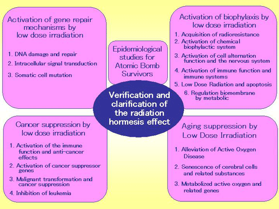
It has been studied in Japan since 1982 in multiple research centers.  The Nuclear Technology Research Center there has published a historical timetable of early events.

Image sourceImage may be NSFW.
Clik here to view.

Among the early studies from Japan, a 1993 publication  even went so far as to suggest that low dose radiation might be used as a protective measure to prevent lung cancer metastasis(ref).

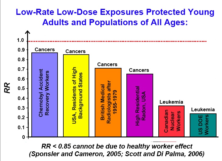
Epidemiological evidence of various kinds exists for the radiation hormesis hypothesis.  For example, studies indicate that mortality of populations varies inversely with altitude and amount of natural background radiation.

The June 2012 publication Cancer mortality, state mean elevations, and other selected predictorsreports: “This ecological inquiry compares cancer mortality rates in the U.S. to the predictor of natural background radiation (via land elevation means) along with eight other predictors thought to be associated with cancer mortality. Age-adjusted cancer mortality in 2006 was compared to the predictors of mean land elevation, percent of smokers, educational attainment, percent of population without health insurance, income, obesity, health perception, physical activity, and diet. Among the six predictors considered appropriate for multiple linear regression, three were found to be statistically significant; from strongest to weakest, these three were: smoking, land elevation, and educational attainment. The predictors of smoking and educational attainment have long been considered associated with cancer mortality. The finding that the predictor of land elevation / natural background radiation is inversely related to cancer mortality is another piece of evidence supporting the theory of radiationhormesis. In this study, land elevation / natural background radiation ranked second in predictive strength regarding cancer mortality, behind smoking and ahead of educational attainment. Since this is an ecological inquiry, no causal inferences can be made.”

Other epidemiological studies related to radiation hormesis have looked at cancer risk.  For example, the December 2010 review publication [Cancer incidence and mortality after low-dose radiation exposure: epidemiological aspects]reports:“Current recommendations for limiting exposure to ionizing radiation are based on the linear-no-threshold (LNT) model for radiation carcinogenesis under which every dose, no matter how low, carries with it some cancer risk. In this review, epidemiological evidences are discussed that the LNT hypothesis is incorrect at low doses. A large set of data was accumulated that showed that cancer risk after ordinarily encountered radiation exposure (natural background radiation, medical X-rays, etc.) is much lower than projections based on the LNT model. The discovery of the low-level radiation hormesis (stimulating effect) implies a non-linear dose-response curve in the low-dose region. The further studies in this field will provide new insights about the mechanisms of radiation carcinogenesis.”

An epidemiological study looking at radon gas exposure risk also points to the presence of radiation hormesis.  The December 2010 publication Epidemiological Evidence for Possible RadiationHormesis from Radon Exposure: A Case-Control Study Conducted in Worcester, MA reported: “Data from a case-control study of lung cancer and residential radon exposure conducted in Worcester County, Massachusetts, are presented. Lung cancer risk was estimated using conditional logistic regression models that controlled for demographic, smoking, and occupational exposure covariates. Preliminary exploratory analyses using lowess smoothing revealed a non-linear association between exposure and the log odds of lung cancer. Radon exposure was considered by using linear spline terms in order to model this nonlinearity. The best fit of this linear spline model to these data predicted a shift from a positive to a negative slope in the log-odds of lung cancer at a radon concentration of 70 Bq m(-3). A statistically significant decrease in cancer risk with increased exposure was found for values ≤ 157 Bq m(-3) normalized to the reference exposure of 4.4 Bq m(-3), the lowest radon concentration measured(adjusted odds ratio (AOR) [95% CI] = 0.42 [0.180, 1.00], p = 0.049). This result is consistent with those reported elsewhere that considered radon exposure with cubic spline terms (Thompson, RE et al. 2008). Furthermore, this model predicts an AOR that is numerically less than 1.0 for radon exposures up to 545 Bq m(-3) versus the above baseline, reference exposure.”

 Image may be NSFW.
Clik here to view.

Graphic depiction of the stochastic hormetic relative risk model as given in Scott et al. (2009).

 Image may be NSFW.
Clik here to view.

Image source

Biological experiments also establish the existence of radiation hormesis.

For example, the January 2012 publication Low-dose-rate, low-dose irradiation delays neurodegeneration in a model of retinitis pigmentosa reports: “The existence of radiation hormesis is controversial. Several stimulatory effects of low-dose (LD) radiation have been reported to date; however, the effects on neural tissue or neurodegeneration remain unknown. Here, we show that LD radiation has a neuroprotective effect in mouse models of retinitis pigmentosa, a hereditary, progressive neurodegenerative disease that leads to blindness. Various LD radiation doses were administered to the eyes in a retinal degeneration mouse model, and their pathological and physiological effects were analyzed. LD gamma radiation in a low-dose-rate (LDR) condition rescues photoreceptor cell apoptosis both morphologically and functionally. The greatest effect was observed in a condition using 650 mGy irradiation and a 26 mGy/minute dose rate. Multiple rounds of irradiation strengthened this neuroprotective effect. A characteristic up-regulation (563%) of antioxidative gene peroxiredoxin-2 (Prdx2) in the LDR-LD-irradiated retina was observed compared to the sham-treated control retina. Silencing the Prdx2 using small-interfering RNA administration reduced the LDR-LD rescue effect on the photoreceptors. Our results demonstrate for the first time that LDR-LD irradiation has a biological effect in neural cells of living animals. The results support that radiation exhibits hormesis, and this effect may be applied as a novel therapeutic concept for retinitis pigmentosa and for other progressive neurodegenerative diseases regardless of the mechanism of degeneration involved.”

Another study showing a radiation hormesis effect on the cell level is reported in the 2011 publication The low-dose ionizing radiation stimulates cell proliferation via activation of the MAPK/ERK pathway in rat cultured mesenchymal stem cells: “Hormesis induced by low-dose ionizing radiation (LDIR) is often mirrored by its stimulation of cell proliferation. The mitogen-activated protein kinases (MAPK)/ extracellular-signal- regulated kinases (ERK) pathway is known to play important roles in cell growth. Therefore, this study was to examine the effects of LDIR on rat mesenchymal stem cell (MSC) proliferation and MAPK/ERK signaling pathway. Rat MSCs were isolated from the bone marrow from 6 to 8-week-old male Wistar rats and cultured in vitro. Exponentially growing cells within 4-5 passages were irradiated with low doses of X-rays at 20, 50, 75 and 100 mGy with a dose rate of 100 mGy/min. Cell proliferation was evaluated by counting total viable cell number with trypan-blue staining and MTT assay. Cell cycle changes were also evaluated by flow cytometry and the activation of MAPK/ERK signaling pathway was assayed by Western blotting. Results showed that LDIR at 50 and 75 mGy significantly stimulated the proliferation of rat MSCs with the most stimulating effect at 75 mGy. There was a significant increase in the proportion of S phase cells in MSCs in response to 75 mGy X-rays. Activation of several members in the MAPK/ERK signaling pathway, including c-Raf, MEK and ERK were observed in the cells exposed to 75 mGy X-rays. To define the role of ERK activation in LDIR-stimulated cell proliferation, LDIR-treated MSCs were pre-incubated with MEK specific inhibitor U0126, which completely abolished LDIR-increased phosphorylation of ERK and cell proliferation. These results suggest that LDIR stimulates MSC proliferations in the in vitro condition via the activation of MAPK/ERK pathway.”

Of course, there are fruit fly studies showing radiation hormesis.  The 2009 publication [Low-dose rate irradiation induced hormesis, hypersensitivity and adaptive response in Drosophila melanogaster of radiosensitive strains] reports: “We have studied the adaptive response after chronic low dose irradiation (2.5 mGy/h) in wild type Drosophila melanogaster strains (Canton-S and Oregon-R), as well as mutant strains on DNA damage sensing (mei-41), DNA repair (mus209, mus210, mus309, rad54) and free radicals detoxification (sod). The effects of irradiation on the prolongation of the larval stage, pupa lethality, and imago whole body weight have been analyzed. The high dose irradiation (30 Gy, 0.05 Gy/s) induced prolongation of the prepupal period and lethality in all wild type and mutant strains under investigation. The chronic low dose irradiation resulted in shortening of the larval development period (hormetic effect) was observed in wild type (Oregon-R) and mutant (mus209) strains (absorbed dose was 20 cGy) of Drosophila. At the same time these strains demonstrated the hormetic effect after chronic low dose irradiation. The hypersensitivity effect was found in sod and rad54 larvae (20 cGy, prolongation of the prepural period), and rad54 and mei-41 pupa (40 cGy, increase of death rate). The larvae of hypersensitive strains and pupa of all strains under investigation did not have the adaptive response. The chronic irradiation in 6 and 60 cGy with the dose rate of 0.25 and 2.5 mGy/h induced the hormetic effect (imago whole body weight enhancement) Canton-S. The obtained results suggest the important role of free radical detoxification, of DNA damage sensing, and of DNA repair mechanisms in the whole organism radiation induced effects. The appearance of the adaptive response depends on the investigated effect and developmental stage of fly.”

The 2008 publication Evidence for radiation hormesis after in vitro exposure of human lymphocytes to low doses of ionizing radiation reports another of several cell-level studies.  “Previous research has demonstrated that adding a very small gamma-ray dose to a small alpha radiation dose can completely suppress lung cancer induction by alpha radiation (a gamma-ray hormetic effect). Here we investigated the possibility of gamma-ray hormesis during low-dose neutron irradiation, since a small contribution to the total radiation dose from neutrons involves gamma rays. Using binucleated cells with micronuclei (micronucleated cells) among in vitro monoenergetic-neutron-irradiated human lymphocytes as a measure of residual damage, we investigated the influence of the small gamma-ray contribution to the dose on suppressing residual damage. We used residual damage data from previous experiments that involved neutrons with five different energies (0.22-, 0.44-, 1.5-, 5.9-, and 13.7-million electron volts [MeV]). Corresponding gamma-ray contributions to the dose were approximately 1%, 1%, 2%, 6%, and 6%, respectively. Total absorbed radiation doses were 0, 10, 50, and 100 mGy for each neutron source. We demonstrate for the first time a protective effect (reduced residual damage) of the small gamma-ray contribution to the neutron dose. Using similar data for exposure to gamma rays only, we also demonstrate a protective effect of 10 mGy (but not 50 or 100 mGy) related to reducing the frequency of micronucleated cells to below the spontaneous level.”

Hormesis effects have been observed in a small study of interventional radiologists.

The February 2012 publication Cellular adaptive response to chronic radiation exposure in interventional cardiologists reports: “Aims: Invasive cardiologists are the most exposed to ionizing radiation among health professionals and show an increased rate of somatic DNA damage. To evaluate the effects of chronic low-dose exposure to ionizing radiation on redox state and apoptotic activation. Methods and results: We enrolled 10 healthy exposed professionals (all interventional cardiologists, Group II, exposed: age = 38 ± 5 years) and 10 age- and gender-matched unexposed controls (Group I, non-exposed). Exposed subjects had a median exposure of 4 mSv/year (range 1-8) by film badge dosimetry (below lead apron). We measured reduced glutathione (GSH, a marker of antioxidant response) in erythrocytes and plasma generation of hydrogen peroxide (a marker of oxyradical stress) by ferrous oxidation-xylenol orange assay in plasma. In both groups, lymphocytes were isolated and caspase-3 activity (a marker of apoptotic response) measured at baseline and following 2 Gy in vitro irradiation. Exposed subjects showed a three-fold increase in hydrogen peroxide (Group I = 2.21 ± 1.03 vs. II = 6.51 ± 1.55 μM H(2)O(2) equivalents) and a 1.7-fold increase in GSH (I = 12.37 ± 1.22 vs. II = 20.61 ± 2.16 mM). Exposed subjects also showed higher values of caspase-3 activity, both at baseline and-more strikingly-following high-dose radiation challenge. Conclusion: In interventional cardiologists, chronic exposure to low-dose radiation is associated with an altered redox balance mirrored by an increase in hydrogen peroxide and with two possibly adaptive cellular responses: (i) an enhanced antioxidant defence (increase in GSH, counteracting increased oxyradical stress) and (ii) an increased susceptibility to apoptotic induction which might efficiently remove genetically damaged cells.”

After 30 years the relevance and even existence of radiation hormesis remains controversial.  Some have argued that the statistical analyses establishing radiation hormesis are faulty.  Other publications suggest that not enough is known about it to justify changing current radiation safety standards..

The 2012 publication A meta-analysis of evidence for hormesis in animal radiation carcinogenesis, including a discussion of potential pitfalls in statistical analyses to detect hormesis is a case in point: “ A database containing 800 datasets on the incidence of specific tumor types from 262 radiation carcinogenicity experiments identified in a comprehensive literature search through September 2000 was analyzed for evidence of hormesis. This database includes lifetime studies of tumorigenic responses in mice, rats, and dogs to exposures to alpha, beta, gamma, neutron, or x-ray radiation. A J-shaped dose response, in the form of a significant decreased response at some low dose followed by a significant increased response at a higher dose, was found in only four datasets from three experiments. Three of these datasets involved the same control animals and two also shared dosed animals; the J shape in the fourth dataset appeared to be the result of an outlier within an otherwise monotonic dose response. A meta-analysis was conducted to determine whether there was an excess of dose groups with decreases in tumor response below that in controls at doses below no-observed-effect levels (NOELs) in individual datasets. Because the probability of a decreased response is generally not equal to the probability of an increased response even in the null case, the meta-analysis focused on comparing the number of statistically significant diminished responses to the number expected, assuming no dose effect below the NOEL. Only 54 dose groups out of the total of 2579 in the database had doses below the dataset-specific NOEL and that satisfied an a priori criterion for sufficient power to detect a reduced response. Among these 54, a liberal criterion for defining a significant decreases identified 15 such decreases, versus 54 × 0.2 = 10.8 expected. The excess in significant reductions was accounted for almost entirely by the excess from neutron experiments (10 observed, 6.2 expected). Nine of these 10 dose groups involved only 2 distinct control groups, and 2 pairs from the 10 even shared dosed animals. Given this high degree of overlap, this small excess did not appear remarkable, although the overlap prevented a formal statistical analysis. A comprehensive post hoc evaluation using a range of NOEL definitions and alternative ways of restricting the data entering the analysis did not produce materially different results. A second meta-analysis found that, in every possible low dose range ([0, d] for every dose, d) of each of the radiation types, the number of dose groups with significantly increased tumorigenic responses was either close to or exceeded the number showing significantly reduced responses. This meta-analysis was considered to be the more definitive one. Not only did it take dose into account by looking for consistent evidence of hormesis throughout defined low-dose ranges, it was also potentially less susceptible to limitations in experimental protocols that would cause individual animals to respond in a non-independent fashion. Overall, this study found little evidence in a comprehensive animal radiation database to support the hormesis hypothesis. However, the ability of the database to detect a hormetic effect was limited both by the small number of dose groups with doses below the range where positive effects have been found in epidemiological studies (≤ 0.1 Gy) and by the limited power of many of these dose groups for detecting a decrease in response.”

Another example, the June 2012 publication Radiationhormesis: Autophagy and other cellular mechanisms reports: “Purpose: To review the cellular mechanisms of hormetic effects induced by low dose and low dose rate ionising radiation in model systems, and to call attention to the possible role of autophagy in some hormetic effects. Results and conclusions: Very low radiation doses stimulate cell proliferation by changing the equilibrium between the phosphorylated and dephosphorylated forms of growth factor receptors. Radioadaptation is induced by various weak stress stimuli and depends on signalling events that ultimately decrease the molecular damage expression at the cellular level upon subsequent exposure to a moderate radiation dose. Ageing and cancer result from oxidative damage under oxidative stress conditions; nevertheless, ROS are also prominent inducers of autophagy, a cellular process that has been shown to be related both to ageing retardation and cancer prevention. A balance between the signalling functions and damaging effects of ROS seems to be the most important factor that decides the fate of the mammalian cell when under oxidative stress conditions, after exposure to ionising radiation. Not enough is yet known on the pre-requirements for maintaining such a balance. Given the present stage of investigation into radiationhormesis, the application of the conclusions from experiments on model systems to the radiation protection regulations would not be justified.”

There has been a Growing understanding of the epigenetic mechanisms through which radiation hormesis works.

An important contribution in this regard was the 2009 publication Radiation-Stimulated Epigenetic Reprogramming of Adaptive-Response Genes in the Lung: An Evolutionary Gift for Mounting Adaptive Protection Against Lung Cancer.  It establishes, among other things, that the hormetic protective effects of low-level gamma ray exposure can protect against lung cancers induced by alpha-ray exposure.  Humans are continuously exposed to low-level ionizing radiation from natural sources. However, harsher radiation environments persisted during our planet’s early years and mammals survived via an evolutionary gift – a system of radiation-induced natural protective measures (adaptive protection). This system includes antioxidants, DNA repair, apoptosis of severely damaged cells, epigenetically regulated apoptosis (epiapoptosis) pathways that selectively remove precancerous and other aberrant cells, and immunity against cancer. We propose a novel model in which the protective system is regulated at least in part via radiation-stress-stimulated epigenetic reprogramming (epireprogramming) of adaptive-response genes. High-dose radiation can promote epigenetically silencing of adaptive-response genes (episilencing), for example via promoter-associated DNA and/or histone methylation and/or histone deacetylation. Evidence is provided for low linear-energy-transfer (LET) radiation-activated natural protection (ANP) against high-LET alpha-radiation-induced lung cancer in plutonium-239 exposed rats and radon-progeny-exposed humans. Using a revised hormetic relative risk model for cancer induction that accounts for both epigenetic activation (epiactivation) and episilencing of genes, we demonstrate that, on average, >80% of alpha-radiation-induced rat lung cancers were prevented by chronic, low-rate gamma-ray ANP. Interestingly, lifetime exposure to residential radon at the Environmental Protection Agency’s action level of 4 pCi L−1 appears to be associated with on average a > 60% reduction in lung cancer cases, rather than an increase. We have used underlined italics to indicate newly introduced terminology.”
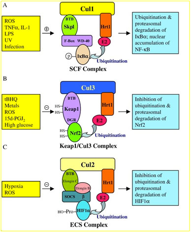
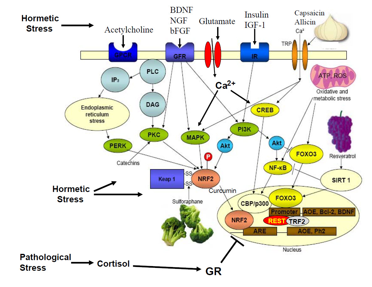
The single most-important biological pathway responsible for radiation hormesis is probably the activation of Nrf2 by radiation-created oxidative stress.

This observation became clear to me as an “of course” matter as soon as I begin digging into the area of radiation hormesis.  I have written extensively about Nrf2 in this blog.  Nrf2 is the master regulator of the body’s response to stresses of most kinds.  Activating hundreds of the body’s natural antioxidant and stress defense genes, the Nrf2/Keap1 pathway provides a central mechanism for most if not all forms of hormesis.  Specifically, you can see the blog entries The pivotal role of Nrf2. Part 1 – a new view on the control of oxidative damage and generation of hormetic effects, The pivotal role of Nrf2. Part 2 – foods, phyto-substances and other substances that turn on Nrf2 and The pivotal role of Nrf2. Part 3– Is promotion of Nrf2 expression a viable strategy for human human healthspan and lifespan extension?. 

Lending credence to this perception is the December 2011 publication Ionizing radiation activates the Nrf2 antioxidant response.  “The transcription factor NF-E2-related factor 2 (Nrf2) binds the antioxidant DNA response element (ARE) to activate important cellular cytoprotective defense systems. Recently several types of cancers have been shown to overexpress Nrf2, but its role in the cellular response to radiation therapy has yet to be fully determined. In this study, we report that single doses of ionizing radiation from 2 to 8 Gy activate ARE-dependent transcription in breast cancer cells in a dose-dependent manner, but only after a delay of five days. Clinically relevant daily dose fractions of radiation also increased ARE-dependent transcription, but again only after five days. Downstream activation of Nrf2-ARE-dependent gene and protein markers, such as heme oxygenase-1, occurred, whereas Nrf2-deficient fibroblasts were incapable of these responses. Compared with wild-type fibroblasts, Nrf2-deficient fibroblasts had relatively high basal levels of reactive oxygen species that increased greatly five days after radiation exposure. Further, in vitro clonogenic survival assays and in vivo sublethal whole body irradiation tests showed that Nrf2 deletion increased radiation sensitivity, whereas Nrf2-inducing drugs did not increase radioresistance. Our results indicate that the Nrf2-ARE pathway is important to maintain resistance to irradiation, but that it operates as a second-tier antioxidant adaptive response system activated by radiation only under specific circumstances, including those that may be highly relevant to tumor response during standard clinical dose-fractionated radiation therapy.”

As a matter if fact, radiation oncologists have been mainly concerned with Nrf2 and its hormetic effects because they could reduce the traditional cancer cell killing impacts of radiation cancer therapies.  Another relevant publication in this regard is the 2010 report Relationship between Radiosensitivity and Nrf2 Target Gene Expression in Human Hematopoietic Stem Cells.  “NFE2-related factor 2 (Nrf2), which belongs to the cap “n” collar family of basic region leucine zipper transcription factors, is a key protein in the coordinated transcriptional induction of expression of various antioxidant genes. The purpose of this study was to analyze the expression of Nrf2 target genes, such as heme oxygenase 1 (HO-1), ferritin heavy polypeptide 1 (FTH1), NAD(P)H dehydrogenase, quinone 1 (NQO1), glutamate-cysteine ligase catalytic subunit, glutamate-cysteine ligase modifier subunit, glutathione reductase (GSR) and thioredoxin reductase 1 (TXNRD1), after X irradiation of CD34+ cells that were prepared from human placental/umbilical cord blood hematopoietic stem cells (HSCs). We evaluated the relationship between radiosensitivity and expression of Nrf2 target genes in HSCs. The number of colony-forming cells derived from 2-Gy-irradiated HSCs decreased to approximately 20% of the nonirradiated control. At the same time, the mRNA expression of HO-1, FTH1, NQO1, GSR and TXNRD1 was significantly increased after X irradiation. A statistically significant negative correlation was observed between the surviving fraction of HSCs and the intrinsic NQO1 mRNA expression, indicating that HSCs in which NQO1 mRNA levels are low may also be radioresistant. The present results suggest that the antioxidant system associated with Nrf2 is involved in the radiosensitivity of HSCs.”

It is interesting though that in the publication of those directly concerned with radiation hormesis, I have yet to come across a mention of Nrf2.

Radiation hormesis is still not a mainline view

There is epidemiological, experimental and genetic pathway evidence for the existence of radiation hormesis.  However, given political and social situations, the radiation safety establishment has chosen to view even low-level radiation as unsafe, despite a growing body of scientific evidence contrary to this view.

This situation is characterized in the April 2012 publication Hormesis: A peep in to the human nature.  “Hormesis is a term to denote biphasic dose response to an agent which reveals a stimulatory or beneficial effects at low dose and an inhibitory or toxic consequence at a higher dose or concentration. Hormesis is a concept which is involved to biphasic dose response effects of environmental agents including ionizing radiation. Proponents of anti-nuclear lobby brand the idea of hormesis as the privilege of the cranks, and the ill informed. UNSECAR in 1958 and 1 CRP in 1959 adopted linear non threshold theory (LNT) in contrast to hormetic or biphasic relation. These organizations based their recommendation by extrapolation of the studies done on atomic bomb survivors who had received higher doses of radiation. According to LNT theory (a) The effect of low doses or radiation can be estimated by linear extrapolation from effects observed by high doses, and (b) There are not any safe doses as even very low doses of ionizing radiation produce some biological effect (Mortazan). — Science dislikes any vacuum of ideas, experiments, and evidence.  – It is generally assumed that science is based on specific results which is independent of personalities; in other words, the interpretation is secular. However, in reality, interpretation of results may to an extent, depend on the position of the interpreter.  The frenzied debates about hormesis in radiation or global warming attest to the above assertion. Jamea Miller observed mutation following radiation of Drosophila. He assumed a linear relation between radiation and mutation though his experiments did not include very low doses of radiation. Catastrophe following nuclear war on Hiroshima and Nagasaki has resulted in radiophobia and an aversion to the concept of hormesis. The evidence for hormesis is accumulating. According to UNSECAR (1994), amongst A-bomb survivors from Hiroshima and Nagasaki, who received doses below 200 mSv, there was no incidence of cancer deaths. And deaths due to leukemia in the sub group who received less than 100 mSv was less than age matched controls. Nambi and Soman in 1987 have demonstrated a significantly reduced death due to cancer in high back ground areas versus areas of low background radiation in Kerala. In a Canadian survey the mortality caused by cancer at nuclear plants was 58% lower than national average. (Abbat 1983). Thus, there is no evidence of harmful effects of radiation at lower doses. Yet, most people tend to go by LNT model including regulatory authorities. — There are instances in the history of medicine where intuitive decision have been disastrous. It took a while to establish a link between administration of saturated oxygen and retrolenticular fibroblasia in neonates. It was felt that life sustaining oxygen can never harm human life even at higher concentration. Thus, establishing a casual link between retrolenticular hyperplasia and saturated oxygen took considerable time by which time innumerable neonates were affected. The biphasic action of chlorpromazine as protector and senstiser at various concentrations is yet another instance of concentration dependent behavior. There are innumerable such examples of biphasic phenomenon. — Hormesis is integral to the normal physiological function of cells and organisms (Mark Mattson) Adaptive responses evolve due to biphasic response to many physical and chemical stimuli. Hormesis also has played a crucial role in the evolution of life. Life had to evolve despite the harsh environmental conditions, including higher cosmic radiation that is seen now. Cellular mechanism including signaling pathways responsible for adaptive pathways are emerging. Now, there is a mechanistic explanation for a possible hermetic response for a wide range of stimulants, including that for ionizing radiation. — Radiophobia has propelled many a nuclear activitists and scientists in to a narrow tunnel vision, obscuring the evidence, as well as disabling a congent thinking. Men are no Drosophila or cells in the Petri dish . Complex biological systems behave much differently than most people concede. Failure to acknowledge hormesis is also a failure to realize essential realities of biological complexity.”

There is a slow broadening of views in the area of radiobiology to where hormesis is being accepted.

The June 2012 publication Changing paradigms in radiobiologysummarizes the nature of this shift: “The last 25 years have seen a major shift in emphasis in the field of radiobiology from a DNA-centric view of how radiation damage occurs to a much more biological view that appreciates the importance of macro-and micro-environments, hierarchical organization, underlying genetics, evolution, adaptation and signaling at all levels from atoms to ecosystems. The new view incorporates concepts of hormesis, nonlinear systems, bioenergy field theory, uncertainty and homeodynamics. While the mechanisms underlying these effects and responses are still far from clear, it is very apparent that their implications are much wider than the field of radiobiology. This reflection discusses the changing views and considers how they are influencing thought in environmental and medical science and systems biology.”

For years, voices have been raised calling for acknowledgement of the existence of radiation hormesis and for revision of the LNT (linear no-threshold) standards for radiation protection which state that radiation exposure at any dose is biologically damaging.  These calls have not gotten anywhere so far. 

The 2008 publication It’s time for a new low-dose-radiation risk assessment paradigm–one that acknowledges hormesis is one of many over the years calling for revision of the LNT.  “The current system of radiation protection for humans is based on the linear-no-threshold (LNT) risk-assessment paradigm. Image may be NSFW.
Clik here to view.
Perceived harm to irradiated nuclear workers and the public is mainly reflected through calculated hypothetical increased cancers. The LNT-based system of protection employs easy-to-implement measures of
radiation exposure. Such measures include the equivalent dose (a biological-damage-potential-weighted measure) and the effective dose (equivalent dose multiplied by a tissue-specific relative sensitivity factor for stochastic effects). These weighted doses have special units such as the sievert (Sv) and millisievert (mSv, one thousandth of a sievert). Radiation-induced harm is controlled via enforcing exposure limits expressed as effective dose. Expected cancer cases can be easily computed based on the summed effective dose (person-sievert) for an irradiated group or population. Yet the current system of radiation protection needs revision because radiation-induced natural protection (hormesis) has been neglected. A novel, nonlinear, hormetic relative risk model for radiation-induced cancers is discussed in the context of establishing new radiation exposure limits for nuclear workers and the public.”

Given that individuals may vary significantly in their hormetic responses to radiation, establishing standards to replace LNT could be a daunting task.

Wrapping it up

It is unfortunate that radiation safety standards and therefore radiation hormesis seem to be so bound up with nuclear energy safety issues, issues of great public concern where most people want to be on the safe side.  It is thought by some that radiation hormesis is mainly an argument put forward by the nuclear industry to lessen the regulatory burden on it, and this may to some extent be so.  However, science is science.

My personal opinion is that radiation hormesis is real and important, first because of the collective research evidence to this effect, second because it is very well established that controlled oxidative stress induces hormesis, so it can be expected that free radicals produced by radiation would produce such stress and induce hormesis, and third, because the key biological pathway, the Nrf2/Keap1 pathway, is now understood. 

Radiation hormesis is ancient and evolutionary but good news, providing us with layers of protection we did not know about.

It also seems clear to me that the LNT standards for radiation safety should be revised given the presence of radiation hormesis, but how much so will require further concerted study and that in turn will require a new public safety initiative.  Fully acknowledging the effects of radiation hormesis conceivably could significantly reduce public concern for radiation safety from x-rays and radon as well as nuclear power plants.

Mitohormesis


By Vince Giuliano

This blog is about Mitohormesis, a different form of hormesis than that discussed in the previous blog entry Radiation Hormesis.  Mitohormesis has to do with cell metabolic pathways and oxidative stress, topics I have discussed in many previous blog entries.  The proper functioning of mitohormesis has to do with multiple aspects of health and with extended organism longevity in many species, quite probably including our own.  And the absence of proper mitohormesis functioning has been shown to be associated with certain disease processes and shortened lifespans in a range of animals including humans.  As is the case for radiation hormesis, the concept of mitohormesis has been around for some time but has not been universally accepted.  It is only recently being widely acknowledged as an important biological phenomenon.  And only relatively recently are its mechanisms of operation being unveiled.

I am grateful for an intensive round of communication with James P Watson, a follower of this blog and stem cell researcher, which helped me to improve this blog entry.  Also I feel free to selectively quote some of Watson’s comments sent me by e-mail while this blog entry was being researched.  This is not to say that Watson, clearly a very brilliant person, will necessarily agree with all I have to say.

In researching this blog entry I encountered a number of interesting recent research results having to do with topics I have discussed previously including mitochondrial metabolism, the OXPHOS system in general, mitochondrial biogenesis, frataxin, the key roles of SIRT3 in controlling ROS stress in mitochondria, and the health roles of PCG1alpha.  And I started to question whether it makes sense to supplement with PQQ.  And there are pathways which seem to produce health benefits independently of motohormesis, like inhibition of TOR.  Because of the great complexity involved I decided to cover most of those topics in subsequent blog entries – even though they all bear on mitohormesis.  So, my intent is to keep this present blog entry focused on mitohormesis itself, what turns it on and off, and the direct health implications of mitohormesis.  In particular, I deal with the important practical topic of mitohormesis and exercise, and how some antioxidants nullify the health benefits of regular exercise.

Hormesis in General

The first section of the previous blog entry Radiation Hormesis is a short introduction to the topic of hormesis in general, and I will not repeat that introduction here.

What is Mitohormesis?

A definition and summary introduction to mitohormesis is found in the abstract of the 2010 publication How increased oxidative stress promotes longevity and metabolic health: The concept of mitochondrial hormesis (mitohormesis):  Recent evidence suggests that calorie restriction and specifically reduced glucose metabolism induces mitochondrial metabolism to extend life span in various model organisms, including Saccharomyces cerevisiae, Drosophila melanogaster, Caenorhabditis elegans and possibly mice. In conflict with Harman’s free radical theory of aging (FRTA), these effects may be due to increased formation of reactive oxygen species (ROS) within the mitochondria causing an adaptive response that culminates in subsequently increased stress resistance assumed to ultimately cause a long-term reduction of oxidative stress. This type of retrograde response has been named mitochondrial hormesis or mitohormesis, and may in addition be applicable to the health-promoting effects of physical exercise in humans and, hypothetically, impaired insulin/IGF-1-signaling in model organisms. Consistently, abrogation of this mitochondrial ROS signal by antioxidants impairs the lifespan-extending and health-promoting capabilities of glucose restriction and physical exercise, respectively. In summary, the findings discussed in this review indicate that ROS are essential signaling molecules which are required to promote health and longevity. Hence, the concept of mitohormesis provides a common mechanistic denominator for the physiological effects of physical exercise, reduced calorie uptake, glucose restriction, and possibly beyond.”

The July 2011 review publication Extending life span by increasing oxidative stress also argues for the existence of mitohormesis across very diverse species: “Various nutritional, behavioral, and pharmacological interventions have been previously shown to extend life span in diverse model organisms, including Saccharomyces cerevisiae, Caenorhabditis elegans, Drosophila melanogaster, mice, and rats, as well as possibly monkeys and humans. This review aims to summarize published evidence that several longevity-promoting interventions may converge by causing an activation of mitochondrial oxygen consumption to promote increased formation of reactive oxygen species (ROS). These serve as molecular signals to exert downstream effects to ultimately induce endogenous defense mechanisms culminating in increased stress resistance and longevity, an adaptive response more specifically named mitochondrial hormesis or mitohormesis.  Consistently, we here summarize findings that antioxidant supplements that prevent these ROS signals interfere with the health-promoting and life-span-extending capabilities of calorie restriction and physical exercise. Taken together and consistent with ample published evidence, the findings summarized here question Harman’s Free Radical Theory of Aging and rather suggest that ROS act as essential signaling molecules to promote metabolic health and longevity.”

My regular readers will discover in these statements many themes discussed in previous blog entries: Reactive Oxygen Species (ROS) are important signaling molecules(ref),  increased ROS expression up to a certain point may be good instead of bad for you(ref)(ref), the stress induced by ROS leads (via the Nrf2 pathway) to activation of multiple endogenous antioxidant and other protective genes(ref)(ref).  And, in fact, suppression of ROS by taking antioxidants can be health-damaging and life-shortening(ref).  These points were also made in the publications related to radiation hormesis cited in the previous blog entry.  They are also articulated in several of the publications that will be cited here as relevant to mitohormesis.

Hormesis, Nrf2 and Antioxidant supplementation – a unifying framework

Before proceeding further with specific findings reported in the literature, I would like to outline a framework for viewing these findings, findings which otherwise sometimes seem inconsistent.

The major points of this framework are:

  1. I hypothesize that the operation of the the Keap1-Nrf2 pathway is a fundamental mechanism of hormesis.  I do that because the operation of the Keap1-Nrf2 pathway is by itself sufficient to explain most if not all the observed phenomena of mitohormesis and hormesis in general. 

In reviewing hundreds of research articles relating  the Nrf2 pathway, I found hormesis mentioned only rarely.  And, in reviewing the literature of hormesis I likewise find that the keap1-Nrf2 pathway is mentioned only rarely.   Yet my research suggests that everything that is known to happen associated with hormesis is completely explainable by the operation of keap1-Nrf2 pathway, without reference to other pathways normally associated with hormesis such as the heat-shock response.  See the subsection entitled The single most-important biological pathway responsible for radiation hormesis is probably the activation of Nrf2 by radiation-created oxidative stress   in the blog entry Radiation Hormesis.   “I have written extensively about Nrf2 in this blog.  Nrf2 is the master regulator of the body’s response to stresses of most kinds.  Activating hundreds of the body’s natural antioxidant and stress defense genes, the Nrf2/Keap1 pathway provides a central mechanism for most if not all forms of hormesis.  Specifically, you can see the blog entries The pivotal role of Nrf2. Part 1 – a new view on the control of oxidative damage and generation of hormetic effects, The pivotal role of Nrf2. Part 2 – foods, phyto-substances and other substances that turn on Nrf2 and The pivotal role of Nrf2. Part 3– Is promotion of Nrf2 expression a viable strategy for human human healthspan and lifespan extension?.”

In stressing the role of the keap1-Nrf2 pathway in hormesis, I of course acknowledge that this is a simplification and far from tells the whole story.  Many other pathways downstream from keap1-Nrf2 are involved in a hormetic response , namely the ones activated by the AREs (antioxidant response genes) after Nrf2 translocates to the cell’s nucleus.  SIRT3and the heat shock proteins like HPS70 play important  roles in hormesis(ref)(ref), along with and possibly due to expression triggered by Nrf2 expression(ref)(ref)(ref).  L-proline catabolism can also be involved(ref).  HDAC inhibition and histone-dependent gene silencing can be involved.  And the whole subject is wrapped up in the vastly complex subjects of ROS dynamics and cell metabolism involving AMPK, PGC1-alpha, PPAR-gamma, SOD2, OXPHOS, P38, etc..  And of course there are other largely independent body stress-response pathways like the UPR (unfolded protein response)(ref) and the various kinds of DNA repair machinery.  But seeing the keap1-Nrf2 pathway as primary can be a very useful one, because research on Nrf2 has been yielding many insights relevant to understanding the scope and relevancy of hormesis  So, the hypothesis is an application of Occam’s Razor, where simplicity is paramount. 

2.     A key to understanding what is going on in hormesis is understanding how the hormetic dose-response curve works, and this can be formulated in terms of NRF2 expression.

 Image may be NSFW.
Clik here to view.


Graphic depiction of the stochastic hormetic relative risk model as given in Scott et al. (2009).

Understanding the typical dose-response curve associated with hormesis is critical for interpreting  seemingly contradictory research. In my interpretation, the horizontal axis depicts level of stress, say as driven by ROS load.  The vertical axis represents relative risk, level of probable pathological organismic response where normal level is 1.  To the left of the first axis crossing in the diagram (point D) the Keap1-Nrf2 pathway is progressively kicking in but not sufficiently so as to overcome the direct negative effects of ROS stress. So, in Transition zone A there is under-expression of the ARE genes and a negative health condition Between stress  levels b and D***  there is hormetic protection compared to what would be expected given a linear model of negative response to stress and better-than-expected health, due at least in part to activation of Nrf2 and the ARE genes.  The zone of maximum protection is between D* and D**.  Starting at D** to D*** the stress load begins to overwhelm the defensive activities of the ARE genes and the protection becomes less and less until at point D*** the hormetic response associated with ARE gene activation becomes negligible.  In the case of radiation damage at least, beyond point D*** the damage according to conventional wisdom is in linear proportion to the stressor, the amount of radiation.  Phantom risk is theoretical risk for low stress levels that would apply if the linear model were extrapolated for low stress dosages.

According to this model, supplementation with radical-scavenging supplements like vitamins C and E inhibits the ROS which triggers the release of Nrf2 which produces hormesis when the stress is in the hormesis range, i.e between b and D*** on the diagram.  The result of such supplementation is a lower health state.  That is, supplementation with such antioxidants when the stress is within the hormesis range of stress can turn off the stress that activates the keap1-Nrf2 pathway therefore turning off hormesis and can do more harm than good.  It appears, for example, that this is what happens with exercise where taking antioxidants eliminates the hormetic health benefits of the exercise.

On the other hand, taking radical-scavenging supplements can be very useful and even highly desirable when the stress at a level is to the right of point D*** The traditional Harman free radical theory aging and the invoking of traditional antioxidant protection is valid in the linear zone beyond point D*** or even possibly beyond point D**.  Some of the earlier research on free radicals and their damaging effects appears to be concerned with high dosages in this beyond-hormesis region.  I am thinking of publications like ß-Cell Glucose Toxicity, Lipotoxicity, and Chronic Oxidative Stress in Type 2 Diabetes,  Pancreatic islet β-cell and oxidative stress : The importance of glutathione peroxidase  (2007), and Feeding acetyl-L-carnitine and lipoic acid to old rats significantly improves metabolic function with decreasing oxidative stress (2002).  So these early anti-ROS and pro-antioxidant papers are valid in their own domains and not in fact contradicted by later papers that show positive health effects associated with lower levels of ROS stress.  I believe serious nuclear plant radiation overexposure and accidental ingestion of heavy metals are excellent reasons for taking heavy doses of radical-scavenging antioxidants.

Many phytochemical substances present in certain foods and sold as supplements are called “antioxidants,” but actually are not free radical scavengers.  They work by activating Nrf2 and triggering a hormesis response which lowers ROS and produces a wide range of positive health effects.  I discuss these and other substances which activate Nrf2 in the blog entry The pivotal role of Nrf2. Part 2 – foods, phyto-substances and other substances that turn on Nrf2.  I believe most of the phytosubstances mentioned there are useful to trigger hormesis or reinforce it within the hormesis stress range (up to point D*** in the diagram), and in no-way inhibit hormesis.  Of course, they can’t overcome massive doses of ROS which correspond to the region to the right of D***on the hormesis response curve.

Now, moving on to some results reported in the literature:

In some lower organisms, inducing enhanced ROS signaling can lead to mitohormesis and significantly extended lifespans.

The April 2012 publication Impaired insulin/IGF1 signaling extends life span by promoting mitochondrial L-proline catabolism to induce a transient ROS signal reports: “Impaired insulin and IGF-1 signaling (iIIS) in C. elegans daf-2 mutants extends life span more than 2-fold. Constitutively, iIIS increases mitochondrial activity and reduces reactive oxygen species (ROS) levels. By contrast, acute impairment of daf-2 in adult C. elegans reduces glucose uptake and transiently increases ROS. Consistent with the concept of mitohormesis, this ROS signal causes an adaptive response by inducing ROS defense enzymes (SOD, catalase), culminating in ultimately reduced ROS levels despite increased mitochondrial activity. Inhibition of this ROS signal by antioxidants reduces iIIS-mediated longevity by up to 60%. Induction of the ROS signal requires AAK-2 (AMPK), while PMK-1 (p38) and SKN-1 (NRF-2) are needed for the retrograde response. IIIS upregulates mitochondrial L-proline catabolism, and impairment of the latter impairs the life span-extending capacity of iIIS while L-proline supplementation extends C. elegans life span. Taken together, iIIS promotes L-proline metabolism to generate a ROS signal for the adaptive induction of endogenous stress defense to extend life span.”

Image may be NSFW.
Clik here to view.

Image source

Many substances can promote mitohormesis at low doses, even poisons which are highly toxic at higher doses.

An example is the highly toxic pesticide rotenone.   The publicationEffects of Low Concentrations of Rotenone Upon Mitohormesis in SH-SY5Y Cells reports: “The mitochondrial toxin rotenone exerts cytotoxicity viaoverproduction of reactive oxygen species (ROS) and depolarization of the mitochondrial membrane. We investigated the effects of rotenone (12.5, 25, 50, 100 nmol/L) on mitochondrial biogenesis and the potential roles of ROS production in SH-SY5Y cells. Mitochondrial biogenesis was assessed by counting the number of mitochondria, determining protein expression of peroxisome proliferator-activated receptor γ coactivator α (PGC1-α) and its regulator, SIRT1, and oxygen consumption. ROS production and levels of reduced glutathione (GSH) and oxidized glutathione(GSSG) were also determined. Compared with controls, rotenone (12.5 nmol/L) significantly increased the quantity of mitochondria and amount of oxygen consumption, whereas rotenone at >12.5 nmol/L decreased the quantity of mitochondria and amount of oxygen consumption. GSH contents and GSH/GSSG were also significantly enhanced by rotenone at 12.5 nmol/L and decreased by rotenone at >12.5 nmol/L. Except for ROS production and SIRT1 protein expression, all concentration-response relationships showed a typical inverted-U shape. ROS production was continually increased in cells treated with rotenone. These data indicate that low concentrations of rotenone can induce mitohormesis, which may be attributed to ROS production.

James P Watson comments about this finding: “This study will not be replicated in humans!  It does show that even mitochondrial poison pesticides can have a beneficial mitohormetic effect at low doses.  The “mitohormetic ROS dose” is 12.5-25 nmol/L whereas the mitochondrial toxic dose is > 25 nmol/L.  PGC-1alpha and SIRT1 increased with “mitohormetic doses” of rotenone, whereas they both decreased with higher doses.  Again, more proof of mitohormetic dose-response curve.”

One of several substances that promotes mitohormesis in nematodes is the phytochemical glaucarubinone.  The 2011 publication The phytochemical glaucarubinone promotes mitochondrial metabolism, reduces body fat, and extends lifespan of Caenorhabditis elegans reports: “Naturally occurring compounds that promote energy expenditure and delay aging in model organisms may be of significant interest, since these substances potentially provide pharmaceutical approaches to tackle obesity and promote healthy lifespan in humans. We aimed to test whether pharmaceutical concentrations of glaucarubinone, a cytotoxic and antimalarial quassinoid known from different species of the plant family Simaroubaceae, are capable of affecting metabolism and/or extending lifespan in a nematodal model organism for aging processes, the roundworm Caenorhabditis elegans. — Adult C. elegans roundworms, maintained on agar plates, were fed with E. coli strain OP50 bacteria, and glaucarubinone was applied to the agar to test (i) whether it alters respiration rates and mitochondrial activity, (ii) whether it affects body fat content, and (iii) whether it may promote longevity by quantifying survival in the presence and absence of the compound. We have found that glaucarubinone induces oxygen consumption and reduces body fat content of C. elegans. Moreover and consistent with the concept of mitohormesis, glaucarubinone extends C. elegans lifespan when applied at a concentration of 1 or 10 nanomolar. Taken together, glaucarubinone is capable of reducing body fat and promoting longevity in C. elegans, tentatively suggesting that this compound may promote metabolic health and lifespan in mammals and possibly humans.”

Yet another substance that appears to induce a hormetic-like effect in muscle tissues is Stanozolol.  The 2011 publication Stanozolol treatment decreases the mitochondrial ROS generation and oxidative stress induced by acute exercise in rat skeletal musclereports: “Anabolic androgenic steroids are used in the sport context to enhance muscle mass and strength and to increase muscle fatigue resistance. Since muscle fatigue has been related to oxidative stress caused by an exercise-linked reactive oxygen species (ROS) production, we investigated the potential effects of a treatment with the anabolic androgenic steroid stanozolol against oxidative damage induced on rat skeletal muscle mitochondria by an acute bout of exhaustive exercise. Mitochondrial ROS generation with complex I- and complex II-linked substrates was increased in exercised control rats, whereas it remained unchanged in the steroid-treated animals. Stanozolol treatment markedly reduced the extent of exercise-induced oxidative damage to mitochondrial proteins, as indicated by the lower levels of the specific markers of protein oxidation, glycoxidation, and lipoxidation, and the preservation of the activity of the superoxide-sensitive enzyme aconitase. This effect was not due to an enhancement of antioxidant enzyme activities. Acute exercise provoked changes in mitochondrial membrane fatty acid composition characterized by an increased content in docosahexaenoic acid. In contrast, the postexercise mitochondrial fatty acid composition was not altered in stanozolol-treated rats. Our results suggest that stanozolol protects against acute exercise-induced oxidative stress by reducing mitochondrial ROS production, in association with a preservation of mitochondrial membrane properties.”

The September 2011 publication Lonidamine extends lifespan of adult Caenorhabditis elegans by increasing the formation of mitochondrial reactive oxygen species reports: “Compounds that delay aging in model organisms may be of significant interest to antiaging medicine, since these substances potentially provide pharmaceutical approaches to promote healthy lifespan in humans. The aim of the study was to test whether pharmaceutical concentrations of the glycolytic inhibitor lonidamine are capable of extending lifespan in a nematodal model organism for aging processes, the roundworm Caenorhabditis elegans. Several hundreds of adult C. elegans roundworms were maintained on agar plates and fed E. coli strain OP50 bacteria. Lonidamine was applied to test whether it may promote longevity by quantifying survival in the presence and absence of the compound. In addition, several biochemical and metabolic assays were performed with nematodes exposed to lonidamine. — Lonidamine significantly extends both median and maximum lifespan of C. elegans when applied at a concentration of 5 micromolar by 8% each. Moreover, the compound increases paraquat stress resistance, and promotes mitochondrial respiration, culminating in increased formation of reactive oxygen species (ROS). Extension of lifespan requires activation of pmk-1, an orthologue of p38 MAP kinase, and is abolished by co-application of an antioxidant, indicating that increased ROS formation is required for the extension of lifespan by lonidamine. Consistent with the concept of mitohormesis, lonidamine is capable of promoting longevity in a pmk-1 sensitive manner by increasing formation of ROS.

Image may be NSFW.
Clik here to view.

Image source

IMitohormesis appears to be a pathway response preserved by evolution over many species, ranging from yeast cells to nematode worms to humans.

An editorial article in the journal publishing the previous-cited report entitled Alternative mitochondrial fuel extends life span had to say: “In this issue of Cell Metabolism, Ristow and colleagues (Zarse et al., 2012) elucidate a conserved mechanism through which reduced insulin-IGF1 signaling activates an AMP-kinase-driven metabolic shift toward oxidative proline metabolism. This, in turn, produces an adaptive mitochondrial ROS signal that extends worm life span. These findings further bolster the concept of mitohormesis as a critical component of conserved aging and longevity pathways.”

The November 2011 publication Mitochondria, reactive oxygen species, and chronological aging: a message from yeast reports: “As a major intracellular source of reactive oxygen species (ROS), mitochondria are involved in aging and lifespan regulation. Using the yeast chronological aging model, researchers have identified conserved signaling pathways that affect lifespan by modulating mitochondrial functions. Caloric restriction and a genetic mimetic with reduced target of rapamycin signaling globally upregulate the mitochondrial proteome and respiratory functions. — Recent discoveries support the notion that an altered mitochondrial proteome induces mitohormesis. Mitohormesis involves a variety of ROS during several growth stages and extends lifespan in yeast and other organisms. Here we recap recent advances in understanding of ROS as signals that decelerate chronological aging in yeast. We also discuss parallels between yeast and worm hypoxic signaling. In sum, this mini-review covers mitochondrial regulation by nutrient-sensing pathways and the complex underlying interactions of ROS, metabolic pathways, and chronological aging.”

Exploitation of mitohormesis could become an important strategy for prevention and control of heart diseases.

The February 2012 publication Mitochondria as a source and target of lipid peroxidation products in healthy and diseased heart reports:  “The heart is a highly oxidativ1e organ in which cardiomyocyte turnover is virtually absent, making it particularly vulnerable to accumulation of lipid peroxidation products (LPP) formed as a result of oxidative damage. 2. Reactive oxygen and nitrogen species are the most common electrophiles formed during lipid peroxidation and lead to the formation of both stable and unstable LPP. Of the LPP formed, highly reactive aldehydes are a well-recognized causative factor in ageing and age-associated diseases, including cardiovascular disease and diabetes. 3. Recent studies have identified that the mitochondria are both a primary source and target of LPP, with specific emphasis on aldehydes in cardiomyocytes and how these affect the electron transport system and Ca(2+) balance. 4. Numerous studies have found that there are functional consequences in the heart following exposure to specific aldehydes (acrolein, trans-2-hexanal, 4-hydroxynonenal and acetaldehyde). Because these LPP are known to form in heart failure, cardiac ischaemia-reperfusion injury and diabetes, they may have an underappreciated role in the pathophysiology of these disease processes. 5. Lipid peroxidation products are involved in the transcriptional regulation of endogenous anti-oxidant systems.  Recent evidence demonstrates that transient increases in LPP may be beneficial in cardioprotection by contributing to mitohormesis (i.e. induction of anti-oxidant systems) in cardiomyocytes. Thus, exploitation of the cardioprotective actions of the LPP may represent a novel therapeutic strategy for future treatment of heart disease.”

The 2010 publication Activation of mitochondrial energy metabolism protects against cardiac failure spoke to the same basic points: “Cardiac failure is the most prevalent cause of death at higher age, and is commonly associated with impaired energy homeostasis in the heart. Mitochondrial metabolism appears critical to sustain cardiac function to counteract aging. In this study, we generated mice transgenically over-expressing the mitochondrial protein frataxin, which promotes mitochondrial energy conversion by controlling iron-sulfur-cluster biogenesis and hereby mitochondrial electron flux.   Hearts of transgenic mice displayed increased mitochondrial energy metabolism and induced stress defense mechanisms, while overall oxidative stress was decreased. Following standardized exposure to doxorubicin to induce experimental cardiomyopathy, cardiac function and survival was significantly improved in the transgenic mice. The insulin/IGF-1 signaling cascade is an important pathway that regulates survival following cytotoxic stress through the downstream targets protein kinase B, Akt, and glycogen synthase kinase 3. Activation of this cascade is markedly inhibited in the hearts of wild-type mice following induction of cardiomyopathy. By contrast, transgenic overexpression of frataxin rescues impaired insulin/IGF-1 signaling and provides a mechanism to explain enhanced cardiac stress resistance in transgenic mice.   Taken together, these findings suggest that increased mitochondrial metabolism elicits an adaptive response due to mildly increased oxidative stress as a consequence of increased oxidative energy conversion, previously named mitohormesis. This in turn activates protective mechanisms which counteract cardiotoxic stress and promote survival in states of experimental cardiomyopathy. Thus, induction of mitochondrial metabolism may be considered part of a generally protective mechanism to prevent cardiomyopathy and cardiac failure.”

A comment on this publication sent me by James P Watson is: “ This is a transgenic mouse model of chemotherapy-induced CHF. The chemotherapy drug is a common FDA approved drug that causes cardiomyopathy when patients are treated with too much of this drug.  The mechanism that causes the doxorubicin-induced cardiomyopathy is down-regulation at 3 points of the intra-cytoplasmic Insulin/IGF-1 pathway (Protein Kinase B, Akt, and glycogen synthetase kinase 3).   In this experiment, they created a transgenic mouse that over-expressed the mitochondrial protein frataxin, which increases expression of a critical rate-limiting complex in the OxPhos pathway (iron-sulphur-cluster, ISC) and in Kreb’s cycle (aconitase, which is also dependent on ISC).  If a human does not produce enough frataxin, this is called Fredrich’s ataxia and you will die in your 30s from heart failure (along with diabetes, muscle wasting, and ataxia).  In the frataxin-over expressed mice, there was an induction of mitochondrial metabolism and ROS defense.  When given doxorubicin, 85% of the frataxin mice survived instead of 55%  in the wild type mice.”

Statins induce mitohormesis, mitochondrial biogenesis and associated positive health effects in cardiac tissue but do not induce mitohormesis and instead induce excessive ROS stress and negative health effects in skeletal muscles.

This interesting observation is reported in the June 2012 publication Opposite effects of statins on mitochondria of cardiac and skeletal muscles: a ‘mitohormesis’ mechanism involving reactive oxygen species and PGC-1.  AIMS: Statins protect against cardiovascular-related mortality but induce skeletal muscle toxicity. To investigate mechanisms of statins, we tested the hypothesis that statins optimized cardiac mitochondrial function but impaired vulnerable skeletal muscle by inducing different level of reactive oxygen species (ROS).  METHODS AND RESULTS: In atrium of patients treated with statins, ROS production was decreased and oxidative capacities were enhanced together with an extensive augmentation of mRNAs expression of peroxisome proliferator-activated receptor gamma co-activator (PGC-1) family. However, in deltoid biopsies from patients with statin-induced muscular myopathy, oxidative capacities were decreased together with ROS increase and a collapse of PGC-1 mRNA expression. Several animal and cell culture experiments were conducted and showed by using ROS scavengers that ROS production was the triggering factor responsible of atorvastatin-induced activation of mitochondrial biogenesis pathway and improvement of antioxidant capacities in heart. Conversely, in skeletal muscle, the large augmentation of ROS production following treatment induced mitochondrial impairments, and reduced mitochondrial biogenesis mechanisms. Quercetin, an antioxidant molecule, was able to counteract skeletal muscle deleterious effects of atorvastatin in rat.  CONCLUSION:  Our findings identify statins as a new activating factor of cardiac mitochondrial biogenesis and antioxidant capacities, and suggest the importance of ROS/PGC-1 signalling pathway as a key element in regulation of mitochondrial function in cardiac as well as skeletal muscles.” 

The report highlights the importance of dosage in determining whether ROS stress is in the range where the positive effects of hormesis are realized (where the hormetic response curve is above the line), or the stress is too great (where the hormetic response curve is below the line), and the net health result is negative.  The dose response curve must be scaled different for cardiac and muscle tissue.  Apparently, a dose of statins with a response above the line for cardiac tissue produces a result below the line for muscle tissue.

Image may be NSFW.
Clik here to view.
 

A second point of the above publication is the importance of the ROS.PGC-1 signaling pathway in regulation of mitochondrial biogenesis and functions.

For background on PGC-1, see the blog entries PGC-1alpha and exercise, AMPK and longevity and PQQ – activator of PGC-1alpha, SIRT3 and mitochondrial biogenesis.  “PGC-1-alpha is a gene co-activator, necessary to turn on the PPARG gene and essential in the metabolic process. PGC-1-alpha (Peroxisome proliferator-activated receptor gamma coactivator 1-alpha) “is a protein that in humans is encoded by the PPARGC1Agene.[1] The protein encoded by this gene is a transcriptional coactivator that regulates the genes involved in energy metabolism. This protein interacts with the nuclear receptorPPAR-gamma, which permits the interaction of this protein with multiple transcription factors. This protein can interact with, and regulate the activities of, cAMP response element binding protein (CREB) and nuclear respiratory factors (NRFs). It provides a direct link between external physiological stimuli and the regulation of mitochondrial biogenesis, and is a major factor that regulates muscle fiber type determination. This protein may be also involved in controlling blood pressure, regulating cellular cholesterol homoeostasis, and the development of obesity(ref).[2]

Regarding this article, James P Watson had some very interesting comments which he communicated to me privately, as follow. 

“This is a very interesting article that basically say that statins are “good for your heart” and “bad for your skeletal muscles”.  They also clearly established  that this effect was mediated by PGC-1alpha and PGC-1beta in the nucleus and also affected mitochondrial gene expression.  IN BOTH NUCLEAR GENES AND MITOCHONDRIAL GENES,  THIS EFFECT WAS ABOLISHED BY ANTIOXIDATNS!  This is very convincing evidence of an ROS-mediated mitohormetic mechanism in the heart for statins and an opposite mitodestructive mechanism in skeletal muscles with statin therapy.

 If true, this is an example of “organ pleiotropy” – i.e. what is good for one organ is bad for another.  In medicine, we call this “drug side effects”.    In pharmacology, they call this  ”off target effects”.  The study included both an animal study and a human study of both cardiac and skeletal muscle  (well done) and also included a statin + quercetin arm to the study.  Here are the results summarized:

  1. 1.     Animals: This was actually a mitochondrial biogenesis study

Nuclear co-activators and gene expression;

statin alone

=> reduced PGC-1alpha and PGC-1beta in skeletal muscle => reduced Cox1 and NRF1 gene expression

=> increased PGC-1beta in heart, no change in PGC-1alpha in the heart => increased Cox1 and NRF1 gene expression

statin + anti-oxidant (quercetin)

=> abolished all effects in both cardiac and skeletal muscle

mitochondrial gene expression

statin alone

=> reduced Cytochrome b and pyruvate kinase gene expression in skeletal muscle mitochondria

=> increased Cytochrome b and pyruvate kinase gene expression in cardiac muscle mitochondria

statin + quercetin (antioxidant)

=> abolished all effects in both cardiac and skeletal mitochondria

ROS production

statin alone

=> increased ROS in skeletal muscle

=> reduced ROS in cardiac muscle

statin + quercetin

=> abolished effect in skeletal muscle, no change in cardiac muscle

2,  Humans: 

This study did not include anti-oxidants.  It was just a skeletal muscle vs cardiac muscle biopsy study, done during open heart surgery.”

statins

=> reduced PGC-1alpha, PGC-1beta, SOD1, and SOD2 in skeletal muscle

=> increased PGC-1alpha, PGC-1beta, SOD1, and SOD2 in cardiac muscle

My comment (James P Watson, continuing) on this article and diagram:
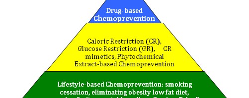
This is the first article that I have seen that shows changes in both nuclear gene expression (of extrinsic mitochondrial genes) and intrinsic mitochondrial gene expression with statins and that this occurs via an ROS-dependent manner.  This study explains why patients on statins live longer than controls and that this may be a mitohormesis effect, and NOT a cholesterol lowering effect of statins.  It also is the first to show “organ pleiotropic” effects of a drug and explains  the known side effect of muscle pain due to statin therapy.  Taking anti-oxidants to prevent this side effect would abolish the beneficial effect of statins on the heart, so anti-oxidant supplementation would NOT be a good idea for patients on statins.

I really like this diagram, because it has one up arrow for ROS production in the heart and four up arrows for ROS production in skeletal muscle. This clearly shows that statins have a “hermetic ROS dose range” for the heart and a “toxic ROS dose range” for skeletal muscle.  This is “proof” that ROS dosing is the clear reason for the difference between hermetic doses and “Denham Harmon doses” of ROS.”

Another publication relating exercise, ROS and the impact of statins is the 2011 publication Atorvastatin treatment reduces exercise capacities in rats: involvement of mitochondrial impairments and oxidative stress.  “Physical exercise exacerbates the cytotoxic effects of statins in skeletal muscle. Mitochondrial impairments may play an important role in the development of muscular symptoms following statin treatment. Our objective was to characterize mitochondrial function and reactive oxygen species (ROS) production in skeletal muscle after exhaustive exercise in atorvastatin-treated rats. — Our results show that exhaustive exercise exacerbated metabolic perturbations and ROS production in skeletal muscle, which may reduce the exercise capacity and promote the muscular symptoms in sedentary atorvastatin-treated animals.” 

By blocking mitohormesis, regular supplementation with antioxidants may block the beneficial effects of regular exercise.

This is a sobering revelation for those who both regularly exercise and take antioxidant supplements, believing that these interventions work synergistically.  A 2009 publication Antioxidants prevent health-promoting effects of physical exercise in humans speaks as to how by blocking mitohormesis, supplementation with Vitamins C and E nullifies the health-producing effects of physical exercise.  “Exercise promotes longevity and ameliorates type 2 diabetes mellitus and insulin resistance. However, exercise also increases mitochondrial formation of presumably harmful reactive oxygen species (ROS). Antioxidants are widely used as supplements but whether they affect the health-promoting effects of exercise is unknown. We evaluated the effects of a combination of vitamin C (1000 mg/day) and vitamin E (400 IU/day) on insulin sensitivity as measured by glucose infusion rates (GIR) during a hyperinsulinemic, euglycemic clamp in previously untrained (n = 19) and pretrained (n = 20) healthy young men. Before and after a 4 week intervention of physical exercise, GIR was determined, and muscle biopsies for gene expression analyses as well as plasma samples were obtained to compare changes over baseline and potential influences of vitamins on exercise effects. Exercise increased parameters of insulin sensitivity (GIR and plasma adiponectin) only in the absence of antioxidants in both previously untrained (P < 0.001) and pretrained (P < 0.001) individuals. This was paralleled by increased expression of ROS-sensitive transcriptional regulators of insulin sensitivity and ROS defense capacity, peroxisome-proliferator-activated receptor gamma (PPARgamma), and PPARgamma coactivators PGC1alpha and PGC1beta only in the absence of antioxidants (P < 0.001 for all). Molecular mediators of endogenous ROS defense (superoxide dismutases 1 and 2; glutathione peroxidase) were also induced by exercise, and this effect too was blocked by antioxidant supplementation. Consistent with the concept of mitohormesis, exercise-induced oxidative stress ameliorates insulin resistance and causes an adaptive response promoting endogenous antioxidant defense capacity. Supplementation with antioxidants may preclude these health-promoting effects of exercise in humans.”

Image may be NSFW.
Clik here to view.

Image source   “Mitohormesis links physical exercise and subsequent formation of reactive oxygen species to insulin sensitivity and antioxidant defense. Physical exercise exerts ameliorating effects on insulin resistance by increasing mitochondrial formation of reactive oxygen species in skeletal muscle to induce expression of PGC1α, PGC1β, and PPARγ as inducers of insulin sensitivity, as well as superoxide dismutases 1 and 2 and glutathione peroxidase 1, key enzymes of ROS defense. Notably, by blocking exercise-dependent formation of reactive oxygen species due to ingestion of antioxidant supplements, health promoting effects of physical exercise are abolished, and physical exercise fails to promote insulin sensitivity and antioxidant defense in the presence of vitamin C and vitamin E.”  I note that this finding seems to be relevant within the hormetic range of the ROS dose-response curve.

The patterns of coupling the taking of antioxidants along with regular physical exercise in the interest of general health is, for many people, deeply entrenched, encouraged by much advertising, and only recently being seriously questioned.  The August 2012 publication Does vitamin C and e supplementation impair the favorable adaptations of regular exercise? does not directly discuss mitohormesis but reports: “The detrimental outcomes associated with unregulated and excessive production of free radicals remains a physiological concern that has implications to health, medicine and performance. Available evidence suggests that physiological adaptations to exercise training can enhance the body’s ability to quench free radicals and circumstantial evidence exists to suggest that key vitamins and nutrients may provide additional support to mitigate the untoward effects associated with increased free radical production. However, controversy has risen regarding the potential outcomes associated with vitamins C and E, two popular antioxidant nutrients. Recent evidence has been put forth suggesting that exogenous administration of these antioxidants may be harmful to performance making interpretations regarding the efficacy of antioxidants challenging. The available studies that employed both animal and human models provided conflicting outcomes regarding the efficacy of vitamin C and E supplementation, at least partly due to methodological differences in assessing oxidative stress and training adaptations. Based on the contradictory evidence regarding the effects of higher intakes of vitamin C and/or E on exercise performance and redox homeostasis, a permanent intake of non-physiological dosages of vitamin C and/or E cannot be recommended to healthy, exercising individuals.” – “The discrepancy among studies described above regarding the influence of vitamin C and/or E supplementation on adaptations in redox homeostasis also applies to the effects of antioxidant supplementation on alterations in redox homeostasis in response to acute exercise. In fact, most of the relevant studies have reported that vitamin C and/or E supplementation does not affect redox status [14, 18, 3640], less studies have reported that attenuates oxidative stress [4145] and there are even reports indicating a pro-oxidant effect [46, 47]. We believe that this “agreement on disagreement” between responses and adaptations to exercise indicates the inherent complexity of redox biochemistry and signifies the difficulty in providing unidirectional predictions after supplementation with antioxidant agents.”  I speculate that these disagreements among studies can be explained by reference to the ROS dose-response curve for hormesis discussed above. .

Long-term antioxidant supplementation, specifically with Vitamin C and alpha-lipoic acid, reduces mitochondrial biogenesis in skeletal muscle tissue by interfering with ROS signaling that triggers mitohormesis.

Antioxidants produce a result similar to that produced by statins as described above, inhibiting mitohormesis,  The June 2012 publication Antioxidant supplementation reduces skeletal muscle mitochondrial biogenesisreports “PURPOSE: Exercise increases the production of reactive oxygen species (ROS) in skeletal muscle, and athletes often consume antioxidant supplements in the belief they will attenuate ROS-related muscle damage and fatigue during exercise. However, exercise-induced ROS may regulate beneficial skeletal muscle adaptations, such as increased mitochondrial biogenesis. We therefore investigated the effects of long-term antioxidant supplementation with vitamin E and α-lipoic acid on changes in markers of mitochondrial biogenesis in the skeletal muscle of exercise-trained and sedentary rats.  METHODS: Male Wistar rats were divided into four groups: 1) sedentary control diet, 2) sedentary antioxidant diet, 3) exercise control diet, and 4) exercise antioxidant diet. Animals ran on a treadmill 4 d · wk at 70%VO2max for up to 90 min · d for 14 wk.  RESULTS: Consistent with the augmentation of skeletal muscle mitochondrial biogenesis and antioxidant defenses, after training there were significant increases in peroxisome proliferator-activated receptor γ coactivator 1α (PGC-1α) messenger RNA (mRNA) and protein, cytochrome C oxidase subunit IV (COX IV) and cytochrome C protein abundance, citrate synthase activity, Nfe2l2, and SOD2 protein (P < 0.05). Antioxidant supplementation reduced PGC-1α mRNA, PGC-1α and COX IV protein, and citrate synthase enzyme activity (P < 0.05) in both sedentary and exercise-trained rats.”

The March 2012 review study Antioxidants and Skeletal Muscle Performance: “Common Knowledge” vs. Experimental Evidence also shows absence of research support for the popular idea that antioxidant supplementation is a good thing to do associated with fitness exercising.  “Antioxidants are assumed to provide numerous benefits, including better health, a reduced rate of aging, and improved exercise performance. Specifically, antioxidants are commonly “prescribed” by the media, supplement industry, and “fitness experts” for individuals prior to training and performance, with assumed benefits of improved fatigue resistance and recovery. This has provoked expansion of the supplement industry which responded by creation of a plethora of products aimed at facilitating the needs of the active individual. However, what does the experimental evidence say about the efficacy of antioxidants on skeletal muscle function? Are antioxidants actually as beneficial as the general populous believes? Or, could they in fact lead to deleterious effects on skeletal muscle function and performance? This Mini Review addresses these questions with an unbiased look at what we know about antioxidant effects on skeletal muscle, and what we still need to know before conclusions can be made.” –  “Conclusion: Experimental evidence does not support the “common knowledge” that antioxidant treatment greatly improves exercise performance and recovery. On the contrary, studies with antioxidant supplementations generally show no effect on muscle function during and after exercise. The exception is NAC treatment, which has been found to improve performance during submaximal exercise. The limited effects of ROS/RNS and antioxidants during exercise are unexpected in that increases in ROS/RNS are likely to occur and these are potentially harmful. It appears that muscle fibers are in some way protected against deleterious effects of oxidants during exercise and fibers are generally much more sensitive to exposure to oxidants in the rested state than during fatigue.”  While mitohormesis is not mentioned, clearly that explains what is happening.

The March 2012 publication Rationale for antioxidant supplementation in sarcopenia sounds a similar note of caution regarding antioxidant supplementation: “Sarcopenia is an age-related clinical condition characterized by the progressive loss of motor units and wasting of muscle fibers resulting in decreased muscle function. The molecular mechanisms leading to sarcopenia are not completely identified, but the increased oxidative damage occurring in muscle cells during the course of aging represents one of the most accepted underlying pathways. In fact, skeletal muscle is a highly oxygenated tissue and the generation of reactive oxygen species is particularly enhanced in both contracting and at rest conditions. It has been suggested that oral antioxidant supplementation may contribute at reducing indices of oxidative stress both in animal and human models by reinforcing the natural endogenous defenses. Aim of the present paper is to discuss present evidence related to possible benefits of oral antioxidants in the prevention and treatment of sarcopenia. – Conclusion: In summary, there is some evidence that oral antioxidant supplementation may reduce muscle damage, but experimental results are largely preliminary and far to be clinically relevant, at least, as suggestive of positive benefits. In fact, a large body of evidence may indicate extreme cautiousness in taking antioxidant supplementation as preventive measures against aging process and age-related conditions. Further studies are needed to support the widespread practice of oral antioxidant supplementation and to determine appropriate recommendations in elderly. — ”  Although this article contains significant discussion on ROS during exercise and the body’s endogenous antioxidant defense system, no mention is made of the key mechanisms of mitohormesis and the actions of Nrf2.  Instead, seemingly puzzled, the authors suggest a need for more research to relate sarcopenia to antioxidant use.

In acknowledgement of the existence of mitohormesis, metabolic researchers are beginning to think in terms not only of oxidative stress but also in terms of antioxidant stress, especially associated with the consumption of antioxidants..

The May 2012 review publication The neglected significance of “antioxidative stress”summarizes the situation “Oxidative stress arises when there is a marked imbalance between the production and removal of reactive oxygen species (ROS) in favor of the prooxidant balance, leading to potential oxidative damage. ROSs were considered traditionally to be only a toxic byproduct of aerobic metabolism. However, recently, it has become apparent that ROS might control many different physiological processes such as induction of stress response, pathogen defense, and systemic signaling. Thus, the imbalance of the increased antioxidant potential, the so-called antioxidative stress, should be as dangerous as well. Here, we synthesize increasing evidence on “antioxidative stress-induced” beneficial versus harmful roles on health, disease, and aging processes. Oxidative stress is not necessarily an un-wanted situation, since its consequences may be beneficial for many physiological reactions in cells. On the other hand, there are potentially harmful effects of “antioxidative stress,” especially in the cases of overconsumption of synthetic antioxidants. Antioxidants can neutralize ROS and decrease oxidative stress; however, this is not always beneficial in regard to disease formation or progression (of, e.g., cancer) or for delaying aging.” 

Another publication making the same point is the February 2012 e-publication review Antioxidant-induced stress: “Antioxidants are among the most popular health-protecting products, sold worldwide without prescription. Indeed, there are many reports showing the benefits of antioxidants but only a few questioning the possible harmful effects of these “drugs”. The normal balance between antioxidants and free radicals in the body is offset when either of these forces prevails. The available evidence on the harmful effects of antioxidants is analyzed in this review. In summary, a hypothesis is presented that “antioxidant-induced stress” results when antioxidants overwhelm the body’s free radicals.”   Clearly, this applies only within the range of normal endogenous ROS loads.

Negative health effects due to inhibition of ROS and consequent inhibition of mitohormesis applies to classical antioxidants, substances like vitamins C and E that act as free radical scavengers.  On the other hand many phytosubstances commonly referred to also as “antioxidants” are not in fact free radical scavengers, do not inhibit mitohormesis and exercise positive health effects.  Instead such phytosubstances act via activating the body’s endogenous antioxidant defense and stress response system. 

Regarding this point, see the blog entry The pivotal role of Nrf2. Part 2 – foods, phyto-substances and other substances that turn on Nrf2.

Speaking also directly to this point is the March 2012 publication Oligomerized lychee fruit extract (OLFE) and a mixture of vitamin C and vitamin E for endurance capacity in a double blind randomized controlled trial.  “Antioxidant supplementations are commonly used as an ergogenic aid for physical exercise despite its limited evidence. The study aimed to investigate the effects of a polyphenol mixture and vitamins on exercise endurance capacity. Seventy regularly exercising male participants were randomly assigned to receive oligomerized lychee fruit extract, a mixture of vitamin C (800 mg) and E (320 IU), or a placebo for 30 consecutive days. The study results showed that oligomerized lychee fruit extract significantly elevated the submaximal running time (p = 0.01). The adjusted mean change was 3.87 min (95% CI: 1.29, 6.46) for oligomerized lychee fruit extract, 1.33 (-1.23, 3.89) for the vitamins, and 1.60 (-1.36, 4.56) for the placebo (p = 0.33 in between groups). Oligomerized lychee fruit extract significantly increased the anaerobic threshold by 7.4% (1.8, 13.0). On the other hand, vitamins significantly attenuated VO(2)max by -3.11 ml/kg/m (-5.35, -0.87). Their effects on plasma free radical amount, however, were similar. Our results suggest that a polyphenol-containing supplement and typical antioxidants may have different mechanisms of action and that the endurance-promoting effect of oligomerized lychee fruit extract may not directly come from the scavenging of free radicals but may be attributed to other non-antioxidant properties of polyphenols, which requires further investigation.”

Consistant with the above discussions regarding mitohormesis, researchers are continuing to discover additional important biological roles played by oxidative stress, even extending to sexual signaling.

The August 2012 e-publication Oxidative stress and condition-dependent sexual signals: more than just seeing red relates: “The links between fitness, health, sexual signals and mate choice are complex and subject to ongoing study. In 1999, von Schantz et al. made the valuable suggestion that oxidative stress may be an important missing piece of this complex puzzle. Their suggestion has been enthusiastically tested, with over 300 studies citing their paper, but most effort has concerned carotenoid-based (and to a lesser extent melanin-based) visual signals, predominantly in birds and fishes. Today, we know a great deal more about oxidative stress and related physiology, in both a pathological and regulatory sense, than we did in 1999. We revisit von Schantz et al.’s predictions and, more importantly, highlight novel mechanisms that could link oxidative stress with a range of energetically demanding signals, greatly increasing the scope from visual signalling systems that are usually discussed and nearly always tested. In particular, we argue that differences between individuals in their ability to regulate physiology related to oxidative stress may be an important factor influencing the production of sexual signals and the costs that are incurred from investment.”

Wrapping it up

Mitohormesis, like radiation hormesis exists and is important.  Here I have been able to discuss some key aspects of mitohormesis, but there is probably much more to come.  My experience in writing this blog entry was the usual one of more questions being raised than answered.  The leading edges of science are never neat and nicely wrapped up.  James P Watson has argued with me privately, for example, that most all scientifically proven lifespan extension phenomena are due to a mitohormetic principle.  And I hope that some combination of he and I will be able to lay that argument out in a blog posting to come.   


Image may be NSFW.
Clik here to view.

Me, working up a little mitohormesis in the gym.  I also benefit from blogohormesis: Working to get my mind around the subject of this blog entry initially produced a similar experience of nearly-impossible weight and stress.  This was slowly replaced by a feeling of wellbeing, one that will be good hopefully until I get deeply into the next blog entry.

Vince


Editorial -Bridging the Great Divide

 Image may be NSFW.
Clik here to view.

 Image may be NSFW.
Clik here to view.
Image may be NSFW.
Clik here to view.

 By Vince Giuliano

There appears to be a Great Divide in the world when it comes to health, manifest most clearly in Western Countries and in the US in particular.  The divide is between two major paradigms of thinking, philosophies, methods and institutions used to maintain health and treat diseases.  On one side of the divide we have what today is popularly called Health Care.  On the other side of the Divide we have the paradigm I will call Wellness - which is actually the current form of Folk Health. 

This blog entry updates and amplifies on the theme of my May 2011 blog entry Shift to the wellness-longevity paradigm. I reiterate some of the key points in that blog entry and focus particularly on the dynamics that keep the Great Divide in place.  I discuss how new scientific knowledge of certain plant-based substances can contribute to bridging the Great Divide.  And I express concern about how that can happen.

This is a long blog entry and if you want to go first to conclusions and suggestions you can scroll down to the final section entitled Wrapping it all up – bridging the Great Divide.

The two paradigms of Health Care and Wellness encompass behavior patterns, educational approaches, major institutions, government policies and agencies.  The divide is to some extent also between old and new, between East and West, and between how a person decides on what to do about his or her wellness and health.  It involves who people trust for health advice, and between what educated people think they are supposed to do and what they actually do. 

Though operating in very different spheres, there is some evidence that these two paradigms are increasingly overlapping and moving together.  I believe this is a very good thing.  In this blog entry I discuss the strengths and weaknesses of each paradigm.  I talk about how the two paradigms complement and require each other, and how the process of bringing them together might be accelerated.  My primary reference here is the situation in the US, although similar patterns exist in other advanced countries.

Health Care

First, let me outline the paradigm of Health Care as I see it.  Health Care is based mainly on Allopathic Medicine and goes on to encompass vast institutions.  Representing about 17% of the gross national product in the US, healthcare and its superstructure include doctors, nurses, dentists, hospitals, medical and nursing schools and clinics, pharmaceutical and biotech companies and institutions like the FDA and the NIH.  At the periphery lie health insurance companies, nursing homes and secondary care facilities, government bureaucracies and countless university and private laboratories and research institutions. 

The numbers involved in US health care are mind-numbing.  The latest CDC report Health United States 2011 lays out some of them.  Cost to the US economy of healthcare was $1.2 trillion in 2000 and $2.1 trillion in 2011 and rising at an unsustainable rate.  Direct cost of care was $4,122 per individual in 2000 and $6,797 in 2011.  And these costs do not include suffering from diseases, lost time from work for individuals and employers, loss of productivity, and care provided by unpaid caregivers.

The Health Care system facilitates progress in many important dimensions.  But the relationships among central players also limits progress in overall public health

What drives or limits progress in Health Care?  What is the source of innovation?  What inhibits innovation? The answers are complex and multi-facetted, but I think they lie mainly in the interaction among three central groups of players:  1.  MD doctors and the medical schools and boards that train and discipline them, 2.  The pharmaceutical, biotech and medical technology industries that convert research knowledge into practical drugs and educate doctors about these, and 3.  Government agencies, especially the FDA which sets the rules for how new drugs are developed and released to the public.  Only doctors and a few other highly licensed practitioners can prescribe legally-sanctioned drugs.  An individual can’t just go to the drugstore and buy them as in some countries.  The doctors and the public are educated about new drugs by advertising, promotional activities, and “detail men” paid for by the pharma companies – activities that may cost hundreds of million dollars for one drug.  Usually, more is spent on marketing and distribution than on the R&D required to develop a drug.   New drugs are developed typically through the course of an 8-15 years period of research and development, culminating with Phase 1, Phase2 and Phase 3 clinical trials.  These trials are overseen by the FDA and alone may cost from one to several hundred million dollars.  Between R&D and marketing the cost of researching, developing and getting out a new drug may exceed a billion dollars.  No wonder that big drug companies aim new drugs at massive markets and the business model is like that for blockbuster movies.  There are “fast track and accelerated” FDA approval provisions for “orphan drugs” aimed at rare diseases, but even getting one of these developed and to market may cost $100 million dollars and it may require over 5 years to do so.

Medical doctors are highly trained, regulated and respected in our society.  They function within practice frameworks prescribed by law and by rule-setting professional associations like the American College of Cardiology, the American Association for Thoracic Surgery, the American Academy of Pediatrics, etc.  Few doctors will administer or prescribe treatments that have not gone through the FDA approval process and that are not mainline in their professional subspecialty.  One reason is that straying beyond FDA and association-approved medications engenders a risk of ruinous lawsuits.  If a treatment prescribed is not an established standard of medical practice and someone claims to be harmed by that treatment, the doctor is at risk of ruin.  Use of standard and government approved drugs provides badly needed legal coverage in case of such commonly-occurring lawsuits.   Another reason, sadly, is that doctors, being extremely busy as they are, may simply not know about highly effective alternative treatments in Wellness which have been highly researched and found effective but which are not FDA sanctioned.  Physicians get much of their drug information not from reading research journals but from pharma company representatives that are constantly calling on them.   As a result, I believe there are numerous effective alternative treatments that are locked out of medical practice.  By-and-large, allopathic physicians do not know about them.  They live in limbo.

Health Care is basically a repair-oriented industry

The main context of Health Care has been repair rather than regular maintenance.  For the most part, patients invoke Health Care to fix a problem when it manifests itself.   You have a sore throat or a serious pain, you see a doctor. 

The most serious problem with the repair-oriented paradigm of Health Care is that by the time a serious degenerative disease problem shows up, it is frequently too late effectively to do anything about it.  Too many aging-related diseases like Alzheimer’s disease, Parkinson’s disease, ALS and many cancers are simply not curable.  And for them a diagnosis is like a death sentence.  Early-detection biomarkers for more and more of these diseases are being identified and there are a number of simple preventative maintenance interventions that can be applied to forestall actual diseases from emerging, even when there is a genetic risk factor for them.  However, the concept of Preventative Medicine is only now starting to receive semi-serious attentions.  For most of us, Preventative Medicine within the Health Care paradigm means relatively superficial checkups.   Blood pressure and pulse is checked at every office visit to a doctor.  An annual physical exam involves a few blood and urine tests, the doctor thumping us and looking in our orifices, asking us a few questions.  Once every few years there might be a cardiogram, chest x-ray, mammogram, colonoscopy or other diagnostic procedure.  For seemingly healthy people however, even aging ones, predictive biomarkers for specific deadly age-related diseases are not looked for.

In the blog entry Shift to the wellness-longevity paradigm, I contrast our Health Care  with healthcare for commercial airliners.  For airliners, healthcare is mainly about constant checkups and preventative maintenance, not about repair-after-breakdown.  An airliner must be inspected before each flight and is subject to a strictly enforced schedule of preventative maintenance required by law.  Imagine what it would be like if airliners were in a healthcare system like us humans are – where repairs are initiated only when a problem becomes clearly manifest.  For an airliner this could mean an engine stopping or catching fire in mid-air or a flat tire that is only discovered when taking off.  The idea is very scary and our airline accident death rate would be many times what it is now. 

In all fairness we can’t do preventative maintenance on the body like done on airliners because our system of biomarker indicators for health problems is too primitive, we can’t be taken apart, and we can’t readily have our parts swapped in and out.  However by moving in the direction of regular preventative maintenance we could do a lot better than we are doing now.

The term Health Care itself is misleading because the paradigm of Health Care actually cares a lot more about trying to cure problems of health than it cares about maintaining individuals or a population in a healthy state.

How well is Health Care doing? 

The answer depends on the perspective.  Health care is responsible for many achievements.  Sophisticated technologies have been brought to bear on a massive scale, such as the use of MRI machines for diagnosis and interventional radiology for performing surgeries without cutting.  A few formerly incurable diseases are now curable, although the treatments are sometimes draconian.   More cancers are being cured.  People who are walking around would have been dead without triple bypass surgeries or the use of special drugs that address certain deadly and rare diseases.  Orthopedic procedures such as knee, hip and shoulder replacements keep many people functional and moving, and there is progress in developing robotic appendages.  Measures of health such as longevity, diseases of all causes, infant mortality and disease incidences and cure rates have generally continued to improve year-to-year .  See ref for details. 

Yet, the US with by far the highest per-capita expenditure for health care, trails many other countries in terms of most key health indicators  The WHO Statistics document tells the story,  Given that we are the bastion of Health Care and spend so much on it, Why does the US rank  as the 49th country in the world in terms of infant mortality(ref)?  Why have we dropped from being ranked 23th in 1960?  Why are we not even in the top 20 countries when it comes to life expectancy when we ranked 7th. in 1960?  And the same pattern exists for most other health statistics related to specific diseases and conditions.  Our performance ranks down with that of third-world countries while, all at the same time that we spend at least twice as much per healthcare per capita than in other countries. 

Despite hundreds of billions spent by the Health Care establishment on research  and ineffective drugs over the years, most of the degenerative diseases of old age like Alzheimer’s Disease, Parkinson’s Disease, solid tumor cancers and diabetes and other metabolic diseases remain mainly incurable.  And as the population ages and lifespans become longer, these diseases become more and more important.

Health Care is not the major factor in determining health of a population.  Wellness appears to be more important.   

The conclusion seems inescapable.   There is great concern today in improving the efficiency of health care, controlling health care costs, speeding up the FDA approval process, eliminating unnecessary medical procedures, developing better medical records, etc..  These could be very good things to do.  However, improving the efficiency of Health Care is not going to be enough.  By itself the Health Care paradigm is incomplete, broken and in key respects obsolete.  Unless other initiatives are taken, health care costs are likely to continue to spiral upwards and our relative health performance as a country is likely to spiral even further downwards. .  We have to do something else, and I believe the place to look for that something else is in the Wellness paradigm.

Wellness – Folk Health

Wellness, Folk Health, has deep historical roots and includes but is not confined to Folk Medicine.  Wellness is not focused on sickness but rather on maintenance of health and longevity. It is comparable to preventive maintenance where steps are taken to prevent debility or sickness sufficiently in advance so that actual debility or sickness becomes relatively scarce. While important aspects of wellness are captured in the traditional Health Care idea of preventative medicine, wellness-longevity goes much further in expectations for ever-enhanced longevity, personal productivity and transformed lifestyles.

As I see it, Wellness encompasses all health-oriented activities that take place outside of the health care (allopathic medicine) establishment.  Wellness includes all the things people do as individuals to keep themselves healthy including eating healthy food, fastening seat belts, watching their weight, exercising regularly, using air and water purifiers in their homes and consuming dietary supplements.  And Wellness includes public health initiatives.

Historically, the most striking gains in health and population longevity have been due to public health initiatives.  In recent history for example, automobile injury deaths and rates of certain cancers have declined due to safer cars and anti-smoking campaigns.  AIDS educations has been another important health-promoting factor, as is the use of mosquito netting in the third world to control malaria.   Over the last 200 years water treatment and sewage systems have been major contributors to longevity.  Some public health gains have been the results of other technological developments.  The replacement of horses with automobiles eliminated fecal materials in the streets and reduced bacterial contamination in cities, and modern oil and gas furnaces eliminates the cancerous clouds of smoke from wood burning fireplaces that once hung over our cities.  Catalytic converters and other emission-control measures are reducing smog in cities that contributes to pulmonary problems.    And today anti-obesity campaigns and public awareness programs about diabetes are kicking in. 

Folk Medicine

Folk medicine, a subset of Wellness, is still the mainly practiced form of medicine in many parts of the world, and is surprisingly vital and growing today, even in the US.  “The term folk medicine refers to healing practices and ideas of body physiology and health preservation known to a limited segment of the population in a culture, transmitted informally as general knowledge, and practiced or applied by anyone in the culture having prior experience.[1]Folk medicine may also be referred to as Traditional medicine, Alternative medicine, Indigenous medicine, Complementary medicine, and Natural medicine(ref).”  It is often called holistic medicine.  Practiced in almost every country in the world, Folk Medicine has very deep roots in ChineseMedicine and Indian Aryuvedic Medicine.  Each of these is thousands of years old and has involved the passage of knowledge from generation to generation primarily by oral means.  These and other forms of folk Medicine tend to look at diseases more in terms of perturbations of whole-body systems than in Health Care which is more reductionist.  Most forms of Folk Medicine practiced throughout the world are heavily focused on herbal treatments.  Folk Medicine is usually passed on by an oral tradition and its practitioners may be trained or be self-declared.  They can be known by such names as healers, shamans, bush doctors and curanderos. 

 Image may be NSFW.
Clik here to view.

Image source.   Dhanvantari (धन्वंतरी), known as an avatar of Vishnu is the Hindu god associated with Ayurveda”Image may be NSFW.
Clik here to view.


Image source.   Contemporary Chinese pharmacy

Folk Health is mainly focused on disease prevention, staying healthy and longevity, and only partially on disease repair.  Folk Medicine tends to be concerned with both health maintenance and treating diseases, in most cases viewing the two as intrinsically related.  Often, Folk Medicine as well as other practices of Folk Health have strong spiritual and mental wellbeing components.  In the US for example, many practitioners of yoga and meditation view themselves as being important contributors to Wellness.

Folk health today – called “wellness”

I believe Wellness is alive and thriving in the US and Western countries with ever-new forms of expression.  Like traditional Folk Health it is largely separate and independent from Health Care.  Like traditional Folk Health the major focus is on wellbeing, general health and longevity. 

One major trend in wellness is that individuals increasingly are taking personal responsibility for their own health.  There is no one strong group of respected healers, curanderos or bush doctors in our US society.  But there are many groups with their individual wellness-related specialties like athletic trainers, specialized non-medical therapists of all kinds, dietary advisors and TV Wellness gurus, practitioners of homeopathy and naturopathy, and teachers of yoga, tai chi, massage, meditation and acupuncture.  Signs of the Wellness paradigm are everywhere in our culture.  For one thing, the value of regular exercise is being widely acknowledged:  exercise and health clubs are thriving, more and more people are regularly “working out,” there are dozens of new forms of exercise and yoga for everyone to participate in. 

Manifestations of the Wellness trend seem to be everywhere.  Public policy and institutions are continuing to evolve to better support the health and Wellness trend.  Cigarette smoking has already been banned in most public places, cigarettes are highly taxed and their use in the first-world is on the decline.  Recently, and New York City has banned large-size sugary drinks in fast-food restaurants.  Bike paths are augmenting roads in cities and old railroad lines are becoming hike-and-bike trails.  There are hundreds of health and wellness apps available at little or no cost for smart cell phones and computer tablets.  New condo buildings are constructed with built-in health clubs. Michelle Obama has launched a crusade against childhood obesity, “junk food” chains like McDonalds have been induced to offer healthier alternatives, and there is a focus on healthier school lunches.  These Wellness activities are all supported  by the Health Care establishment.

Dietary Supplements in Wellness

Another trend in Wellness is broader recognition of the importance of dietary intake for health and longevity.  Supermarkets are offering organic and other forms of “healthier” foods and chains that offer premium healthier foods are thriving. 

Dietary health-producing supplements is a large and growing business.  The global nutraceuticals (health-producing dietary supplements) market is projected to exceed US$243 billion by 2015, owing to consumer desire for leading a healthy life and increasing scientific evidence supporting health foods(ref).” According to a CDC report, at least 54% of Americans consume dietary supplements in 2006 (the last year available)(ref).  There is belief in the industry that the number is now hovering around 70%.  And I suspect that more often than not, this consumption of pills is without the consent or even knowledge of individual’s medical doctors.  The US continues to be the largest market for nutrients & supplements in the world in terms of retail sales. In 2010, the US nutrition and dietary supplements market reached USD 28.04 billion compared to USD 25.00 billion of 2009(ref).”

Herbal substances and Wellness

Although Folk Medicine can be practiced in hundreds of different forms and traditions, a common denominator appears to be usages of plant-based substances for healing and health maintenance.  And I believe it is exactly these plant-based substances that can provides a needed bridge between Wellness and  Health Care.   

Remarkably, many of the most important medicinal herbs are found in many very different traditions of Folk Medicine.  They have passed not clinical trials but instead multiple trials-by-culture over thousands of years.   And for many categories of these Phyto (plant-based) substances, science has recently been discovering how and why they work in terms of gene activation. 

These substance do not exist by accident.  They have been carefully crafted by millions of years of plant evolution.

They are signaling molecules that govern multiple survival pathways in plants, regulating responses to light and dark, temperature, moisture, resistance to insects, etc.  .  In many cases the same molecular pathways are evolutionarily conserved in humans.  So the substances can produce important gene-activation signaling and health benefits in humans too.  Finally, many of the most important health-producing phytosubstances are readily available in foods we can opt to eat and in dietary supplements we can purchase.

One example of a universal health-producing phytosubstances is Bitter Melon.  See the blog entry Focus on Bitter melon.  This plant (Momordica charantia) “is known as bitter melon, bitter gourd or bitter squash in English, and  has many other local names. (including “which include kugua (Chinese: 苦瓜, pinyin: kǔguā, “bitter gourd”); parya (Ilokano), pare or pare ayam (Javanese and Indonesian), pavayka or kayppayka (Malayalam:പാവയ്ക്ക, കയ്പ്പക്ക ), goya (Okinawan: ゴーヤー) or nigauri (Japanese: 苦瓜; although the Okinawan word goya is also used in Japanese), paakharkaai (Tamil: பாகற்காய்), hāgalakāyi (Kannada: ಹಾಗಲಕಾಯಿ), ma’reah (Khmer: ម្រះ), kaakarakaya(Telugu: కాకరకాయ), করলা (korola) (Bengali), ampalaya (Tagalog), muop dang (Vietnamese: mưp đng) or kho qua (Vietnamese: kh qua). It is also known as caraille or carilley on Trinidad and Tobago, carilla in Guyana, cundeamor is a small variety very common in Puerto Rico (actually is the Momordica balsamina), “asorosi” or assorosie” in Haiti, and cerasee or cerasse in Jamaica and elsewhere in the Caribbean, including parts of South America (although is known in Portuguese as melão de São Caetano – and Spanish-speaking areas, however is known by the Okinawan or Japanese names in others regions). It is karela in Hindi – and Urdu-speaking areas, कारले (karle) in Marathi. It is known as तीतेकरेला (tite karela) in Nepali. In Suriname, it is known as sopropo. The fruit is called kudhreth narhy (kudret narı) in Turkey, faaga in Maldives, and karavila in Sri Lankan(Sinhalese). Additional local names include hagala kayi(ಹಾಗಲಕಾಯಿ) in Kannada, karla in Bengali and Marathi, kakarakaya in Telugu, and paakal-kaai (பாகற்க்காய்) in Tamil.)(ref).    . 

How could the same plant show up in so many different traditions of health and medicine ?.  World commerce?  To some extent perhaps.  But, I think mainly not so since usages of the herb appear to predates significant international commerce.  And the cultural forms of Folk Health that use the same herb can be very different.  I think bitter melon and many other traditional herbs were discovered and incorporated into multiple folk traditions of health and medicine for one simple reason, they actually work to produce health.  The most popular medicinal herbs happened to grow wild in a particular area, were consumed randomly by people who lived in that area, they were found to have particular health benefits, and this knowledge was passed down orally from generation to generation.

“Good” supplements and “bad” ones: Not all dietary supplements are equal, either according to research studies or large population tests.  This leads some in the Health Care paradigm mistakenly to paint them all as bad.

Although in the public’s mind antioxidants, vitamins and plant and herbal substances may be part of the same mix of little green and brown bottles on the shelf of a drugstore or health food store, studies are showing that they are very different. 

  • Entries in this blog have gone into detail as to why indiscriminant consumption of classical antioxidants and vitamins like vitamins C and E, beta-carotene and vitamin A and folic acid may do nothing or even have negative health effects – they can interfere with operation of the body’s natural anti-oxidant defense system.  And the free radicals that classical antioxidants go after are not necessarily bad; they are essential biological signaling molecules(ref)(ref).  While at the same time a great many plant-based substances sold as supplements like curcumin and resveratrol have a hormesis-producing health effect due to their activation of Nrf2 which turns on hundred of natural anti-oxidant and health promoting genes(ref)(ref)(ref)(ref) (ref).
  • Large-scale population research supports the same conclusion.  But some scientists in the Health Care establishment fail to recognize the critical distinctions between types of supplements and paint them all as bad.  For example, a October 2012 article in Gastroenterology and Endoscopy News reported: Use of Dietary Supplements May Raise Cancer Risk: by George Ochoa    “Dietary supplements have little to no effect in preventing cancer and may actually increase cancer risk, according to a review published in the Journal of the National Cancer Institute (Martínez ME et al. 2012;104:732-739).  Restricting their review to supplements that have been researched in sufficiently powered clinical trials or large observational studies, the authors focused on antioxidants, folate and folic acid, vitamin D and calcium.  – Despite early evidence suggesting an anticancer benefit from antioxidants, clinical studies have not borne out that promise, the authors said. For example, b-carotene does not prevent recurrence of non-melanoma skin cancer (Greenberg ER et al. N Engl J Med 1990;323:789-795); b-carotene and vitamin A do not protect against lung cancer (Omenn GS et al. N Engl J Med 1996;334:1150-1155); vitamins C and E do not protect against total cancer incidence (Gaziano JM et al. JAMA 2009;301:52-62); and a-tocopherol, vitamin C, and b-carotene do not protect against total cancer or cancer mortality (Lin J et al. J Natl Cancer Inst 2009;101:14-23). — However, several trials have shown evidence of an increased cancer risk from antioxidants, the review authors, led by María Elena Martínez, PhD, at the University of California, San Diego, reported. One such study, conducted in a population at high risk for lung cancer, found a 39% increase in lung cancer incidence in the b-carotene arm compared with the placebo arm (Omenn GS et al. N Engl J Med 1996;334:1150-1155). — Similarly, the authors did not find evidence that folic acid and folate protect against cancer, whereas they did note evidence of increased risk for cancer from long-term folic acid supplementation. They found insufficient evidence to draw conclusions about vitamin D, and “diverse results” regarding calcium. — Many expert groups have reached a “general consensus” that “nutritional supplements have little to no benefit in preventing cancer,” the authors wrote.”  

The quoted “general consensus” statement paints all nutritional substances as equal and is not at all justified by the research cited.  This statement can be viewed as propaganda in a war the pharma industry is waging against the dietary supplement industry, a war I describe below.  The public could benefit considerably from better and more authoritative information highlighting the distinctions among dietary supplements and their health benefits based on current research.  Unfortunately, large numbers of Internet sites concerned with dietary supplements, particularly sites selling popular health books or supplements, completely blur those distinctions and promulgate now-obsolete research results as the truth.  They irresponsibly continue to try to scare people about free radicals and taut conventional antioxidants as well as phytosubstances. 

Much additional research is needed about dietary supplements

A great deal of knowledge now exists as to how a number of dietary substances work, what they do in terms of gene activation, and what their health benefits are.  We now know, for example, that broad classes of polyphenols work via hormesis, activate the Nrf2 pathway which activates hundreds of anti-oxidant and other protective genes, and turn off NF-kappaB and its pro-inflammatory activities.  See my blog entries cited below.  But, unlike many proprietary drugs, plant extracts usually consist of many compounds which activate multiple biological channels.  We are missing reliable systematic information as to which polyphenols are superior to others, which are short acting and which are long-acting, and what their hormetic range of dosage is, the range where health effects are maximized.  We know that many such substances have poor bioavailability and we don’t really have good dose-response curves for humans. 

When the question is asked “What dietary supplements should I take when; what is the best combination?” the answers can at best be roughly estimated.  Finally, there is a serious issue of herb-drug interactions(ref) which also can benefit from additional research.  So, much additional research on dietary supplements is required.

The dietary supplement industry can’t pick up the slack and do the required research on its products.  By itself, it cannot develop and bring to market new innovative Wellness products based on research it has done on plant substances.  It must depend on research of others.

The companies in this industry are not well-positioned to do the required research.  It is mainly a low-margin consumer commodity product industry comprising tens of thousands of manufacturers marketing more than 29,000 products.  Companies are mainly focused on manufacturing,  marketing and distribution of  me-too products.  They mostly cannot afford and do not do any scientific research and only little product-oriented research.  The idea of such a company spending $300,000 to a million dollars for a clinical trial of a single product is unthinkable. 

Further, a supplement company cannot obtain major venture funding to bring a new plant-based product to market.  Because it cannot patent such a product, it can’t protect itself from competitors sweeping in and selling the same product cheaper.  What a few supplement companies have done, however, is develop proprietary counterparts of commodity phytosubstances like resveratrol and curcumin that are –or at least claimed to be –more bioactive.  And they often package several substances together which they market under such names as “Men’s Formula,” “Power Weight Loss Formula,” or “Immune Strengthener.” 

Natural substances, the mainstays of Wellness, cannot make the transition into becoming medicines in Health Care because they can’t be patented and owned by any company. 

Therefore, no pharma company will spend the hundreds of million dollars required to put such substances through clinical trials.  It’s that simple.  And not being FDA-approved medicines, most doctor’s shy away from them

Supplement manufacturer trade associations have focused primarily on regulatory issues and legislation, though they are aware of and are interested in addressing some of the key issues facing this industry.

The IADSA (International Alliance of Dietary/Food Supplement Associations) is an umbrella association of food supplement associations with member associations in six continents.  IADSA is structured to play an integrative role in a widely dispersed industry.  IADSA is the leading international expert association regarding the globalisation of food supplement markets and increasing regulatory challenges. Bringing together food supplement associations from 6 continents, IADSA aims to build an international platform for debate and a sound legislative and political environment for the development of the food supplement sector worldwide.” –IADSA is primarily composed of member associations in the food supplement sector representing over 20,000 companies worldwide. As a member of one of these associations you become part of the global network, receiving information and documentation developed by IADSA and able to play a part in shaping future global regulation(ref).”  Besides legislative activities, IDSA sponsors research and publications on major issues confronting the industry 

For example, regarding the issue of arriving at standards for dosage, IADSA sponsored a 2011 publication Bioactive Food Components: Changing the Scientific Basis for Intake Recommendations. October 2011:  “This publication proposes a new framework for recommended intake, enabling the incorporation of aspects of basic, pre-clinical and clinical research – including the Evidence Based Medicine approach of Randomized Controlled Trials (RCT) – but allowing for decision-making based not primarily on RCT but on the totality of the evidence. It suggests the need for human intervention studies of a smaller scale than those used to evaluate drug efficacy and safety, arguing that RCT used to establish the safety and efficacy of drugs is, alone, not an appropriate method for establishing recommended intakes for nutrients and other bioactive substances.

Peace and War between Health Care and Wellness

There is peace and co-operation

In important respects Health Care and Folk Health (Wellness) are at peace and cooperate with each other.  In other respects they are at war.  Example of the former are schools of public health and growing  emphasis in medical schools and some clinics on preventative medicine, holistic medicine and complementary medicine..More Doctors are advising exercise and good diets, though many do not know what exactly to recommend.  And some doctors are at the forefronts of public health initiatives. Finally, some insurance companies pay for and some HMOs offer access to “complementary medicine” treatments by non-MD practitioners.

It is slowly dawning on the US government and insurance companies that one dollar spent on wellness education may pay off ten-to-one or better in terms of reduced medical costs.  An intestinal transplant is likely to cost $1,121,800, a heart transplant $787,000, and a bone morrow transplant $676,800(ref).  And the figure for open heart surgery is $324,000.  And these figures do not include costs of personal suffering or lost time, or costs to family members.  These amounts, just for one procedure on one individual, can buy substantial programs of Wellness education that affect large numbers of people.

And then, there is war

An example of ongoing war between Medical Care and Wellness/Folk Medicine is the Pharma industry’s efforts to see laws enacted that establish much tighter FDA regulation over the dietary supplement industry – even going so far as to having many familiar herbal substances reclassified as drugs that would require clinical trials before they are made available to the public.  I agree that the supplement industry in the US is currently under-regulated and that indiscriminant use of the substances it provides can lead to negative health results..  There is currently no requirement that effectiveness be established for any natural-product dietary supplement.  Labeling requirements for dietary supplements are quite lax, and people often do not know what they are getting.  Nobody checks.  Supplement dosages or ingredients may be incorrectly labeled and not reveal contaminants such as heavy metals. 

Further, certain supplements relating to athletic performance and weight loss have been shown to be outright dangerous.  Scarcely a day passes without some news item about negative effects of dietary supplements.

The Durbin Bill

However, in my opinion some of the efforts to regulate the supplement industry like the Durbin Bill (the “Dietary Supplement Labeling Act of 2011)  have been over-reaching.  They are threatening to put the supplement industry squarely under the FDA’s heavy thumb so this industry becomes tamed and reshaped as yet-another appendage of Health Care.  Currently, there is no requirement for pre-market testing for safety and efficacy for dietary supplements, and dietary supplement manufacturers are not required to register with the FDA or to report adverse events from product usage.[10] that may change if Senator Durbin’s proposed bill becomes law(ref)” The FDA already has some authority to assure product safety.  The bill led to concern that some familiar substances like curcumin could be reclassified as drugs, handed to pharma companies for clinical trials, eventually packaged in proprietary formulations owned by drug companies, and then sold to the public by prescription for 20-50  times the current prices.  All this would be enforced by law.  This possible scenario is frightening to the supplement industry and also frightening of takers of dietary supplements who cannot afford to pay pharmaceutical-level prices for them.  Is this scenario a serious possibility?  I think it possibly is. In Germany, France, Sweden and Australia, herbs are classified and regulated as medicines, while in the US these herbs are almost all unregulated and freely available to the public(ref).  So, there has been controversy over whether the Durbin Bill was a serious attempt to protect consumers or whether it is a power grab by the pharma industry(ref).  Possibly it is both.  All the natural product dietary supplement trade associations rallied against the bill and critical amendment to the bill was defeated May 24, 2012.  Yet the legislative struggle between the pharma and dietary supplement industries goes on and is not likely to abate.    

Wrapping it all up – Bridging the Great Divide

In summary, several forces are working to bridge the practical divide between Health Care and Folk Health/Wellness.  I see the most important ones as:

  • Wellness is in the air and manifesting itself everywhere, part of the evolution of our social fabric of our time. 
  • Elements of Wellness are slowly transforming Health Care.  And there is a clear economic incentive for this to happen
  • People who also rely on doctors are increasingly taking responsibility for their own wellness – be this via some form of exercise, diet, mental discipline, etc  The Internet makes a wealth of wellness and medical information available.  Individuals are taking all kinds of Wellness initiatives independently of their doctors.
  • The unquestionable authority of medical doctors is increasingly being challenged.  Lay people are learning more and more about wellness.  Lay people are starting to research their own illnesses on Internet.  They use social media to learn about the experiences of others.  They sometimes look to exercise trainers and others more than to their doctors for wellness information.  And they are likely to barrage their doctor’s with hard questions or confront them with information that the doctor may not know about.  Asking for second opinions is increasingly common.
  • This blog, focused on health and wellness-research results that have largely not been embraced yet by Health Care, is another manifestation of the trend.
  • Big pharma is increasingly turning to plant-based products for discovering chemicals that can be the basis of new drugs.   
  • Health Care used to have the advantage of pointing out that it had science on its side and that Traditional Folk Health had only folk tales and anecdotal evidence that could not be really trusted.  That is no longer the case as shown by a great many entries in this blog which focus on the science behind herbal substances.  It is quite the opposite.  It appears that most practitioners of Health Care either do not know about or ignore in practice the science behind Health and Wellness.

For me personally, the most exciting shift relates to an important theme of this blog – hard scientific evidence that elements of Folk Health, particularly the use of certain foods and dietary supplements are supported by a wealth of current scientific research.  Many traditional herbs have been extensively studied using the latest knowledge and tools of science.  We now know not only that many substances work as shown by the test of time in multiple countries and social contexts, but we are clarifying how and why they work in terms of molecular biology and genetics.  I have written blog entries reviewing a number of lesser known traditional herbal substances like curcumin, resveratrol, ginger, cat’s claw (ucaria tomentosa or Uncaria guianensis), bitter melon (Momordica charantia), epimedium and icarin, andrographis, rhodiola,  dashen root (salvia miltiorrhiza bunge), fucoidan, cordyceps militaris, rosmarinic acid, caffeic acid, gambogenic and gambogic acid and grape seed extract. I have written a number of blog entries explaining how these substances work in terms of hormesis and Nrf2 activation(ref)(ref)(ref)(ref) (ref).  I have written extensively about the advantages of phytosubstances(ref)(ref)(ref).  And I have written about healthy foods like extra-virgin olive oil, blueberries, and dark chocolate.

There are many challenges still

  • While Health Care is well organized and tightly regulated, Wellness is wild and open to everybody, including snake oil salesmen and practitioners that offer quack cures.  And today the quest for Wellness sometimes leads people to trust offshore clinics that offer dangerous stem cell “therapies” as cures for just about everything.  And, unregulated purveyors of dietary supplements often make misleading claims that certain products have research-proven health benefits when the opposite may be the case.  
  • Many available herbal substances may offer significant health benefits, but they are eschewed by Health Care and live in a limbo state as part of Wellness.  As far as their use for our personal Wellness goes, we continue to be largely on our own.
  • The general public is quite ignorant about the distinctions between classical vitamins and antioxidants, and plant-based substances.  And this critical distinction remains blurred for commercial reasons and because of ignorance.  So the public is barraged and confused by news reports and infomercials that say supplements are good for you and ones that say that supplements are bad for you.
  • Much further research is needed to verify safety and efficacy and proper dosages of certain herbal substances, ones which clearly promote wellness and may have specific therapeutic values.
  • Governments and foundations must pick up the tab for the needed research on phytosubstances; the supplement industry can’t do it and the pharma industry won’t.  At present, there is no clear indication that that is going to happen on the needed scale. 
  • The public can benefit from additional forms of regulation of the supplement industry, probably along the lines suggested by the IADSA.
  • Most people respect their medical doctors and look to them as a source of wellness advice.  Doctors need to be better prepared and supported to fulfill this role by moving more heavily into Wellness.  This process has been started but needs to be carried much further.
  • It would be good if Health Care seriously embraced herbal medicine as a specialty.  Elements of herbal medicine should to be taught in medical and nursing schools.   Family doctors should be made familiar with a small subset of herbal substances for which research establishes safety and efficacy

Health Care and Wellness are both needed and can work better together

In a nutshell, Wellness is needed to insure general health and longevity; this cannot be achieved by Health Care alone.  Health Care is needed to treat diseases and ailments and provide care for the sick; tasks far beyond the scope of Wellness.  It is ridiculous for the two paradigms to be at war with one another and balances need to be established between them.  Health care insurance companies, government and businesses that bear the costs of health care have enormous economic stakes in further integration of Health Care and Wellness.  So do we as individuals.  Let’s do it!

I am grateful to Melody Winnig for significant help in assembling the ideas for this document and creating the document itself.

The Hormetic Wild Animal “Zoo” and Their “Zookeepers”

By James P Watson

There are 2 “Mantras” Here in the Zoo – Hormesis and Nrf2 inducible Isoforms

Preface – By Vince Giuliano

I want to introduce and welcome James P Watson as a senior researcher and writer for this blog.  James, a surgeon and stem cell researcher, has been responsible for contributing very astute comments in this blog    James (Jim) and I have been in intensive interaction by e-mail and phone over the past few of weeks on a number of interesting topics relating to longevity science, including epigenetics, stem cell senescence, hormesis and the roles of Nrf2.  I have been learning a lot in the course of this dialog and have come to deeply respect James depth in molecular biology as well as his breadth of his knowledge relative to the multiple scientific disciplines bearing on aging.  I am honored that Dr. Watson  has decided to join me as a researcher and writer for this blog.

This blog entry is a natural extension to the blog entries on hormesis and those on Nrf2 expression(ref)(ref)(ref)(ref) (ref).  It is based on an e-mail sent me by Jim and our phone interactions.  I have edited it a bit and added a few remarks  for context.  And I added the pictures.

Introduction

I have discovered a zoo in our cells!  The “animals” are gases (CO, NO, H2S) and non- gases (HCN, O2-, H2O2, etc.), reactive species which are signaling molecules that act as 2nd messengers at hormetic doses.  (“Second messengers are molecules that relay signals from receptors on the cell surface to target molecules inside the cell, in the cytoplasm or nucleus. They relay the signals of hormones like epinephrine (adrenaline), growth factors, and others, and cause some kind of change in the activity of the cell. They greatly amplify the strength of the signal.[1][2] Secondary messengers are a component of signal transduction cascades(ref).”)    The zoo animals are definitely wild and some like carbon monoxide and hydrogen cyanide have very nasty reputations.  Although they are  deadly poisons, they are NOT toxins at hormetic doses!  They are ACTIVELY produced and destroyed by enzymes in our bodies.  Nrf2 controls the production of the enzymes that create and destroy these 2nd messengers, at least in most known cases.  The regulation of these 2nd messengers is very tightly controlled by both synthesis and degradation.  I suspect that these zoo animals most have half-lives of less than a second


Nitric Oxide (NOS)      Image may be NSFW.
Clik here to view.

  • This is a small gaseous molecule enzymatically produced physiologically in minute quantities. 
  • At small concentrations it has major beneficial physiologic effects such as vasodilatation – ie. controlling blood flow.
  • At higher concentrations, it has major detrimental effects, such as death!  “It is the proximate cause of septic shock and may play a role in many diseases with an autoimmune etiology(ref)”
  • NOS is being intensively studied as a signaling molecule and plays key roles in neuroinflammation and neurogenesis(ref)
  • NOS is produced by nitric oxide synthethases (NOS) – “(NOSs) are a family of enzymes that catalyze the production of nitric oxide (NO) from L-arginine(ref).”
  • Isoforms of NOS (forms of the protein) are regulated differently:

1. Nrf2 inducible isoform – NOS-2 – regulated by cellular stress

2. non-inducible isoform – eNOS – controlled by endothelial cell stress, blood flow turbulence, etc.

  • Transcription factor controlling NOS-2: Nrf2
  • how to non-invasively measure NO: expired air (i.e. in your breath!): machine: multichannel gas analyzer: Logan 2500, Logan Research Limited, Kent, UK.  reproducible? – yes. sensitivity – 0.1%.  time taken to measure this – 6-13 minutes

The wild animal is NO and the zoo keeper keeping it under control is NOS-2, via a NRF2 mechanisms.

Carbon monoxide (CO)      Image may be NSFW.
Clik here to view.

  • This is a small gaseous molecule enzymatically produced for signaling purposes in physiologically minute quantities
  • It has major beneficial physiologic effects such as bronchodilation.  
  • At higher concentrations, it has major detrimental effects, such as competitive binding with oxygen on hemoglobin, which results in death!
  • CO is produced by heme oxygenases (HO) – which again exist in different isoforms which are regulated differently.

1. NRF2 Inducible isoform – HO-1 – regulated by cellular stress

2. non-inducible isoform – HO – I don’t know what triggers this form

  • Transcription factor controlling HO-1:  NRF2
  • how to non-invasively measure CO:  expired air (i.e. in your breath!): machine: multichannel gas analyzer: Logan 2500, Logan Research Limited, Kent, UK.  reproducible? – yes.  Time taken to measure this – 6-13 min

The wild animal is CO and the zoo keeper keeping it under control is HO-1, via NRF2 mechanisms

Reference: Ryter SW, Otterbein LE, Morse D, Choi AM  Heme oxygenase/carbon monoxide signaling pathways: regulation and functional significance  Mol Cell Biochem. 2002 May-Jun;234-235(1-2):249-63. 

See also Rochette L, Cottin Y, Zeller M, Vergely CCarbon monoxide: Mechanisms of action and potential clinical implicationsPharmacol Ther. 2012 Sep 29. pii: S0163-7258(12)00209-4. doi: 10.1016/j.pharmthera.2012.09.007. [Epub ahead of print]

Hydrogen sulfide (H2S)-   Image may be NSFW.
Clik here to view.

  • H2S is again a small gaseous molecule, enzymatically produced in physiologic minute quantities
  • It has major beneficial physiologic effects such as vasodilation – i.e. controlling blood vessel caliber and flow and involved in inflammation and in the regulation of cardiac response to ischemia and reperfusion injury.
  • The nice thing about H2S is that it does not have the capacity to form a toxic metabolite (i.e. it is different in this regard from CO and NO), even though it smells very bad.
  • H2S is produced by cystathionine γ-lyase (CSE) and cystathionine β-synthase (CBS) (ref)(ref), and probably by other naturally occurring enzymes.

1. Nrf2 Inducible isoform: CSE

2. non-inducible isoform -

  • Transcription factor controlling CSE: Nrf2
  • How to non-invasively measure H2S:  no one has described this yet – we could win the Nobel prize for this! 

The wild animal is H2S and the zoo keeper keeping it under control is CSE, via NRF2 mechanisms

Reference: Ling L, Rose P, Moore P, Hydrogen Sulfide and Cell Signaling, Annual Review of Pharmacology and Toxicology, Vol 51, pp 169-187, February, 2011

Hydrogen Cyanide (HCN)  –   Image may be NSFW.
Clik here to view.

  • HCN is a small molecule , that is enzymatically produced in physiologic minute quantities.
  • HCN has major beneficial effects. (I am not sure what they all are yet). 
  • HCN signaling appears to mainly have been studied in plants.  We know that HCN plays a signaling role in the germination of and development apple seedlings(ref)(ref)
  • I also don’t know if HCN is under the control of Nrf2.
  • What I do know is that at higher concentrations, HCN has major detrimental effects such as death!  

Superoxide (O2)      Image may be NSFW.
Clik here to view.

  • O2 is a  small molecule (NOT a gas), that is enzymatically produced in physiologic minute quantities.
  • O2  tat has major beneficial physiologic effects as a growth signal via the ras-NAD(P)H oxidase-MAPK signaling pathway,

- peroxisome proliferator-activator-activated receptor gamma (PPAR-gamma) pathway (involved with PGC-1alpha), etc.

  • At higher concentrations, superoxide has major detrimental effects, such as death!
  • Production: There are several inducible systems and several non-inducible systems

1. NRF2 Inducible O2- systems: NAD(P)H oxidase

2. Non-inducible O2- systems: mitochondria

  • Scavengers:SOD1 (cytoplasm), SOD2 (mitochondria), SOD3 (plasma, extracellular space)
  • Transcription factor controlling SODs: Nrf2
  • How to non-invasively measure O2?  No one has described this yet!  We could win our 2nd Nobel prize for this

The wild animal is O2- and here the zoo keeper controls both the production of the wild animal (NADPH oxidase) and the destruction of the wild animal (SOD), via Nrf2

Reference: Buetler T, Krauskopf A, Ruegg U, Role of Superoxide as a Signaling Molecule, Physiology,Vol 19(3), pp 120-123, June 1, 2004


Hydrogen Peroxide (H2O2)   Image may be NSFW.
Clik here to view.

  • H202 is a small molecule (NOT a gas), that is enzymatically produced in physiologic minute quantities.
  • H202  has major beneficial physiologic effects as a cell signaling molecule.  
  • It does this via interaction with redox sensitive proteins with a cystein-sulfhydryl group on them with a low pKa at their active sites.
  • Here is a list of them (i.e. redox sensitive cell signaling molecules:

Cysteine Residue proteins:

- Thioredoxin

- Disulfide isomerase

- Cysteine proteases – Ex: caspaces

- peroxiredoxin -

- serine/threonine kinases – cAMP-dependent kinase, S6Kinase, cGMP-dependent kinase, etc.

  • What molecules trigger H2O2 as a signaling molecule?   PDGF 
  • At high levels, H2O2 can have detrimental effects such as death!
  • Scavenger for H2O2:catalase, GST, etc.
  • Transcription factor controlling scavenger system: Nrf2
  • How to non-invasively measure H2O2?  No one has described this. We could win our 3rd Nobel prize!
  • In this case I don’t know if the zookeeper controls the production of the wild animal

The wild animal is H2O2 and here the zookeeper Nrf2 controls the destruction of the wild animal (catalase).

Reference: Rhee SG, Redox signaling: hydrogen peroxide as intracellular messengerExperimental and Molecular Medicine, 

Vol 31(2), pp 53-59, June, 1999

 Summary:  All of these small molecules function as intracellular 2nd messengers in some aspect, but do not work via the classic lock-and-key, enzyme pocket interactions with proteins.  They are way too “reactive” to communicate this way!  They are wild animals!  They attack!  They don’t shake hands and sing Kumbaya!.  The way they communicate as 2nd messengers is to react with cysteine sulfhydryl groups on their targets which affects the protein they interact with, via some conformational change in shape of the protein.  And they don’t live very long at all.

It was once thought that in cells these zoo animals were by-products of metabolism.  No more.  They are messengers which can bend proteins out of shape.

There are probably many more “wild animals in the zoo.  Here are the ones I have not studied: peroxynitrate (ONOO-), thiyl peroxyl radical (RSOO), ferry radical (FeO2++), and the hydroxyl radical (OH).  These may all interact with cystein residues with pKa of < 7.0.   I think we have quite a zoo here!

One implication of all this is that we must bury the FRTA (Free radical theory of aging) as soon as possible!  Free radicals and stress are essential in the fabric of life (ref)(ref)(ref).  Another is that we have to stop thinking of substances as good and bad.  It all depends on the dose and context and the very worst stuff like ccarbon monoxide gas – well – it is made by us, used by us and we need it.  Hormesis rules the day! (ref)(ref)(ref).  And oh yes, we recall that the master zookeeper Nrf2 is regulated by Keap1, which is the zoo keaper.  A final thought relative to taking conventional antioxidants which kill free radicals:  Don’t shoot the 2nd messengers.

Plant polyphenols – six epigenetic knockout punches against cancers

Certain plant polyphenols not only exercise general positive health effects but also inhibit oncogenic transformation or the proliferation of cancer cells.  Or, they outright kill cancer cells.  Included are a substantial number of cancer-fighting plant polyphenols, some of which are found in familiar substances like green tea, turmeric, ginger, garlic, broccoli and bitter melon.  This blog entry is about the biological pathways and epigenetic transformations through which these cancer-fighting effects take place.  I start with short descriptions of six semi-independent modes of epigenetic action through which certain polyphenols attack  cancer cells – the six knockout punches delivered by these polyphenols.  Then I cite a selection of research publications supporting this general picture and filling in the details.

Image may be NSFW.
Clik here to view.
        Image may be NSFW.
Clik here to view.

Picture source                     Image source
Cancer-fighting polyphenols (CFPs) exert their actions through at least six mechanisms:

First knockout punch mechanism – downregulation of expression of NF-kappaB

CFPs downregulate the expression of the pro-inflammatory intermediary NF-kappaB.  Cancer cells proliferate most readily in a pro-inflammatory environment and find anti-inflammatory environments hostile.

Second knockout punch mechanism – CFPs creates stress and stress signaling in cells

In normal cells, the stress generated by normal dietary intake of CFPs is within the hormetic range of response.  Nrf2 translocates into the nucleus, ARE genes are activated including anti-oxidant and Phase 2 detoxifying genes.  The net result is health-producing for the cell.  The same is true when the stress is due to normal doses of CFP supplements.   In cancer cells which are relatively deregulated to start with, the stress generated by CFP intake can be beyond the hormetic range, with the result being damage or death to the cancer cell.

Third knockout punch mechanism – HDAC inhibition which re-activates tumor suppressor genes.

CFPs act as HDAC inhibitors or HATs (histone deacetylase inhibitors or histone acetyl transferases).  Expression of protective pro-apoptotic genes like P53 and P21 is turned on in acetylated cancer cells while it is usually turned off in deacetylated cancer cells.  So, sensing self-damage, acetylated cancer cells are more likely to terminate themselves via apoptotic death.

Fourth knockout punch mechanism – impact on DNA repair machinery

For the same reasons related to both non-hormetic oxidative stress and histone acetylation, cancer cells in an acetylated state are more likely to experience damage to their DNA repair machinery.

Fifth knockout punch – impact of CFPs on mRNAs

CFPs act on mRNAs so as to induce tumor suppressor mRNAs and block oncogenic mRNAs

Sixth knockout punch mechanism – – CFPs inhibit DNA methyltransferases (DNMT) in cancer cells, thus blocking the methylation and inactivation of tumor suppressor genes in cancers.

These mechanisms are further elucidated in the research publications cited below.

What are the major CFPs?

A listing of many of them is found in the abstract for the publication Molecular targets of dietary agents for prevention and therapy of cancer, although in 2006, the time of that publication, the epigenetic mechanisms were only partially understood.  “While fruits and vegetables are recommended for prevention of cancer and other diseases, their active ingredients (at the molecular level) and their mechanisms of action less well understood. Extensive research during the last half century has identified various molecular targets that can potentially be used not only for the prevention of cancer but also for treatment. However, lack of success with targeted monotherapy resulting from bypass mechanisms has forced researchers to employ either combination therapy or agents that interfere with multiple cell-signaling pathways. In this review, we present evidence that numerous agents identified from fruits and vegetables can interfere with several cell-signaling pathways. The agents include curcumin (turmeric), resveratrol (red grapes, peanuts and berries), genistein (soybean), diallyl sulfide (allium), S-allyl cysteine (allium), allicin (garlic), lycopene (tomato), capsaicin (red chilli), diosgenin (fenugreek), 6-gingerol (ginger), ellagic acid (pomegranate), ursolic acid (apple, pears, prunes), silymarin (milk thistle), anethol (anise, camphor, and fennel), catechins (green tea), eugenol (cloves), indole-3-carbinol (cruciferous vegetables), limonene (citrus fruits), beta carotene (carrots), and dietary fiber. For instance, the cell-signaling pathways inhibited by curcumin alone include NF-kappaB, AP-1, STAT3, Akt, Bcl-2, Bcl-X(L), caspases, PARP, IKK, EGFR, HER2, JNK, MAPK, COX2, and 5-LOX. The active principle identified in fruit and vegetables and the molecular targets modulated may be the basis for how these dietary agents not only prevent but also treat cancer and other diseases. This work reaffirms what Hippocrates said 25 centuries ago, let food be thy medicine and medicine be thy food.”

The overall epigenetic actions of CFPs on regulating cancers is summarized in the Sept 2012 publiction Cancer Chemoprevention and Nutri-Epigenetics: State of the Art and Future Challenges: “The term “epigenetics” refers to modifications in gene expression caused by heritable, but potentially reversible, changes in DNA methylation and chromatin structure. Epigenetic alterations have been identified as promising new targets for cancer prevention strategies as they occur early during carcinogenesis and represent potentially initiating events for cancer development. Over the past few years, nutri-epigenetics – the influence of dietary components on mechanisms influencing the epigenome – has emerged as an exciting new field in current epigenetic research. During carcinogenesis, major cellular functions and pathways, including drug metabolism, cell cycle regulation, potential to repair DNA damage or to induce apoptosis, response to inflammatory stimuli, cell signalling, and cell growth control and differentiation become deregulated. Recent evidence now indicates that epigenetic alterations contribute to these cellular defects, for example epigenetic silencing of detoxifying enzymes, tumor suppressor genes, cell cycle regulators, apoptosis-inducing and DNA repair genes, nuclear receptors, signal transducers and transcription factors by promoter methylation, and modifications of histones and non-histone proteins such as p53, NF-κB, and the chaperone HSP90 by acetylation or methylation.The present review will summarize the potential of natural chemopreventive agents to counteract these cancer-related epigenetic alterations by influencing the activity or expression of DNA methyltransferases and histone modifying enzymes. Chemopreventive agents that target the epigenome include micronutrients (folate, retinoic acid, and selenium compounds), butyrate, polyphenols from green tea, apples, coffee, black raspberries, and other dietary sources, genistein and soy isoflavones, curcumin, resveratrol, dihydrocoumarin, nordihydroguaiaretic acid (NDGA), lycopene, anacardic acid, garcinol, constituents of Allium species and cruciferous vegetables, including indol-3-carbinol (I3C), diindolylmethane (DIM), sulforaphane, phenylethyl isothiocyanate (PEITC), phenylhexyl isothiocyanate (PHI), diallyldisulfide (DADS) and its metabolite allyl mercaptan (AM), cambinol, and relatively unexplored modulators of histone lysine methylation (chaetocin, polyamine analogs). So far, data are still mainly derived from in vitro investigations, and results of animal models or human intervention studies are limited that demonstrate the functional relevance of epigenetic mechanisms for health promoting or cancer preventive efficacy of natural products. Also, most studies have focused on single candidate genes or mechanisms research has the potential to explore nutri-epigenomics at a genome-wide level to understand better the importance of epigenetic mechanisms for gene regulation in cancer chemoprevention.”. With the emergence of novel technologies such as next-generation sequencing, future

Background for first knockout punch mechanism – downregulation of expression of NF-kappaB

Regarding cancer-fighting polyphenols: I have discussed the anti-cancer activities of CFPs in a number of blog entries.  In the blog entry New, emerging and potential treatments for cancers: Part 3 – selected less-known phytochemicals that have long been used in traditional Chinese medicine – focus on gambogic and gambogenic acids I related “ Many traditional Chinese medicines have been extensively studied in China during the last 10-20 years using the current tools and intellectual frameworks of modern Western science. These medicines have been looked at in terms of their detailed chemical structures, their proteomic properties, the molecular biological pathways through which they work, their gene activation and epigenetic properties, their pharmacological properties, etc, This work has generally been of high quality and has resulted in thousands or tens of thousands of research reports, many of them published in highly respected Western journals. Abstracts to these publications can be found in the definitive US National Library of Medicine database pubmed.org.  – A good place to start is with the July 2011 e-publication Anti-cancer natural products isolated from chinese medicinal herbs, as to be expected written by a team of Chinese researchers. “In recent years, a number of natural products isolated from Chinese herbs have been found to inhibit proliferation, induce apoptosis, suppress angiogenesis, retard metastasis and enhance chemotherapy, exhibiting anti-cancer potential both in vitro and in vivo. This article summarizes recent advances in in vitro and in vivo research on the anti-cancer effects and related mechanisms of some promising natural products. These natural products are also reviewed for their therapeutic potentials, including flavonoids (gambogic acid, curcumin, wogonin and silibinin), alkaloids (berberine), terpenes (artemisinin, β-elemene, oridonin, triptolide, and ursolic acid), quinones (shikonin and emodin) and saponins (ginsenoside Rg3), which are isolated from Chinese medicinal herbs. In particular, the discovery of the new use of artemisinin derivatives as excellent anti-cancer drugs is also reviewed.”

Regarding expression of NF-kappaB: From my Treatise: “An important line of epigenomic research relates to NF-kappaB signaling. NF-kappaB is a nuclear transcription factor involved in cell signaling, i.e. a protein that binds to a specific sequence of DNA. It is present in a latent (non-activated) form in many cell types. On the one hand, expression of NF-kB appears to be one of the body’s regulatory means for handling situations of stress, cancer, damage or disease. In eukaryotic cells NF-kB is an important regulator of genes that control cell proliferation and cell survival. NF-kB regulates anti-apoptotic genes that protect healthy cells from cell death and activates the expression of genes that keep cells proliferating. On the other hand, activated NF-kB binding to genes has long been known to play a central role in promoting runaway inflammation and inflammation’s negative consequences. — These consequences include promotion of angiogenesis, proliferation, metastasis and invasiveness in cancer tumors, autoimmune diseases, neurodegenerative diseases and contributing to the activation of human immunodeficiency virus (HIV) leading to AIDS.  – There appears to be increasing evidence that inhibition of expression of NF-kB could be a key approach for fighting cancers, controlling inflammatory diseases, AIDS, neurodegenerative conditions like Parkinson’s Disease and a number of other significant age-related maladies. — Recent studies position NF-kB even more centrally with respect to longevity. It is likely that NF-kB expression is central to a programmed set of changes which we call aging. One study(ref1) confirms that that in multiple mammalian tissues (including skin fibroblasts, kidney, cortex, kidney medulla, abdominal muscle, skeletal muscle, and brain), aging involves continuing changes in expression of hundreds of genes. And, further, NF-kB signaling appears to be a major regulator of gene expression related to the aging progress. In fact, by inhibiting NF-kB cell signaling the researchers were able to cause the epidermal tissue of old mice to revert to the state of very young mouse tissue, both in observable characteristics and in genetic expression profile. The authors show that NF-kB cell signaling is a meta-factor for determining aging of nine other key cell types as well, and they argue that the results should apply equally to humans and other mammals.”  The relationship of NF-kappaB expression to oncogenic processes.  cancer proliferation and metastasis runs deep and wide.  See for example The activated NF-kappaB-Snail-RKIP circuitry in cancer regulates both the metastatic cascade and resistance to apoptosis by cytotoxic drugs.

 Regarding CFPs and the expression on NF-kappaB:   Discussion in my treatise as well as many blog entries point to how CFPs inhibit the expression of NF-kappaB.

“Regarding proinflammatory environments and cancer susceptibility:  It has long been known that a pro-inflammatory environment can significantly enhance cancer susceptibilities.  The 2002 publication Inflammation and cancer pointed out: “Recent data have expanded the concept that inflammation is a critical component of tumour progression. Many cancers arise from sites of infection, chronic irritation and inflammation. It is now becoming clear that the tumour microenvironment, which is largely orchestrated by inflammatory cells, is an indispensable participant in the neoplastic process, fostering proliferation, survival and migration. In addition, tumour cells have co-opted some of the signalling molecules of the innate immune system, such as selectins, chemokines and their receptors for invasion, migration and metastasis. These insights are fostering new anti-inflammatory therapeutic approaches to cancer development.  — The functional relationship between inflammation and cancer is not new. In 1863, Virchow hypothesized that the origin of cancer was at sites of chronic inflammation, in part based on his hypothesis that some classes of irritants, together with the tissue injury and ensuing inflammation they cause, enhance cell proliferation1. Although it is now clear that proliferation of cells alone does not cause cancer, sustained cell proliferation in an environment rich in inflammatory cells, growth factors, activated stroma, and DNA-damage-promoting agents, certainly potentiates and/or promotes neoplastic risk.”  Inflammation ,au contribute to cancers via engendering higher rate of DNA mutations.  An author of a 2011 study reported “”Our study shows that miR-155 is upregulated by inflammatory stimuli and that overexpression of miR-155 increases the spontaneous mutation rate, which can contribute to tumorigenesis.”

Together, points a. – d. above characterize the first mechanism through which  CFPs combat cancers: CFPs downregulate the expression of the pro-inflammatory intermediary NF-kappaB.  Cancer cells proliferate most readily in a pro-inflammatory environment and find anti-inflammatory environments hostile.

Background for the second knockout punch mechanism – CFPs creates stress and stress signaling in cells

In normal cells, the stress associated with CFP intake is within the hormetic range of response, Nrf2 translocates into the nucleus, ARE genes are activated including anti-oxidant and Phase 2 detoxifying genes.  The net result is health-producing for the cell.  See the blog entries Nrf2: Part 1 – a new view on the control of oxidative damage and generation of hormetic effectsThe pivotal role of Nrf2. Part 3 – Part 3 – Is promotion of Nrf2 expression a viable strategy for human human healthspan and lifespan extension?, and Part 2 – foods, phyto-substances and other substances that turn on Nrf2.

However, in cancer cells which are relatively deregulated to start with, the stress created by ingestion of CFPs can be beyond the hormetic range, with the result being damage or death to the cancer cell. See the blog entries Mitohormesis and Radiation Hormesis for discussion of hormetic range.  For example, for human head and neck squamous cell carcinoma cells, DNA damage has been shown to be caused by resveratrol(ref).  Grape seed extract likewise causes DNA damage to squamous cell carcinoma cells (ref).  Grape seed extract leads to apoptotic death of human prostate carcinoma DU145 cells via caspases activation accompanied by dissipation of mitochondrial membrane potential and cytochrome c release(ref).  Sulphoraphane, a naturally occurring isothiocyanate induces apoptosis in breast cancer cells by targeting heat shock proteins(ref).

Background for the third knockout punch mechanism – HDAC inhibition which re-activates tumor suppressor genes

Cancer cells tend to be epigenetically dysregulated.   This can be seen in several epigenetic dimensions, for example that of gene methylation profiles.  The just-published (October 2012) publication  LRpath analysis reveals common pathways dysregulated via DNA methylation across cancer types  reports: “BACKGROUND: The relative contribution of epigenetic mechanisms to carcinogenesis is not well understood, including the extent to which epigenetic dysregulation and somatic mutations target similar genes and pathways. We hypothesize that during carcinogenesis, certain pathways or biological gene sets are commonly dysregulated via DNA methylation across cancer types. The ability of our logistic regression-based gene set enrichment method to implicate important biological pathways in high-throughput data is well established. RESULTS: We developed a web-based gene set enrichment application called LRpath with clustering functionality that allows for identification and comparison of pathway signatures across multiple studies. Here, we employed LRpath analysis to unravel the commonly altered pathways and other gene sets across ten cancer studies employing DNA methylation data profiled with the Illumina HumanMethylation27 BeadChip. We observed a surprising level of concordance in differential methylation across multiple cancer types. For example, among commonly hypomethylated groups, we identified immune-related functions, peptidase activity, and epidermis/keratinocyte development and differentiation. Commonly hypermethylated groups included homeobox and other DNA-binding genes, nervous system and embryonic development, and voltage-gated potassium channels. For many gene sets, we observed significant overlap in the specific subset of differentially methylated genes. Interestingly, fewer DNA repair genes were differentially methylated than expected by chance.  CONCLUSIONS: Clustering analysis performed with LRpath revealed tightly clustered concepts enriched for differential methylation. Several well-known cancer-related pathways were significantly affected, while others were depleted in differential methylation. We conclude that DNA methylation changes in cancer tend to target a subset of the known cancer pathways affected by genetic aberrations.”

Histone deacetylase inhibition (HDACi) is an important emerging approach to cancer therapy.

I have made this point before(ref).  HDAC inhibitors are of interest for treating many medical conditions besides cancers including targeting Alzheimer’s disease(ref), other neurodegenerative conditions(ref), and diabetes(ref). While many substances are HDAC inhibitors including a number of CFPs, the drug panobinostat is being particularly studied and experimentally used in the cancer research community. “Panobinostat (LBH-589) is an experimental drug developed by Novartis for the treatment of various cancers. It is a hydroxamic acid[1] and acts as a non-selective histone deacetylase inhibitor (HDAC inhibitor) — Panobinostat inhibits multiple histone deacetylase enzymes, a mechanism leading to apoptosis of malignant cells via multiple pathways.[1] (ref).[2]

Going back to a 2004 publication Role of histone deacetylase inhibitors in the treatment of cancer (Review):  “Acetylation and deacetylation of nucleosomal histones play an important role in the modulation of chromatin structure, chromatin function and in the regulation of gene expression. Histone acetyltransferases (HATs) and histone deacetylases (HDACs) are two opposing classes of enzymes, which tightly control the equilibrium of histone acetylation. An imbalance in the equilibrium of histone acetylation has been associated with carcinogenesis and cancer progression. So far, a number of structurally distinct classes of compounds have been identified as HDAC inhibitors including the short-chain fatty acids, hydroxamates, cyclic tetrapeptides and benzamides. These compounds lead to an accumulation of acetylated histone proteins both in tumor cells and in normal tissues. HDAC inhibitors are able to activate differentiation, to arrest the cell cycle in G1 and/or G2, and to induce apoptosis in transformed or cancer cells. Attention is currently being drawn to molecular mechanisms involving histone deacetylases. An induction of p21(WAF/CIP1) and a suppression of angiogenic stimulating factors have been observed in tumor cells following exposure to HDAC inhibitors. In xenograft models, several HDAC inhibitors have demonstrated antitumor activity with only few side effects. Several clinical trials showed that HDAC inhibitors in well tolerated doses have significant antitumoral activities. A combination of HDAC inhibitors with differentiation-inducing agents and cytotoxic drugs is an innovative therapeutic strategy that carries the potential for significant improvements in the treatment of cancer.”

The 2010 publication Histone deacetylase inhibitors: mechanisms and clinical significance in cancer: HDAC inhibitor-induced apoptosis tells a part of the story.  “Epigenic modifications, mainly DNA methylation and acetylation, are recognized as the main mechanisms contributing to the malignant phenotype. Acetylation and deacetylation are catalyzed by specific enzymes, histone acetyltransferases (HATs) and histone deacetylases (HDACs), respectively. While histones represent a primary target for the physiological function of HDACs, the antitumor effect of HDAC inhibitors might also be attributed to transcription-independent mechanisms by modulating the acetylation status of a series of non-histone proteins. HDAC inhibitors may act through the transcriptional reactivation of dormant tumor suppressor genes. They also modulate expression of several other genes related to cell cycle, apoptosis, and angiogenesis. Several HDAC inhibitors are currently in clinical trials both for solid and hematologic malignancies. Thus, HDAC inhibitors, in combination with DNA-demethylating agents, chemopreventive, or classical chemotherapeutic drugs, could be promising candidates for cancer therapy.”

HDAC-inhibiting drugs have been approved for cancer indications by the FDA and are in clinical use.  A list of HDAC-inhibiting drugs for cancers and other disease indications in clinical trials in 2010 can be found here.   As reported in the 2011 publication Rational therapeutic combinations with histone deacetylase inhibitors for the treatment of cancer, HDAC-inhibiting drugs have been approved by the FDA and are used generally in combination with other drugs.  ”Histone deacetylases (HDACs) regulate the acetylation of a variety of histone and nonhistone proteins, controlling the transcription and regulation of genes involved in cell cycle control, proliferation, survival, DNA repair and differentiation. Unsurprisingly, HDAC expression is frequently altered in hematologic and solid tumor malignancies. Two HDAC inhibitors (vorinostat and romidepsin) have been approved by the US FDA for the treatment of cutaneous T-cell lymphoma. As single agents, treatment with HDAC inhibitors has demonstrated limited clinical benefit for patients with solid tumors, prompting the investigation of novel treatment combinations with other cancer therapeutics. In this article, the rationales and clinical progress of several combinations with HDAC inhibitors are presented, including DNA-damaging chemotherapeutic agents, radiotherapy, hormonal therapies, DNA methyltransferase inhibitors and various small-molecule inhibitors. The future application of HDAC inhibitors as a treatment for cancer is discussed, examining current hurdles to overcome before realizing the potential of this new approach.”

Going beyond the above, it is possible now to surface deeper mechanisms through which HDAC inhibitors fight cancers

There are at least two mechanisms through which HDAC inhibitors fight cancers: 1. turning on pro-apoptotic tumor suppresssor genes like P53 and P21 in cancer cells and 2. downregulates the cancer cell’s DNA repair machinery.

It has long been known that histone deacytlases negatively regulate tumor suppressor genes thus promoting carcinogenesis(ref).  Similarly HDAC inhibitors can cause re-expression of tumor suppressor genes like P53 which are turned off in cancer cells.  For example, benzyl isothiocyanate (BITC), a constituent of edible cruciferous vegetables, decreases viability of cancer cells by causing apoptosis.  Its mode of operation appears to involve upregulation of P53(ref), likely to be due to HDAC inhibitory activity.

Image may be NSFW.
Clik here to view.

Image source  “Effects of dietary polyphenols on the DNA methylation and histone modifications  -  Simplified scheme demonstrates a number of epigenetic changes that occur during carcinogenesis. In cancers, tumor suppressor genes become “inactivated” (shown as red circles) while oncogenes are “activated” (green circles). Epigenetic gene expression regulation is a complex process and several key enzymes play crucial roles. DNA methyltransferase (DNMT) is responsible for transfer of methyl group to 5′-cytosine. Histone acetylases (HAT) and histone deacetylases (HDAC) are responsible for the acetylation and de-acetylation of lysine residues within histone tails, respectively. Because of these histone modifications, conformational changes in chromatin structure lead to changes in DNA accessibility for transcription regulators and polymerases. Polyphenols can impact these enzymes in specific ways induces reversibility of epigenetic dysregulation in cancer cells(ref).”

The 2010 publication Downregulation of Homologous Recombination DNA Repair Genes by HDAC Inhibition in Prostate Cancer Is Mediated through the E2F1 Transcription Factor discusses the second mechanism in the case of prostate cancer: “Background Histone deacetylase inhibitors (HDACis) re-express silenced tumor suppressor genes and are currently undergoing clinical trials. Although HDACis have been known to induce gene expression, an equal number of genes are downregulated upon HDAC inhibition. The mechanism behind this downregulation remains unclear. Here we provide evidence that several DNA repair genes are downregulated by HDAC inhibition and provide a mechanism involving the E2F1 transcription factor in the process. Methodology/Principal Findings Applying Analysis of Functional Annotation (AFA) on microarray data of prostate cancer cells treated with HDACis, we found a number of genes of the DNA damage response and repair pathways are downregulated by HDACis. AFA revealed enrichment of homologous recombination (HR) DNA repair genes of the BRCA1 pathway, as well as genes regulated by the E2F1 transcription factor. Prostate cancer cells demonstrated a decreased DNA repair capacity and an increased sensitization to chemical- and radio-DNA damaging agents upon HDAC inhibition. Recruitment of key HR repair proteins to the site of DNA damage, as well as HR repair capacity was compromised upon HDACi treatment. Based on our AFA data, we hypothesized that the E2F transcription factors may play a role in the downregulation of key repair genes upon HDAC inhibition in prostate cancer cells. ChIP analysis and luciferase assays reveal that the downregulation of key repair genes is mediated through decreased recruitment of the E2F1 transcription factor and not through active repression by repressive E2Fs. Conclusions/Significance Our study indicates that several genes in the DNA repair pathway are affected upon HDAC inhibition. Downregulation of the repair genes is on account of a decrease in amount and promoter recruitment of the E2F1 transcription factor. Since HDAC inhibition affects several pathways that could potentially have an impact on DNA repair, compromised DNA repair upon HDAC inhibition could also be attributed to several other pathways besides the ones investigated in this study. However, our study does provide insights into the mechanism that governs downregulation of HR DNA repair genes upon HDAC inhibition, which can lead to rationale usage of HDACis in the clinics.

Background: Histone deacetylase inhibitors (HDACis) re-express silenced tumor suppressor genes and are currently undergoing clinical trials. Although HDACis have been known to induce gene expression, an equal number of genes are downregulated upon HDAC inhibition. The mechanism behind this downregulation remains unclear. Here we provide evidence that several DNA repair genes are downregulated by HDAC inhibition and provide a mechanism involving the E2F1 transcription factor in the process. Methodology/Principal Findings: Applying Analysis of Functional Annotation (AFA) on microarray data of prostate cancer cells treated with HDACis, we found a number of genes of the DNA damage response and repair pathways are downregulated by HDACis. AFA revealed enrichment of homologous recombination (HR) DNA repair genes of the BRCA1 pathway, as well as genes regulated by the E2F1 transcription factor. Prostate cancer cells demonstrated a decreased DNA repair capacity and an increased sensitization to chemical- and radio-DNA damaging agents upon HDAC inhibition. Recruitment of key HR repair proteins to the site of DNA damage, as well as HR repair capacity was compromised upon HDACi treatment. Based on our AFA data, we hypothesized that the E2F transcription factors may play a role in the downregulation of key repair genes upon HDAC inhibition in prostate cancer cells. ChIP analysis and luciferase assays reveal that the downregulation of key repair genes is mediated through decreased recruitment of the E2F1 transcription factor and not through active repression by repressive E2Fs.  Conclusions/Significance: Our study indicates that several genes in the DNA repair pathway are affected upon HDAC inhibition. Downregulation of the repair genes is on account of a decrease in amount and promoter recruitment of the E2F1 transcription factor. Since HDAC inhibition affects several pathways that could potentially have an impact on DNA repair, compromised DNA repair upon HDAC inhibition could also be attributed to several other pathways besides the ones investigated in this study. However, our study does provide insights into the mechanism that governs downregulation of HR DNA repair genes upon HDAC inhibition, which can lead to rationale usage of HDACis in the clinics.” 

Now for the fourth knockout punch mechanism impact on DNA repair machinery

Very specifically, CFPs can inhibit cancers via damage to their DNA repair machinery inflicted due to the HDAC activity of the polyphenols.

This is the knockout point 4 mentioned above.  The October 2011 publication Dietary phytochemicals, HDAC inhibition, and DNA damage/repair defects in cancer cells reports: “Genomic instability is a common feature of cancer etiology. This provides an avenue for therapeutic intervention, since cancer cells are more susceptible than normal cells to DNA damaging agents. However, there is growing evidence that the epigenetic mechanisms that impact DNA methylation and histone status also contribute to genomic instability. The DNA damage response, for example, is modulated by the acetylation status of histone and non-histone proteins, and by the opposing activities of histone acetyltransferase and histone deacetylase (HDAC) enzymes. Many HDACs overexpressed in cancer cells have been implicated in protecting such cells from genotoxic insults. Thus, HDAC inhibitors, in addition to unsilencing tumor suppressor genes, also can silence DNA repair pathways, inactivate non-histone proteins that are required for DNA stability, and induce reactive oxygen species and DNA double-strand breaks. This review summarizes how dietary phytochemicals that affect the epigenome also can trigger DNA damage and repair mechanisms. Where such data is available, examples are cited from studies in vitro and in vivo of polyphenols, organosulfur/organoselenium compounds, indoles, sesquiterpene lactones, and miscellaneous agents such as anacardic acid. Finally, by virtue of their genetic and epigenetic mechanisms, cancer chemopreventive agents are being redefined as chemo- or radio-sensitizers. A sustained DNA damage response coupled with insufficient repair may be a pivotal mechanism for apoptosis induction in cancer cells exposed to dietary phytochemicals. Future research, including appropriate clinical investigation, should clarify these emerging concepts in the context of both genetic and epigenetic mechanisms dysregulated in cancer, and the pros and cons of specific dietary intervention strategies.”

Next, the fifth knockout punch impact of CFPs on mRNAs which promote tumor suppressor  gene expression and block expression of oncogenes

In the last few years there has been increasing focus on the roles of mRNAs in epigenetic regulation(ref)(ref) The operation of this knockout punch is illustrated in this diagram.

Image may be NSFW.
Clik here to view.

Image source

CFPs are HDAC inhibitors or HAT promoters

“Effect of dietary polyphenols on microRNA (miRNA) expression — miRNA are transcribed in the nucleus into primary miRNA (pri-miRNA) which is further cleaved by Drosha into precursor miRNA (pre-miRNA). Pre-miRNA is exported from nucleus to the cytoplasm and further processed by Dicer into miRNA duplex. Single strand of miRNA duplex (also called mature miRNA) leads this complex to mRNA cleavage or translation repression, which is dependent on miRNA:mRNA complementarity. Dependent on various factors, miRNA can have either an oncogenic role (called onco-miRNAs) if the target mRNA is a tumor suppressor gene, or a tumor suppressive role (tumor-suppressor miRNAs) if the target molecule is an oncogene. Dietary polyphenols can impact expression level of miRNAs and participate in gene expression regulation(ref)”.

Finally, the sixth knockout punch – CFPs inhibit DNA methyltransferases (DNMT) in cancer cells, thus blocking the methylation and inactivation of tumor suppressor genes in cancers.

CFPs and DNA methylation

DNA methylation also plays a major role in the epigenetic regulation of cancer processes.  For example, tumor-protective genes like P53 become methylated in cancers resulting in loss of gene expression. “DNA methylation of genes in most human cancers is now believed to be a most frequent mechanism for the transcriptional silencing of tumor suppressor genes (23). Several detailed and informative reviews on the association between DNA methylation and cancer are available, but these are beyond the scope of this review (11;19;2427).(ref).” There is evidence that CFPs may reverse such methylation.  See the 2009 blog entry DNA demethylation – a new way of coming at cancersA number of other publications support the hypothesis that CFPs inhibit DNA methyltransferases.   See this list.

Image may be NSFW.
Clik here to view.

Image source

Interactions among the pathways and possible additional knockout blows

As invariably so when it comes to cellular processes, there is interaction among the pathways involved in the various knockout punches.  Specifically, it is not clear how independently HDAC inhibition and DNA methyltransferase inhibition  operate.  It appears that histone deacetylation inhibition can lead to demethylation and unsilancing of the same tumor suppressor genes.  Therefore I have decided to frame the discussion of anti-cancer knockout blows primarily in terms of histones and HDAC inhibition, recognizing that there is another layer of complexity involving gene promoter methylation.  Although some CFPs affect both methylation and histone acetylation, not all do so.  There is some literature suggesting that combining DNA methyltransferase inhibitors with HDAC inhibitors might make for more effective cancer tharapies(ref)(ref).  The area of DNA methylation is complex, and that is one reason why I avoid delving further into it here.

With further regard to interactions among the knockout punches, histone acetylation impacts the nuclear translocation of NF-kappaB, the expression of certain mRNA and the expression of P53.  From the 2012 publication Histone deacetylase modulators provided by Mother Nature:  “HDAC activity is involved, due to the large range of substrates, in many cellular processes (Table 1). Indeed, gene expression is modulated by the acetylation status of histones and transcription factors. Acetylation of histone lysine residues modulates histone-DNA and histone-protein interactions as well as chromatin remodeling. Thus, hyperacetylation of histones is associated with a relaxed state of chromatin and active gene expression (Minucci and Pelicci 2006). Moreover, acetylation of transcription factors affects their cellular localization. Indeed, NF-κB, as well as STAT1 (signal transducer and activator of transcription 1) and STAT3, is translocated into the nucleus following acetylation of specific lysine residues, where they activate the transcription of target genes. Interestingly, the transactivation function of other transcription factors, such as p53 and FOXO proteins, is also positively regulated by acetylation. In addition, the activity of the Rb protein is modulated by the presence of acetyl groups blocking its cyclin E-CDK (cyclin-dependent kinase) 2-dependent phosphorylation. This acetylation-dependent hypophosphorylation leads to cell cycle arrest. Moreover, the acetylation of factors involved in maturation, stability and translation of messenger RNA can have an impact on their function. Indeed, the acetylation of hnRPA1 (heterogeneous nuclear ribonucleoprotein A1) and PAP (poly-[A]-polymerase) modifies the interaction between these proteins with pre-mRNA. Protein stability can also be affected by acetylation of proteins destined to the proteasome. Indeed, the acetylation of p53, p73, Smad7 (mothers against decapentaplegic homolog 7) and c-myc prevents their ubiquitination and thus their degradation. In contrast, acetylation of HIF-1α (hypoxia-inducible factor-1α) facilitates its interaction with ubiquitin-ligase E3 and transit to the proteasome. Interestingly, cell mobility is dependent on α-tubulin and cortactin acetylation status. Indeed, HDAC6- and SIRT2-mediated deacetylation of α-tubulin promotes microtubule depolymerization and therefore increases microtubule dynamics and cell mobility (Hubbert et al. 2002). In addition, deacetylated cortactin promotes cell motility by interacting with F-actin, leading to actin polymerization (Aldana-Masangkay and Sakamoto 2011).”

And I have also not discussed how CFPs may operate via non-coding miRNAs to regulate gene expression.  It could well be that examining mechanisms more deeply a seventh or even seventh knockout punch against cancers could be identified.

Why are CFP plant polyphenols so active against cancers?

Of all the many classes of chemical compounds, why are CFPs so singled out for their anti-cancer activities?  In one of our phone conversations, James P Watson suggested an excellent answer based on tens of millions of years of plant evolution.  Unlike animals, plants only rarely get cancers.  Think of them as ancient living laboratories, constantly evolving and creating new defenses to further their survival and propagation.  These defenses include protection against all kind of threats and stresses including extremes of temperature and humidity, seasonal changes, attacks of insects, bacteria, mold and fungi, and cancers of all kinds.  The defenses include the creation of signaling molecules that trigger responses to such stresses.  The CFPs are among those molecules.  They were evolved to fulfill highly specific purposes – cancer protection among them.  Because we humans share so many biological pathways with our plant cousins, the CFPs fortunately work to some extent for us as well.

As a thought flight, think of it this way.  Throughout the world there are dozens or even hundreds of gleaming glass buildings full of white-coated scientists working for drug companies or universities trying to discover drug treatments or cures for cancers.  They have been screening millions of compounds looking for simple magic-bullet solutions.  This kind of screening has been going on for some 40 years now, and for the effort put in we have generally not gotten far in eliminating cancer as a threat to humans.  On the other hand, trillions of plants throughout the world, each an evolutionary laboratory have been working for tens or hundreds of millions of years and have pretty much solved the problem of cancer for plants.  They did this by creating very complex compounds with multiple signal functioning, including the CFPs.  The CFPs are very sophisticated substances.  It is probably time now to shift our major cancer-fighting drug efforts from looking for kinases or other simple drug solutions to comprehending the actions of CFPs and adapting them to satisfy our health and longevity-creating intentions.

Again, I point out that a social problem must be solved if we are going to do what I just suggested in a responsible manner.  The business models of pharmaceutical companies requires that they traffic only in highly proprietary patented substances, and this excludes natural substances like the CFPs.  Well and good that several pharma companies have started drug discovery processes by screening leading to a CFP, and then trying to find analogs that are more powerful or more directed to a specific disease processes.  Such an analog could be patented, put through clinical trials and then sold as a proprietary drug.  Many drugs have in fact been created that way.  But what if a CFP is already highly optimized in its evolution-created form?  Does it make sense for a drug company to create an analog at great expense that is no better but that is FDA legitimized as a drug, that this drug is promoted and adopted by mainline medicine and sells for 30 times the cost of the equally effective CFP?  Probably not, but I am afraid that is what is likely to be happening.  I discuss this issue more extensively in the recent blog entry Editorial -Bridging the Great Divide.

MEDICAL DISCLAIMER

FROM TIME TO TIME, THIS BLOG DISCUSSES DISEASE PROCESSES. THE INTENTION OF THOSE DISCUSSIONS IS TO CONVEY CURRENT RESEARCH FINDINGS AND OPINIONS, NOT TO GIVE MEDICAL ADVICE. THE INFORMATION IN POSTS IN THIS BLOG IS NOT A SUBSTITUTE FOR A LICENSED PHYSICIAN’S MEDICAL ADVICE. IF ANY ADVICE, OPINIONS, OR INSTRUCTIONS HEREIN CONFLICT WITH THAT OF A TREATING LICENSED PHYSICIAN, DEFER TO THE OPINION OF THE PHYSICIAN. THIS INFORMATION IS INTENDED FOR PEOPLE IN GOOD HEALTH. IT IS THE READER’S RESPONSIBILITY TO KNOW HIS OR HER MEDICAL HISTORY AND ENSURE THAT ACTIONS OR SUPPLEMENTS HE OR SHE TAKES DO NOT CREATE AN ADVERSE REACTION


Buckyballs, health and longevity – state of knowledge

Image may be NSFW.
Clik here to view.
By Vince Giuliano 
 

Image source

The popular “life extension” blogs have been lit up recently with exchanges related to a recent publication that reports that a homogenized solution of olive oil and C60 carbon buckyballs fed to middle age rats extends their lifespans by an average of 90%.  If this result stands up it is truly amazing.   Compared with other longevity interventions such as rapamycin feeding or calorie restriction which at best extend lifespans by 15-20%, the 90% figure is off the scale.  So I decided to delve into the research literature to clarify what is known and what is not known about C60 carbon fullerenes as related to biological impacts and health.  I report on this expedition here.  I also chime in with my own hypotheses about the mechanisms through which the C60-olive oil cocktail extends rats lifespans, assuming it really does.  The main points I will be documenting are:

  1. There is a substantial research literature related to C60 buckyballs and their health-producing biological impacts.  The research goes back some 30 years and Pubmed.org shows some 1089 publications related to C60 fullerenes.
  2. The recent rat study stands on the shoulders of many prior research results and, like all good science, is carefully documented though of too-small a scale to lend certitude that the longevity results will stand up on a much larger scale. 
  3. Remarkably, of the super-aged rats that had consumed the C60-olive oil mixture and experienced the 90% life extension effect in this study, none contracted a cancer.
  4. An earlier study also indicates that C60 extends the life of mice.
  5. For it to be biologically active and not toxic, the form of administration of C60 is critical.  It tends to clump and dissolves only very poorly in water.  Consumed orally in water or olive-oil solutions, C60 appears to be nontoxic in rats and is excreted by the body within days.
  6. C60 being a nanomaterial readily penetrates the blood-brain barrier and into cell interiors, mitochondria and the nucleus.  It is believed to exercise major effects in mitochondria.
  7. C60 can be very biologically active and a body of responsible research indicates it exercises powerful antioxidant, anti-cancer, immunomodulatory, neuromodulatory, antiamyloid, and other health-producing effects.  It is being actively investigated as an anti-cancer agent.
  8. C60 can have multiple effects in cells: it binds to DNA and can affect gene expression; it can affect protein shapes and cell morphology and geometry; it binds to microtubules and can affect their forms and multiple functions including tubulin polymerization, and nuclear envelope shape.
  9. Fullerenes can be employed as targeted drug and gene delivery vehicles.
  10. C60 buckyballs are small enough to fit within a physical cavity in the HIV-1 virus.  And once inside that cavity the C60 blocks replication of the virus.  Thus, C60 is seen as an important potential weapon in the battle against HIV and AIDS.
  11. Among the other interesting biological properties of fullerenes is inhibiting the allergic response,  affecting cells involved on phagocytosis, affecting platelet aggregation, affecting the native structure of DNA, impacting gene expression, affecting microtubules in cells, influencing cell mitosis and of course the really important one – potentiating  hair growth.  Mechanisms of action are generally poorly understood or not understood at all, although antioxidant action is given the biggest credit.
  12. The major context for studying C60 has been the development of new structural or semiconductor materials with extraordinary properties.  For this reason, C60 is being manufactured in ever-increasing quantities.  And there is concern that it is being released into the environment with unknown consequences.
  13. These factors have led many scientists to caution about possible biological impacts of C60.  One known negative impact is that C60 cannot be used as a basis of a skin cream because when it is photo-excited it produces destructive singlet oxygen radicals. In general, there seems to be mixed evidence and little agreement about overall negative effects of C60. 
  14. The C60-olive oil cocktail used in the rat longevity study is available commercially from several sources.  It appears from popular blog comments that there is an emerging cohort of people who are buying this cocktail and using themselves as human lab rats.  We don’t know whether these people will in the future be viewed as as long-lived personal health and longevity innovators or as reckless risk-takers who paid heavily for their self-experimentation.   
  15. Most research publications attribute the health and longevity producing effects of C60 to its powerful antioxidant and membrane penetration qualities.  I disagree.  I doubt that antioxidant protection alone could lend 90% life extension in rats.  Further, given that the super-aged rats lived long beyond the period of months when they were treated with the C60-olive oil cocktail and that C60 rapidly clears from the body, some long-lasting shifts in cell and organs must have been created.  I hypothesize that other more-fundamental mechanisms are critically involved, such as C60 impacts on strengthening DNA, effects on microtubules or in the mitochondria and epigenetic impacts.  These may well involve quantum-level phenomena largely ignored in biology up to this point. 
  16. I believe we are so far fairly ignorant of how C60 exercises its health and longevity effects.  If this is so, we may be on the verge of surfacing new biological mechanisms critical to health and longevity – ones unknown and unimagined until now.

  Image may be NSFW.
Clik here to view.
      Image may be NSFW.
Clik here to view.
Image source

C60 buckyball Image source      Buckminister Fuller

 What is a C60 fullerene?

From wickipedia: A fullerene is any molecule composed entirely of carbon, in the form of a hollow sphere, ellipsoid or tube. Spherical fullerenes are also called buckyballs, and they resemble the balls used in football (association football). Cylindrical ones are called carbon nanotubes or buckytubes. Fullerenes are similar in structure to graphite, which is composed of stacked graphene sheets of linked hexagonal rings; but they may also contain pentagonal (or sometimes heptagonal) rings.[1] – The first fullerene molecule to be discovered, and the family’s namesake, buckminsterfullerene (C60), was prepared in 1985 by Richard Smalley, Robert Curl, James Heath, Sean O’Brien, and Harold Kroto at Rice University. The name was an homage to Buckminster Fuller, whose geodesic domes it resembles. The structure was also identified some five years earlier by Sumio Iijima, from an electron microscope image, where it formed the core of a “bucky onion.”[2] Fullerenes have since been found to occur in nature.[3] More recently, fullerenes have been detected in outer space.[4] According to astronomer Letizia Stanghellini, “It’s possible that buckyballs from outer space provided seeds for life on Earth.”[5]

C60 buckyballs and longevity of rats, mice and other lower species

I start out with the publication that has initiated the current buz, The prolongation of the lifespan of rats by repeated oral administration of [60] fullerene published in August 2011. The study reported was designed to assess the toxicity of C60 in an olive oil suspension, not to assess impact on lifespans of rats.  The result that not only was the suspension not toxic but radically increased the livespans of the rats was a surprise to the researchers. “Countless studies showed that [60]fullerene (C60) and derivatives could have many potential biomedical applications. However, while several independent research groups showed that C60 has no acute or subacute toxicity in various experimental models, more than 25 years after its discovery the in vivo fate and the chronic effects of this fullerene remain unknown. If the potential of C60 and derivatives in the biomedical field have to be fulfilled these issues must be addressed. Here we show that oral administration of C60 dissolved in olive oil (0.8 mg/ml) at reiterated doses (1.7 mg/kg of body weight) to rats not only does not entail chronic toxicity but it almost doubles their lifespan. The effects of C60-olive oil solutions in an experimental model of CCl4 intoxication in rat strongly suggest that the effect on lifespan is mainly due to the attenuation of age-associated increases in oxidative stress. Pharmacokinetic studies show that dissolved C60 is absorbed by the gastro-intestinal tract and eliminated in a few tens of hours.  These results of importance in the fields of medicine and toxicology should open the way for the many possible -and waited for- biomedical applications of C60 including cancer therapy, neurodegenerative disorders, and ageing.”

A nice thing about this publication is that it describes the experimental conditions in meticulous detail.  For example, getting a good solvent vector for administration of C60 to animals has been a serious problem.  Unlike many other studies which employed water-based solutions of C60 with poor or uncertain bioavailability and toxic effects, this study used an olive oil brew.  “Fifty mg of C60 were dissolved in 10 ml of olive oil by stirring for 2 weeks at was increased to 60% for 10 min and then hold constant for the remaining 7 min of ambient temperature in the dark. The resulting mixturewas centrifugedat 5.000gfor each sample run. At least 10 column volumes of the initial composition were flushed 1 h and the supernatant was filtered through a Millipore filter with 0.25 mmporosity.”

There were several sub-studies reported in this paper.  In the chronic toxicity and longevity sub-study, only 18 rats middle-aged were involved divided into three cohorts of six rats each: a) a control cohort fed normal rat chow, b) a cohort fed food plus olive oil by gavage, and c) a cohort feed the C60-olive oil brew by gavage (forced feeding).  “The rats were housed three per cage and acclimated for 14 days, before dosing.  Three groups of 6 rats (10 months old, weighing 465.31(10 months old,  were administered daily for one week, then weekly until the end of the second month and then every two weeks until the end of the 7th month, by gavages with 1 ml of water or olive oil or C60 dissolved in olive oil (0.8 mg/ml), respectively.”  All rats in cohort (a) were alive until week 18 of the experiment and all were dead by week 38.  All rats in cohort (b). were alive until week 36 and all were dead by week 58.  In cohort (c). all rats were alive until week 60 and all dead by week 66 (the last one being sacrificed at week 66).  Between weeks 38 and 60 all the control rats were dead and all the C60-fed rats were alive and well.  Olive oil alone produced a weighted average of 18% life extension while the weighted average for the C60-olive oil brew was 90%.  Remarkably, no rats in cohort (c) contracted cancers.

In the sub-study of oxidative stress, the C60-olive oil mix almost completely protected against carbon tetrachloride oxidative liver damage.  “Sixty rats randomly divided into 10 groups of 6 rats were pre-treated daily for 7 days by oral gavages (og groups) or by i.p. injection (ip groups). Groups A (GAog and Groups B and C (GBog, GCog and GBip, GCip) were pre-treated with 1 ml of olive oil while groups D and E (GDog, GEog and GDip, GEip) were pre-treated with 1 ml of C60-olive oil Twenty-four hours before sacrifice,  groups GA,  GC and GE were i.p.  injected with a single dose of CCl4 (1 ml/kg bw) while GB and GD, used as controls, were administered with a 0.9% NaCl aqueous solution under the same conditions.”  The animals were subsequently sacrificed and their livers examined. “– the liver sections of GA and GC animals co-treated with water and CCl4 or with olive respectively,  showed important damage including many inflammatory areas as well as large necrotic areas with ballooning necrotic cells associated with an important steatosis (Fig. 4). In contrast, microscopic examination of the liver sections of GE animals co-treated with C60-olive oil and CCl4, revealed few necrotic areas with some ballooning cells without apoptosis limited to some cords of hepatocytes (Fig. 4).”

The study also investigated the pharmacodynamics and pharmacokinetics of C60 administration.   “The results of this pharmacokinetic study show for the first time that C60 is absorbed by the gastro-intestinal tract (Fig. 1). — In the case of highly hydrophobic drugs (Log P > 5) it is well known that the absorption of the molecules by the gastro-intestinal tract occurs via the mesenteric lymphatic system after association with developing lipoproteins in the enterocytes rather than via the portal blood [40]. Therefore, as the octanol/water partition coefficient of C60 is estimated to be 6.67 [41], the absorption of C60 occursvia the mesenteric lymphatic system rather than via the portal blood.” – “The elimination half-lives indicate that C60 is completely eliminated from blood 97 h after administration irrespective of the route of administration.” – “The elimination process follows a non-urinary route because unmodified C60 was not detected in urine samples taken up 48 h after administration.  Previous investigations showed that C60 is mainly eliminated through the bile ducts [21] –. 

“Conclusion:  The effect of pristine C60 on lifespan emphasizes the absence of chronic toxicity.  These results obtained with a small sample of animals with an exploratory protocol ask for a more extensive studies to optimize the intestinal absorption of C60 as well as the different parameters of the administration protocol: dose, posology, and treatment duration. In the present case, the treatment was stopped when a control  rat died at M17,  which proves that the effects of  the C60 treatment are long-lasting as the estimated median lifespan for C60-treated rats is 42 months. It can be thought that a longer treatment could have generated even longer lifespans.  Anyway, this work should open the road towards the development of the considerable potential  of  C60  in the biomedical  field, including cancer therapy, neurodegenerative disorders and ageing.  Furthermore, in the field of ageing, as C60 can be administered orally and as it is now produced in tons, it is no longer necessary to resort to its water-soluble derivatives, which are difficult to purify and in contrast to pristine C60 may be toxic”

A 2008 publication also indicated that a fullerene is capable of extending the lifespans of mice: A carboxyfullerene SOD mimetic improves cognition and extends the lifespan of mice. “In lower organisms, such as Caenorhabditis elegans and Drosophila, many genes identified as key regulators of aging are involved in either detoxification of reactive oxygen species or the cellular response to oxidatively-damaged macromolecules. Transgenic mice have been generated to study these genes in mammalian aging, but have not in general exhibited the expected lifespan extension or beneficial behavioral effects, possibly reflecting compensatory changes during development. We administered a small-molecule synthetic enzyme superoxide dismutase (SOD) mimetic to wild-type (i.e. non-transgenic, non-senescence accelerated) mice starting at middle age. Chronic treatment not only reduced age-associated oxidative stress and mitochondrial radical production, but significantly extended lifespan. Treated mice also exhibited improved performance on the Morris water maze learning and memory task. This is to our knowledge the first demonstration that an administered antioxidant with mitochondrial activity and nervous system penetration not only increases lifespan, but rescues age-related cognitive impairment in mammals. SOD mimetics with such characteristics may provide unique complements to genetic strategies to study the contribution of oxidative processes to nervous system aging.”

Another 2011 publication Polyhydroxy Fullerenes (Fullerols or Fullerenols): Beneficial Effects on Growth and Lifespan in Diverse Biological Models indicates that fullerenes can extend the lifespans of certain more primitive organisms.  The publication reports “Recent toxicological studies on carbon nanomaterials, including fullerenes, have led to concerns about their safety. Functionalized fullerenes, such as polyhydroxy fullerenes (PHF, fullerols, or fullerenols), have attracted particular attention due to their water solubility and toxicity. Here, we report surprisingly beneficial and/or specific effects of PHF on model organisms representing four kingdoms, including the green algae Pseudokirchneriella subcapitata, the plant Arabidopsis thaliana, the fungus Aspergillus niger, and the invertebrate Ceriodaphnia dubia. The results showed that PHF had no acute or chronic negative effects on the freshwater organisms. Conversely, PHF could surprisingly increase the algal culture density over controls at higher concentrations (i.e., 72% increase by 1 and 5 mg/L of PHF) and extend the lifespan and stimulate the reproduction of Daphnia (e.g. about 38% by 20 mg/L of PHF). We also show that at certain PHF concentrations fungal growth can be enhanced and Arabidopsis thaliana seedlings exhibit longer hypocotyls, while other complex physiological processes remain unaffected. These findings may open new research fields in the potential applications of PHF, e.g., in biofuel production and aquaculture. These results will form the basis of further research into the mechanisms of growth stimulation and life extension by PHF.”

C60 is a powerful antioxidant

This point is long known and confirmed in a number of studies.  From (2007) Medicinal applications of fullerenes: “Results published in 1999 have shown that fullerenes have a potential as biological antioxidants. The antioxidant property is based on the fact that fullerenes possess large amount of conjugated double bonds and low lying lowest unoccupied molecular orbital (LUMO) which can easily take up an electron, making an attack of radical species highly possible. It has been reported that up to 34 methyl radicals have been added onto a single C60 molecule. This quenching process appears to be catalytic. In other words the fullerene can react with many superoxides without being consumed. Due to this feature fullerenes are considered to be the world’s most efficient radical scavenger and are described as radical sponges (Krusic et al 1991). The major advantage of using fullerenes as medical antioxidant is their ability to localize within the cell to mitochondria and other cell compartment sites, where in diseased states, the production of free radicals takes place. — Experiments on rats done by Najla Gharbi and coworkers proved this remarkable trait. They showed that aqueous C60 suspensions prepared without using any polar organic solvent, not only have no acute or sub acute toxicity in rodents, but also protect their livers against free-radical damage (Gharbi et al 2005).

The 2005 publication [60]fullerene is a powerful antioxidant in vivo with no acute or subacute toxicity confirms both this and another point made in the recent rat study: C60 does not engender toxicity in rodents.  “In the present work, we report the effects of C(60)-pretreatments on acute carbon tetrachloride intoxication in rats, a classical model for studying free-radical-mediated liver injury. Our results show that aqueous C(60) suspensions prepared without using any polar organic solvent not only have no acute or subacute toxicity in rodents but they also protect their livers in a dose-dependent manner against free-radical damage. To be sure, according to histopathological examinations and biological tests, pristine C(60) can be considered as a powerful liver-protective agent.”

The 2011 report Antioxidant activity of fullerene C60 against OH free radicals: A Quantum Chemistry and Computational Kinetics Studyreports “Fullerenes are considered to be the world’s most efficient radical scavenger, and represents an attractive tool for biological applications. Indeed, it have been demonstrated in vivo and in vitro, that fullerenes and related structures reduce the toxicity of free radical assault on neuronal tissue, reacting readily and at a high rate with free radicals, which are often the cause of cell damage or death. Although there is strong evidence that antioxidant activity is an intrinsec property of fullerenes, the mechanism of radical scavenging and neuroprotection are still unclear. In this work, we have studied the reaction between fullerene C60 and hydroxyl radicals, using high level quantum chemistry and computational kinetics methods. Energy profiles are calculated using different basis sets, and reaction rate constant are reported for the first time. The presence of nonpolar environments seems to enhance the reactivity of fullerene molecule toward OH radicals, compared to the gas phase. Energetic considerations show that, once a first radical is attached to the fullerene cage, further additions are increasingly feasible, suggesting that fullerene can act as OH radical sponges.  They also protect their livers in a dose-dependent manner against free-radical damage. To be sure, according to histopathological examinations and biological tests, pristine C(60) can be considered as a powerful liver-protective agent.”

C60 has low toxicity and can cross the blood-brain barrier and may lead to many medical applications.

The 2012 publication C60 fullerene derivatized nanoparticles and their application to therapeutics reports “Fullerenes can be formed into many new materials and devices. They have a wide range of applications in medicine, electronics, biomaterials, and energy production. An overview of the nanostructure and the physical and chemical characteristics of fullerene-drug derivatives is given. The biological behavior of fullerene derivatives shows their potential to medical application fields because C(60) is rapidly absorbed by tissues and is excreted through urinary tract and enterons, which reveals low toxicity in vitro and in vivo studies. Nanomedicine has become one of the most promising areas of nanotechnology, while many have claimed its therapeutic use against cancer, human immunodeficiency virus (HIV), and neurodegenerative disorders. Water-soluble C(60) fullerene derivatives that come from chemical modification largely enhance the biological efficacy. The blood-brain barrier (BBB) is a physical barrier composed of endothelial tight junctions that restrict the paracellular permeability. A major challenge facing neuropharmacology is to find compounds that can be delivered into the brain through the bloodstream. Fullerene C(60) was demonstratively able to cross the BBB by hybridizing a biologically active moiety dyad, which provides a promising clue as a pharmacological therapy of neural disorders.”

Fullerene C60 is neuroprotective

The 2001 publication Fullerene-based antioxidants and neurodegenerative disorders reports: “Water-soluble derivatives of buckminsterfullerene (C60) derivatives are a unique class of compounds with potent antioxidant properties.  Studies on one class of these compounds, the malonic acid C60 derivatives (carboxyfullerenes), indicated that they are capable of eliminating both superoxide anion and H2O2, and were effective inhibitors of lipid peroxidation, as well. Carboxyfullerenes demonstrated robust neuroprotection against excitotoxic, apoptotic and metabolic insults in cortical cell cultures. They were also capable of rescuing mesence-phalic dopaminergic neurons from both MPP1 and 6-hydroxydopamine-induced degeneration. Although there is limited in vivo data on these compounds to date, we have previously reported that systemic administration of the C3 carboxyfullerene isomer delayed motor deterioration and death in a mouse model of familial amyotrophic lateral sclerosis (FALS). Ongoing studies in other animal models of CNS disease states suggest that these novel antioxidants are potential neuroprotective agents for other neurodegenerative disorders, including Parkinson’s disease.”

C60 derivative and hybrid structure compounds are also being studied for their neurprotective as well as other medical properties.  See for example [Study of the neuroprotective action of hybrid structures based on fullerene C60]. “The neuroprotective action of hybrid structures based on fullerene C60 with attached proline amino acid has been studied. Hybrid structures contained natural antioxidant carnosine or addends with one or two nitrate groups. It has been shown that all studied compounds had antioxidant activity and decreased the concentration of malondialdehyde in homogenates of the rat brain.”

Fullerene C60 might be useful for the treatment of Alzheimer’s disease.

The 2012 publication Fullerene C60 prevents neurotoxicity induced by intrahippocampal microinjection of amyloid-beta peptide reports: “The dynamics of the state of hippocampal pyramidal neurons after intrahippocampal microinjections of (1) amyloid-beta25-35 (1.6 nmol/1 microl), (2) an aqueous molecule-colloidal solution of C60 (0.46 nmol/1 microl) and (3) an aqueous molecule-colloidal solution of C60 before amyloid-beta25-35 administration were analysed in rats. This model allowed us to study the role of amyloid-beta25-35 in the pathogenesis of Alzheimer’s disease and to test anti-amyloid substances. Methods of fluorescent (acridine orange) and brightfield (cresyl violet and immunohistochemistry) microscopy were used. Acridine orange staining indicated changes in protein synthesis intensity due to alterations in the rRNA state of neuron ribosomes. One day after administration of amyloid-beta25-35, the intensity of protein synthesis in the population of morphologically intact cells decreased by 45%. By day 14, degeneration occurred in the majority of pyramidal cells, and amyloid-beta25-35 deposits were observed in the neuronal cytoplasm. In necrotic cells, acridine orange staining of the cytoplasm was drastically increased as a result of RNA degradation rather than due to an increase in protein synthesis. Because amyloid-beta25-35 administration provoked oxidative stress, we assumed that an aqueous molecule-colloidal solution of C60 administered before amyloid-beta25-35 prevented protein synthesis changes on day 1, while acting as an antioxidant, and by day 14 it inhibited neurodegeneration and amyloid-beta25-35 accumulation. Based on the data that an aqueous molecule-colloidal solution of C60 prevented amyloid-beta25-35 aggregation in in vitro experiments and based on our present evidence on the antitoxicity of an aqueous molecule-colloidal solution of C60, we suggest that functionalised C60 prevents/diminishes amyloid-beta25-35 aggregation in vivo as well. Thus, an aqueous molecule-colloidal solution of C60 administered at a low concentration before amyloid-beta2-35, prevented disturbances in protein synthesis, neurodegeneration and formation amyloid-beta25-35 deposits in hippocampal pyramidal neurons in vivo. This evidence gives promise that functionalised C60 can be used to develop anti-amyloid drugs combining antioxidant and anti-aggregative properties.”

The 2012 publication [Antiamyloid properties of fullerene C60 derivatives]reports “A comparative estimation of the ability of complexes of fullerene C60 with polyvinylpyrrolidone and fullerene C60 derivatives (the sodium salt of the polycarboxylic derivative of fullerene C60, sodium fullerenolate), has been carried out. The fullerenes destroyed amyloid fibrils of the Abeta(1-42) peptide of the brain and the muscle X-protein. A study of the effect of fullerenes on muscle actin showed that complexes of fullerene C60 with polyvinylpyrrolidone and sodium fullerenolate did not prevent the filament formation of actin, nor did they destroy its filaments in vitro. Conversely, sodium salt of the polycarboxylic derivative of fullerene C60 destroyed actin filaments and prevented their formation. It was concluded that sodium fullerenolate and complexes of fullerene C60 with polyvinylpyrrolidone are the most effective antiamyloid compounds among the fullerenes examined.”

Fullerenes may enable new anticancer therapies via various mechanisms: one is as a carrier for conventional anticancer drugs; another is enhancing cytotoxic effects of chemotherapy drugs; another yet is based on the anti-cancer activities of the fullerene molecules themselves.

With respect to the first role, as a potential carrier of conventional anti-cancer drugs, the new (November 2012) publication Water-Dispersible Fullerene Aggregates as a Targeted Anticancer Prodrug with both Chemo- and Photodynamic Therapeutic Actions reports” “Prodrug therapy is one strategy to deliver anticancer drugs in a less reactive manner to reduce nonspecific cytotoxicity. A new multifunctional anticancer prodrug system based on water-dispersible fullerene (C60) aggregates is introduced; this prodrug system demonstrates active targeting, pH-responsive chemotherapy, and photodynamic therapeutic (PDT) properties. Incorporating (via a cleavable bond) an anticancer drug, which is doxorubicin (DOX) in this study, and a targeting ligand (folic acid) onto fullerene while maintaining an overall size of approximately 135 nm produces a more specific anticancer prodrug. This prodrug can enter folate receptor (FR)-positive cancer cells and kill the cells via intracellular release of the active drug form. Moreover, the fullerene aggregate carrier exhibits PDT action; the cytotoxicity of the system towards FR-positive cancer cells is increased in response to light irradiation. As the DOX drug molecules are conjugated onto fullerene, the DOX fluorescence is significantly quenched by the strong electron-accepting capability of fullerene. The fluorescence restores upon release from fullerene, so this fluorescence quenching-restoring feature can be used to track intracellular DOX release. The combined effect of chemotherapy and PDT increases the therapeutic efficacy of the DOX-fullerene aggregate prodrug. This study provides useful insights into designing and improving the applicability of fullerene for other targeted cancer prodrug systems.” 

Another publication, dated 2013, related to use of fullerenes for anti-cancer drug delivery is PEI-derivatized fullerene drug delivery using folate as a homing device targeting to tumor.You can also see (1007) Nanotubes, Nanorods, Nanofibers and Fullerenes for Nanoscale Drug Delivery.

C60 compounds are also promising as delivery vehicles for drugs.

For example, related to myocardial treatments the 2010 publicationThe C60-fullerene porphyrin adducts for prevention of the doxorubicin-induced acute cardiotoxicity in rat myocardial cellsreports:  This is a fullerene-based low toxic nanocationite designed for targeted delivery of the paramagnetic stable isotope of magnesium to the doxorubicin (DXR)-induced damaged heart muscle providing a prominent effect close to about 80% recovery of the tissue hypoxia symptoms in less than 24 hrs after a single injection (0.03 – 0.1 LD50). Magnesium magnetic isotope effect selectively stimulates the ATP formation in the oxygen-depleted cells due to a creatine kinase (CK) and mitochondrial respiratory chain-focusing “attack” of 25Mg2+ released by nanoparticles. These “smart nanoparticles” with membranotropic properties release the overactivating cations only in response to the intracellular acidosis. The resulting positive changes in the energy metabolism of heart cell may help to prevent local myocardial hypoxic (ischemic) disorders and, hence, to protect the heart muscle from a serious damage in a vast variety of the hypoxia-induced clinical situations including DXR side effects.”

C60 can enhance the cytotoxic action of chemotherapeutic agents against cancer through autophagy.

The 2009 publication Autophagy-mediated chemosensitization in cancer cells by fullerene C60 nanocrystalreports: “Autophagy may represent a common cellular response to nanomaterials, and modulation of autophagy holds great promise for improving the efficacy of cancer therapy. Fullerene C60 possesses potent anti-cancer activities, but its considerable toxicity towards normal cells may hinder its practical applications. It has been reported that fullerene C60 induces certain hallmarks of autophagy in cancer cells. Here we show that the water-dispersed nanocrystal of underivatized fullerene C60 (Nano-C60) at noncytotoxic concentrations caused authentic autophagy and sensitized chemotherapeutic killing of both normal and drug-resistant cancer cells in a reactive oxygen species (ROS)-dependent and photo-enhanced fashion. We further demonstrated that the chemosensitization effect of Nano-C60 was autophagy-mediated and required a functional Atg5, a key gene in the autophagy signaling pathway. Our results revealed a novel biological function for Nano-C60 in enhancing the cytotoxic action of chemotherapeutic agents through autophagy modulation and may point to the potential application of Nano-C60 in adjunct chemotherapy.”

With respect to the direct anticancer activities of C60 molecules, you can check our the 2011 publicationPristine C 60  Fullerenes Inhibit The Rate Of Tumor Growth And Metastasis.  “AIM: To estimate the impact of C(60) fullerene aqueous solution (C(60)FAS) on the rate of transplanted malignant tumor growth and metastasis.  METHODS: Lewis lung carcinoma was transplanted into С57Bl/6J male mice. Conventional methods for the evaluation of antitumor and antimetastatic effects have been used. RESULTS: The C(60)FAS at low single therapeutic dose of 5 mg/kg inhibited the growth of transplanted malignant tumor (antitumor effect) and metastasis (antimetastatic effect): the maximum therapeutic effect was found to be of 76.5% for the tumor growth inhibition; the increase of animal life span by 22% was found; the metastasis inhibition index was estimated as 48%.  CONCLUSION: It was found that water-soluble pristine С(60) fullerenes efficiently inhibit the transplanted malignant tumor growth and metastasis.”

C60 protects against radiation-induced cell damage

The 2010 publication Dendro[C(60)]fullerene DF-1 provides radioprotection to radiosensitive mammalian cells reports: “In this study, the ability of the C(60) fullerene derivative DF-1 to protect radiosensitive cells from the effects of high doses of gamma irradiation was examined. Earlier reports of DF-1′s lack of toxicity in these cells were confirmed, and DF-1 was also observed to protect both human lymphocytes and rat intestinal crypt cells against radiation-induced cell death. We determined that DF-1 protected both cell types against radiation-induced DNA damage, as measured by inhibition of micronucleus formation. DF-1 also reduced the levels of reactive oxygen species in the crypt cells, a unique capability of fullerenes because of their enhanced reactivity toward electron-rich species. The ability of DF-1 to protect against the cytotoxic effects of radiation was comparable to that of amifostine, another ROS-scavenging radioprotector. Interestingly, localization of fluorescently labeled DF-1 in fibroblast was observed throughout the cell. Taken together, these results suggest that DF-1 provides powerful protection against several deleterious cellular consequences of irradiation in mammalian systems including oxidative stress, DNA damage, and cell death.”

See also the 2010 publication The polyhydroxylated fullerene derivative C60(OH)24 protects mice from ionizing-radiation-induced immune and mitochondrial dysfunction.

C60 fullerenes have anti-viral properties and might be useful for preventing or delaying the onset of AIDS.

From (2007) Medicinal applications of fullerenes:“Compounds with antiviral activity are generally of great medical interest and different modes of pharmaceutical actions have been described. Replication of the human immunodeficiency virus (HIV) can be suppressed by several antiviral compounds, which are effective in preventing or delaying the onset of acquired immunodeficiency syndrome (AIDS). Fullerenes (C60) and their derivatives have potential antiviral activity, which has strong implications on the treatment of HIV-infection. The antiviral activity of fullerene derivatives is based on several biological properties including their unique molecular architecture and antioxidant activity. It has been shown that fullerenes derivatives can inhibit and make complex with HIV protease (HIV-P) (Friedman et al 1993; Sijbesma et al 1993). Dendrofullerene 1 (Figure 1) has shown the highest anti-protease activity (Brettreich and Hirsch 1998; Schuster et al 2000). Derivative 2, the trans-2 isomer (Figure 1), is a strong inhibitor of HIV-1 replication. The study suggests that relative position (trans-2) of substituents on fullerenes and positive charges near to fullerenes cage provide an antiviral structural activity.”  Also “Amino acid derivatives of fullerene C60 (ADF) are found to inhibit HIV and human cytomegalovirus replication (Kotelnikova et al 2003).”

Fullerenes inhibit the allergic response

The 2007 publication Fullerene nanomaterials inhibit the allergic response reports “Fullerenes are a class of novel carbon allotropes that may have practical applications in biotechnology and medicine. Human mast cells (MC) and peripheral blood basophils are critical cells involved in the initiation and propagation of several inflammatory conditions, mainly type I hypersensitivity. We report an unanticipated role of fullerenes as a negative regulator of allergic mediator release that suppresses Ag-driven type I hypersensitivity. Human MC and peripheral blood basophils exhibited a significant inhibition of IgE dependent mediator release when preincubated with C(60) fullerenes. Protein microarray demonstrated that inhibition of mediator release involves profound reductions in the activation of signaling molecules involved in mediator release and oxidative stress. Follow-up studies demonstrated that the tyrosine phosphorylation of Syk was dramatically inhibited in Ag-challenged cells first incubated with fullerenes. In addition, fullerene preincubation significantly inhibited IgE-induced elevation in cytoplasmic reactive oxygen species levels. Furthermore, fullerenes prevented the in vivo release of histamine and drop in core body temperature in vivo using a MC-dependent model of anaphylaxis. These findings identify a new biological function for fullerenes and may represent a novel way to control MC-dependent diseases including asthma, inflammatory arthritis, heart disease, and multiple sclerosis.”

C60 fullerenes exercise immunomodulary effects.

The 2012 publication [The condition of lipid peroxidation in mice and the effect of fullerene C60 during immune response] reports: “The aim of this study was to assess the influence of fullerene C60 on lipid peroxidation (POL) and antioxidant protection during the induction of the immune response to heteroantigen. Balb/c mice were immunized intraperitoneal (i.p.) with sheep erythrocytes for the primary immunization. Water dispersion of fullerene C60 was injected i.p. once at the dose 50 ng to mice on first, third and sixth days after immunization. During immune response, the increment ofmalonic dialdehide (MDA) was enhanced in liver, kidneys and heart tissues. Fullerene C60 induced POL during the latent phase of immune response, but inhibited this process during progression of immune response. Activities of superoxide dismutase (SOD) and catalase in liver and spleen tissues were induced after injection of fullerene C60 to intact mice. After immunization, high level of activity of antioxidant enzymes and low level of organs mass factor were determined. Injection of fullerene C60 reduced the activities of SOD and catalase in spleen tissues. The results of our study indicate that fullerene C60 can display positive effect on POL processes and antioxidant enzymes activity which is probably due to membrane’s stabilization action or the ability of fullerene C60 to bind free radicals independently.”

Another 2012 publication that demonstrates anti-arthritis immunomodulatory activity in rats is [Fullerene C60 exhibits immunomodulatory activity during adjuvant-induced arthritis in rats].“The effect of fullerene C60 (FC60) on the immune processes during experimental adjuvant-induced arthritis (AA) in rats has been studied. The results indicate the inhibitory action of FN60 during AA on cellular splenocyte proliferation, neutrophil phagocytic and oxygen-stimulatory activities in the NBT test, and humoral immune mechanisms involved in the production of antinuclear antibodies, formation of circulating immune complexes, and restoration of morphological structure of spleen. Taken together, these results allow FC60 to be considered as a new potential pharmacological agent that can realize its effects mainly through anti-inflammatory and immunomodulatory activity.”

C60 fullerenes appear to affect the innate immune system

An august 2012 publication Effect of buckminsterfullerenes o cells of the innate and adaptive immune system: an in vitro study with human peripheral blood mononuclear cellsreported: “C60 nanoparticles, the so-called buckminsterfullerenes, have attracted great attention for medical applications as carriers, enzyme inhibitors or radical scavengers. However, publications evaluating their immunological mechanisms are still rather limited. Therefore, we aimed to analyze systematically the in vitro influence of polyhydroxy-C60 (poly-C60) and N-ethyl-polyamino-C60 (nepo-C60) on peripheral blood mononuclear cells (PBMC) from healthy individuals, angling their effect on proliferation, expression of surface markers, and cytokine production. We isolated PBMC from 20 healthy subjects and incubated them in a first step only with poly-C60 or nepo-C60, and in a second step together with recall antigens (purified protein derivative, tetanus toxoid, bacillus Calmette-Guérin). Proliferation was determined by (3)H-thymidine incorporation, activation of PBMC-subpopulations by flow cytometry by measurement of the activation marker CD69, and secretion of T helper cell type 1 (TH1)- (interferon-gamma [IFN-γ], tumor necrosis factor beta [TNF-β]), TH2- (interleukin-5 [IL-5], -13, -10) and macrophage/monocyte-related cytokines (IL-1, IL-6, TNF-α) into the supernatants by enzyme-linked immunosorbent assay. Both fullerenes did not influence T cell reactivity, with no enhanced expression of CD69 and production of T cell cytokines observed, the CD4/CD8 ratio remaining unaffected. In contrast, they significantly enhanced the release of IL-6 and CD69-expression by CD56 positive natural killer cells. PBMC, which had been cultured together with the three recall antigens were not affected by both fullerenes at all. These data indicate that fullerenes do not interact with T cell reactivity but may activate cells of the innate immune system. Furthermore, they seem to act only on ‘naïve’ cells, which have not been prestimulated with recall antigens, there are however, large inter individual differences.

C60 may affect platelet aggregation

A 2012 Russian publicationEffects of fullerene C60 nanocomposites on human platelet aggregationREPORTS: “The effects of fullerene C(60) nanocomposites on human platelet aggregation induced by ADP, ristocetin, and collagen were studied. The nanocomposite containing fullerene C(60) in polyvinyl pyrrolidone solution did not change platelet aggregation, while fullerene C(60) in crown ether and Twin-80 solutions inhibited ADP-induced platelet aggregation by 20 and 30%, respectively.”  I do not know if the study was controlled to take account the effects of the solvents used. 

Fullerenes can potentiate hair growth

The 2009 publicationFullerene nanomaterials potentiate hair growthreports “Hair loss is a common symptom resulting from a wide range of disease processes and can lead to stress in affected individuals. The purpose of this study was to examine the effect of fullerene nanomaterials on hair growth. We used shaved mice as well as SKH-1 “bald” mice to determine if fullerene-based compounds could affect hair growth and hair follicle numbers. In shaved mice, fullerenes increase the rate of hair growth as compared with mice receiving vehicle only. In SKH-1 hairless mice fullerene derivatives given topically or subdermally markedly increased hair growth. This was paralleled by a significant increase in the number of hair follicles in fullerene-treated mice as compared with those mice treated with vehicle only. The fullerenes also increased hair growth in human skin sections maintained in culture. These studies have wide-ranging implications for those conditions leading to hair loss, including alopecia, chemotherapy, and reactions to various chemicals.”

Less perspective be lost, it is important to keep in mind that the major interests in C60 relate to developing new structural materials and electronic applications.

For these reasons C60 is currently being manufactured in industrial quantities measured in tons and there has been considerable concern about the biological impact of C60 and other fullerenes being released into the environment.

Literature related to the toxicity of C60 comes to mixed conclusions.  One the one hand, there has been much general concern about toxicities and long-term biological impacts of fullerenes.  And theoretical studies strongly suggest toxic actions of C60 against DNA and other cell components.  On the other hand, specific studies of C60 show few or no toxic effects on whole animals.  Researchers caution against possible yet-unobserved long-term effects.

The rat longevity study mentioned earlier was basically conducted to measure C60 toxicity, and found little or none.   Another 2012 study Sub-acute oral toxicity study with fullerene C60 in ratsreports: “To obtain initial information on the possible repeated-dose oral toxicity of fullerene C60, Crl:CD(SD) rats were administered fullerene C60 by gavage once daily at 0 (vehicle: corn oil), 1, 10, 100, or 1,000 mg/kg/day for 29 days, followed by a 14-day recovery period. No deaths occurred in any groups, and there were no changes from controls in detailed clinical observations, body weights, and food consumption in any treatment groups. Moreover, no treatment-related histopathological changes were found in any organs examined at the end of the administration period and at the end of the recovery period. Blackish feces and black contents of the stomach and large intestine were observed in males and females at 1,000 mg/kg/day in the treatment group. There were no changes from controls in the liver and spleen weights at the end of the administration period, but those weights in males in the 1,000 mg/kg/day group increased at the end of the recovery period. Using liquid chromatography-tandem mass spectrometry, fullerene C60 were not detected in the liver, spleen or kidney at the end of the administration period and also at the end of the recovery period. In conclusion, the present study revealed no toxicological effects of fullerene C60; however, the slight increases in liver and spleen weights after the 14-day recovery period may be because of the influence of fullerene C60 oral administration. In the future, it will be necessary to conduct a long-term examination because the effects of fullerene C60 cannot be ruled out.” 

More on the cautious side is the 2009 book chapter Cytotoxicity and Genotoxicity of Carbon Nanomaterials:  With the recent development in nanoscience and nanotechnology, there is a pressing demand for assessment of the potential hazards of carbon nanomaterials to humans and other biological systems. This chapter summarizes our recent in vitro cytotoxicity and genotoxicity studies on carbon nanomaterials with an emphasis on carbon nanotubes and nanodiamonds. The studies summarized in this chapter demonstrate that carbon nanomaterials exhibit material-specific and cell-specific cytotoxicity with the general trend for biocompatibility: nanodiamonds > carbon black powders > multiwalled carbon nanotubes > single-walled carbon nanotubes, with macrophages being much more sensitive to the cytotoxicity of these carbon nanomaterials than neuroblastoma cells. However, the cytotoxicity to carbon nanomaterials could be tuned by functionalizing the nanomaterials with different surface groups. Multiwalled carbon nanotubes and nanodiamonds, albeit to a less extend, can accumulate in mouse embryonic stem (ES) cells to cause DNA damage through reactive oxygen species (ROS) generation and to increase the mutation frequency in mouse ES cells. These results point out the great need for careful scrutiny of the toxicity of nanomaterials at the molecular level, or genotoxicity, even for those materials like multiwalled carbon nanotubes and nanodiamonds that have been demonstrated to cause limited or no toxicity at the cellular level.”

Despite its apparent benevolence when ingested by rats, C60 and its derivatives solutions when photo-activated can produce singlet oxygen radicals which are biologically damaging.

For example, see Photo-Induced Damages of Cytoplasmic and Mitochondrial Membranes by a [C60]Fullerene Malonic Acid Derivative.  On the one hand, the photo-activation properties of C60 appear to make it toxic and dangerous for some aquatic species(ref)(ref)(ref).  So, there is serious concern about release of manufactured C60 into natural aquatic environments.  On the other hand, there has been thought of exploiting these properties in photo-based anticancer therapies(ref). “–fullerenes can effectively photoinactivate either or both pathogenic microbial cells and malignant cancer cells. The mechanism appears to involve superoxide anion as well as singlet oxygen, and under the right conditions fullerenes may have advantages over clinically applied photosensitizers for mediating photodynamic therapy of certain diseases(ref).”  Photo-responsiveness of cells exposed to C60 can be fairly complex(ref).    

I strongly suspect that a deeper biological mechanism is involved in the health and longevity-producing effects of C60 despite the prevailing wisdom.  As I see it the candidates for these deeper effects of C60 are (1) effects exercised on DNA including impacts on structural configuration, epigenetic gene activation effects, histones and nuclear envelope shape, (2) effects exercised on microtubule structures in cells, (3) effects on mitochondria, and (4) epigenetic impacts such as on histones and DNA methylation.

I cannot prove this suspicion; that will require further research.  However I can cite arguments that tend to confirm my suspicion.

(1)  C60 is known to bind to and have impact on DNA.  While the results of modeling studies indicate toxic effects on DNA, certain effects could possibly be beneficial.

That C60 binds to and deforms DNA has been known for some time.  A 2005 publication C60 binds to and deforms nucleotides reported: “Atomistic molecular dynamics simulations are performed for up to 20 ns to monitor the formation and the stability of complexes composed of single- or double-strand DNA molecules and C60 in aqueous solution. Despite the hydrophobic nature of C60, our results show that fullerenes strongly bind to nucleotides. The binding energies are in the range -27 to -42 kcal/mol; by contrast, the binding energy of two fullerenes in aqueous solution is only -7.5 kcal/mol. We observe the displacement of water molecules from the region between the nucleotides and the fullerenes and we attribute the large favorable interaction energies to hydrophobic interactions. The features of the DNA-C60 complexes depend on the nature of the nucleotides: C60 binds to double-strand DNA, either at the hydrophobic ends or at the minor groove of the nucleotide. C60 binds to single-strand DNA and deforms the nucleotides significantly. Unexpectedly, when the double-strand DNA is in the A-form, fullerenes penetrate into the double helix from the end, form stable hybrids, and frustrate the hydrogen bonds between end-group basepairs in the nucleotide. When the DNA molecule is damaged (specifically, a gap was created by removing a piece of the nucleotide from one helix), fullerenes can stably occupy the damaged site. We speculate that this strong association may negatively impact the self-repairing process of the double-strand DNA. Our results clearly indicate that the association between C60 and DNA is stronger and more favorable than that between two C60 molecules in water. Therefore, our simulation results suggest that C60 molecules have potentially negative impact on the structure, stability, and biological functions of DNA molecules.”

The recent 2012 publicationA large-scale association study for nanoparticle C60 uncovers mechanisms of nanotoxicity disrupting the native conformations of DNA/RNA,a modeling study, reports: “Nano-scale particles have attracted a lot of attention for its potential use in medical studies, in particular for the diagnostic and therapeutic purposes. However, the toxicity and other side effects caused by the undesired interaction between nanoparticles and DNA/RNA are not clear. To address this problem, a model to evaluate the general rules governing how nanoparticles interact with DNA/RNA is demanded. Here by, use of an examination of 2254 native nucleotides with molecular dynamics simulation and thermodynamic analysis, we demonstrate how the DNA/RNA native structures are disrupted by the fullerene (C60) in a physiological condition. The nanoparticle was found to bind with the minor grooves of double-stranded DNA and trigger unwinding and disrupting of the DNA helix, which indicates C60 can potentially inhibit the DNA replication and induce potential side effects. In contrast to that of DNA, C60 only binds to the major grooves of RNA helix, which stabilizes the RNA structure or transforms the configuration from stretch to curl. This finding sheds new light on how C60 inhibits reverse transcription as HIV replicates. In addition, the binding of C60 stabilizes the structures of RNA riboswitch, indicating that C60 might regulate the gene expression. The binding energies of C60 with different genomic fragments varies in the range of -56 to -10 kcal mol(-1), which further verifies the role of nanoparticle in DNA/RNA damage. Our findings reveal a general mode by which C60 causes DNA/RNA damage or other toxic effects at a systematic level, suggesting it should be cautious to handle these nanomaterials in various medical applications.”

A 2011 publication DNA Exposure to Buckminsterfullerene (C60): Toward DNA Stability, Reactivity, and Replicationconveys a somewhat different story, indicating that fullernols not only have major impacts on the structures and biological properties of DNA, but also that they can contribute remarkably to DNA stability against thermal degredation.

“Buckminsterfullerene (C60) has received great research interest due to its extraordinary properties and increasing applications in manufacturing industry and biomedical technology. We recently reported C60 could enter bacterial cells and bind to DNA molecules. This study was to further determine how the DNA–C60 binding affected the thermal stability and enzymatic digestion of DNA molecules, and DNA mutations. Nano-C60 aggregates and water-soluble fullerenols were synthesized and their impact on DNA biochemical and microbial activity was investigated. Our results revealed that water-soluble fullerenols could bind to lambda DNA and improve DNA stability remarkably against thermal degradation at 70–85 °C in a dose-dependent manner. DNase I and HindIII restriction endonuclease activities were inhibited after interacting with fullerenols at a high dose. Experimental results also showed the different influence of fullerenol and nano-C60 on their antibacterial mechanisms, where fullerenols contributed considerable impact on cell damage and mutation rate. This preliminary study indicated that the application of fullerenols results in significant changes in the physical structures and biochemical functions of DNA molecules.”

The general topic of nanopartucles binding is covered in a 2012 review publication Prospects of nanoparticle–DNA binding and its implications in medical biotechnology. This remains a very new and immature area of research. 

Right now it seems fair to conclude that C60 is very likely to bind to and interact with DNA/RNA, but the macroscopic outcomes of such interactions are unknown.  There does seem to be contradictions between rodent studies that suggest no overall toxic effects of C60 and the molecular-chemical studies which suggest that C60 could play havoc with DNA.

(2)  C60 is known to affect the formation and durability of microtubules.

First of all, a little on microtubules for those not familiar with them.  Although almost never mentioned in the longevity literature they are critical to health and longevity.  According to Wikipedia, “Microtubules are a component of the cytoskeleton. These cylindrical polymers of tubulin can grow as long as 25 micrometers and are highly dynamic. The outer diameter of microtubule is about 25 nm. Microtubules are important for maintaining cell structure, providing platforms for intracellular transport, forming the mitotic spindle, as well as other cellular processes.[1] There are many proteins that bind to microtubules, including motor proteins such as kinesin and dynein, severing proteins like katanin, and other proteins important for regulating microtubule dynamics — Microtubules are part of a structural network (the cytoskeleton) within the cell’s cytoplasm. The primary role of the microtubule cytoskeleton is mechanical. However, in addition to structural support, microtubules also take part in many other processes. A microtubule is capable of growing and shrinking in order to generate force, and there are also motor proteins that allow organelles and other cellular factors to be carried along a microtubule. This combination of roles makes microtubules important for organising cell layout. — A notable structure involving microtubules is the mitotic spindle used by most eukaryotic cells to segregate their chromosomes correctly during cell division. — The process of mitosis is facilitated by a subgroup of microtubules known as astral microtubules, defined as a microtubule originating from the centrosome that does not connect to a kinetochore. Astral microtubules develop in the actin skeleton and interact with the cell cortex to aid in spindle orientation. They are organized into radial arrays around the centrosomes. The turn-over rate of this population of microtubules is higher than that of any other population. Astral microtubules function in concert with specialized dynein motors, which are oriented with the light chain portion attached to the cell membrane and the dynamic portion attached to the microtubule. This allows for dynein contraction to pull the centrosome toward the cell membrane, thus assisting in cytokinesis. — Astral microtubules are not required for the progression of mitosis, but they are required to ensure the fidelity of the process; they are required for the correct positioning and orientation of the mitotic spindle apparatus. They are also involved in determination of cell division site based on the geometry and polarity of the cells (ref).[2][3]

Image may be NSFW.
Clik here to view.
Image source

Microtubules and microfiliments

I first discussed microtubules in my blog entry Quantum Biology.  There I pointed out how some quantum biologists argue that there is yet-another role for microtubules – they are quantum computers possibly exercising command and control functions for cell processes.  In fact it is known that microtubules are semiconductors as are certain arrays of fullerenes.  However, the quantum computer role for microtubules remains controversial.  For now, it is enough to know that microtubules are important for cell structure and are main rail lines for transport of molecules within cells.

The 2004 publication In Vitro and In Vivo Investigation of Collagen – C60(OH)24 Interactionargues that fullerole affects intermolecular communications from collegen fibers through integrines and microtubules to cell nucleus. 

A 2011 publication In vitro polymerization of microtubules with a fullerene derivative reports that a fullerene C60 derivative inhibits the polymerization of tubulin and therefore inhibits the formation of new microtubules.  “Fullerene derivative C(60)(OH)(20) inhibited microtubule polymerization at low micromolar concentrations. The inhibition was mainly attributed to the formation of hydrogen bonding between the nanoparticle and the tubulin heterodimer, the building block of the microtubule, as evidenced by docking and molecular dynamics simulations. Our circular dichroism spectroscopy measurement indicated changes in the tubulin secondary structures, while our guanosine-5′-triphosphate hydrolysis assay showed hindered release of inorganic phosphate by the nanoparticle. Isothermal titration calorimetry revealed that C(60)(OH)(20) binds to tubulin at a molar ratio of 9:1 and with a binding constant of 1.3 ± 0.16 × 10(6) M(-1), which was substantiated by the binding site and binding energy analysis using docking and molecular dynamics simulations. Our simulations further suggested that occupancy by the nanoparticles at the longitudinal contacts between tubulin dimers within a protofilament or at the lateral contacts of the M-loop and H5 and H12 helices of neighboring tubulins could also influence the polymerization process. This study offered a new molecular-level insight on how nanoparticles may reshape the assembly of cytoskeletal proteins, a topic of essential importance for illuminating cell response to engineered nanoparticles and for the advancement of nanomedicine.”  An in-vitro result, it suggests the opposite of a health-producing effect of C60 on microtubules.

Again, the interactions of C60 with cell microtubules and their creation and destruction appear to be not well understood.  It seems such interactions do exist.  Although modeling studies suggest that the macroscopic results of such interactions may be toxic rather than health-producing, we just don’t know for sure.

(3)  C60 buckballs cross cell barriers and preferentially localize themselves in mitochondria.  There, they exercise powerful antioxidant effects and possibly other effects as well.

“When fullerene is derivatized with polar groups, as in case of polyhydroxylated fullerenes (fullerenol) and C60 tris(malonic)acid, they become water soluble enabling them to cross the cell membrane and localize preferentially to mitochondria (Foley et al 2002; Youle and Karbowski 2005), which generate great masses of cellular oxygen free radicals. This phenomenon makes them useful for a variety of medical applications (Tsai et al 1997; Lotharius et al 1999; Bisaglia et al 2000). These radical scavengers have shown to protect cell growth from various toxins that can induce apoptotic injuries in vitro (Lin et al 1999; Lin et al 2002; Chen et al 2004) in different cell types such as neuronal cells (Dugan et al 1997; Bisaglia et al 2000), hepatoma cells (Huang et al 1998), or epithelial cells (Straface et al 1999).(ref)” 

Does C60 do more in the microchondria than act as a super anti-oxidant?  Or does the super antioxidant power of C60 create permanent changes in the mitochondria?  If the research literature is indicative, no one has so far grappled with these questions or even asked them for that matter.

Final comments

I could quote and discuss here only a small but hopefully representative sample of the unfolding literature related to C60 and its biological impacts.  The rodent longevity studies are tantalizing but tiny and hopefully will be soon followed by much larger ones.  There appear to be some basic contradictions and many more basic questions are raised than those answered.  For rodents at least, far from being toxic pure C60 appears to be not only benevolent but life-extending.  On the other hand, mostly-theoretical studies of the likely impacts of C60 on DNA and on microtubules and cell morphology suggest that C60 may generate all kinds of havoc on the cell level.  Without question C60 is a powerful antioxidant.  However it tends to generate permanent longevity-enhancing changes and it is not at all clear how an antioxidant could do that?  How does it work to so grossly extend longevity?  Are there other means through which C60 works its health and longevity benefits, and if so, what are they? 

The literature references I have been able to surface seemed to focus on the lipid membrane and antioxidant and other chemical properties of C60 – mostly 1990s ways of looking at biological mechanisms which are valid but limited.  The research literature so far seems to be remarkably silent on certain issues that could turn out to be key:  C60 and DNA methylation, impacts of C60 on histones, C60 and the DNA repair machinery, C60 as related to stem cells, C60 and siRNAs, and C60 as related to key known aging pathways.   It the longevity impacts of C60 hold up, there are important layers of knowledge here yet to be revealed.  If this were an archeological dig, we have so far only gone down a foot or two.

Mitochondria in health and aging, and possibilities for life prolongation – Part 1:basics

Written by Vince Giuliano, with major contributions by James P Watson

This is the first of a series of blog entries examining the roles of mitochondria in key biological activities.  The goal of the series is to shed light on whether focusing on mitochondria can lead to new approaches for averting or curing neurological, cardiac and other diseases, and for practical interventions that extend lifespans.  In my treatise ANTI-AGING FIREWALLS – THE SCIENCE AND TECHNOLOGY OF LONGEVITY originally drafted in May 2008, I discussed Mitochondrial Damage as the third-most important of some 18 theories of aging.  I have also written blog entries related to mitochondrial biogenesis(ref)(ref) and to miotohormesis(ref).  These have all remained highly relevant.

My recent refocusing on mitochondria is due to two factors: 1. the incredible life-extension results in rats reported in the previous blog entry Buckyballs, health and longevity – state of knowledge thought to be due to a mitochondrial antioxidant intervention, and 2. an incredibly creative series of phone and e-mail exchanges over the ten days with James P Watson (Jim).  Many of the bolder ideas that will put forward in this series are Jim’s. And a number of the diagrams that appear in this series were put together by Jim.  When I use “we” here, I think I am speaking for both Jim and I.

Our purpose in this and following blog entries in this series is to delve more deeply into exactly what is known and unknown and see what answers can be found to such questions as:

  • Just how important is mitochondrial signaling in driving disease and aging processes that may play out and manifest through DNA genetic and epigenetic processes?  Have we been looking in the right places in our search for basic causes of neurological diseases?
  • In our focus on stem cells, regenerative medicine, iPSCs and cellular senescence, have we paid enough attention to mitochondrial DNA?  In ignoring mitochondrial DNA senescence, are we perhaps missing where basic action is?
  • Is there a solid theoretical basis for seeing a mitochondrial origin to neurological diseases like Parkinson and Alzheimer’s diseases and ALS?  If so, what needs to be done to further explore new therapeutic possibilities?  Further, what practical interventions might be available right now?
  • Is there a solid theoretical basis for believing that mitochondrial interventions, antioxidant or otherwise, might lead to extension of lifespans?  If so, what is that evidence?
  • To what extent has lifespan extension through mitochondrial interventions in lifeforms actually been demonstrated?  What about experiments in yeast, nematodes, mice and rats?  How does this possibility as applied to humans require further testing?
  • Are mitochondria subject to replicative or stress-related senescence?  If so what are the key implications of that?  Can such senescence be reversed?
  • What roles do mitochondria play in cell senescence; can a mitochondrial intervention affect the senescence process or lead to apoptosis in senescent cells?
  • What about the roles mitochondria play in various key processes and pathways such as the unfolded protein response, apoptosis in normal and cancer cells, autophagy, etc.?
  • What about human antioxidant supplementation?  Does it make sense given what is known about mitohormesis?  When might it be particularly useful or harmful?   What are its limits likely to be?  Does the body change and adapt to increasing doses of antioxidants/
  • Could strategies for reducing mitochondrial electron chain leakage be better ones for health and longevity than focusing on mitochondrial-active antioxidants?  Do buckyballs affect electron chain leakage?
  • What experimental work is needed to resolve outstanding questions?  What new biomarkers are needed?

To answer these and related questions, we draw on research from many different perspectives, in many cases on work done by researchers who do not appear to be in communication with each other.  These blog entries will blog will summarize information from several independent streams of publications, collectively a large number of them.   Entire schools of researchers are studying closely related processes but seem to either not know of or to ignore each other’s work.  Trying to put together the pieces of the jigsaw puzzle of health, longevity and aging, sometimes we wonder if all the pieces are from the same puzzle.  We nonetheless try to fit those pieces together, and in doing so hope that in doing that we will be able to surface new important insights and perspectives.  In some cases we speculate on how results from different streams are related, our own efforts to contribute.  It is not at all an easy process and some of our conjectures could be off-base, but the process is a very exciting one for us.

This Part 1 blog reviews basic facts about mitochondria, discusses the electron transport chain and electron leakage in mitochondria.  We discuss the sroles of mitochondria in aging and  genetic mutational diseases for important insights they might lead us to.  And we review and expand upon some of the material covered in previous blogs related to mitochondrial biogenesis and mitohormesis.  Some of this material is basic and well known and some is derived from fairly new research.  The next blog entry in this series, Mitochondria Part 2: mitochondrial pathways, diseases and aging will dig deeper and examine underlying pathway mechanisms for observed disease and aging effects.  We will touch upon mitochondrial signaling and the mitochondrial UPR (unfolded protein response), into the roles of mitochondria in UPR in the endoplasmic reticulum, some of the pathways between mitochondria and the nucleus, and into signaling transport mechanisms such as microtubules and motor proteins.  And we will speculate on a possible underlying mitochondrial cause common to aging and age-related neurological, cardiac and other diseases.  As I now see it, a Part 3 blog entry will focus on practical interventions.  It will review what is known about the impact of administration of key mitochondrial antioxidants, alpha-lipoic acid, acetyl-l-carnitine, melatonin and buckyballs in particular. And it will speculate on how research needs to be directed to answer critical outstanding questions.

Basics about Mitochondria

Those of you who already know about mitochondria might want to skip this section.  First, as to the relevance of mitochondria from my treatise: “Mitochondria are tiny organelles within cells responsible for producing the cell’s source of chemical energy known as adenosine triphosphate (ATP). Many researchers believe that decline in the integrity of mitochondrial DNA is a root cause of aging and that maintaining mitochondrial health is central for longevity. Mitochondria, have their own DNA which is extremely subject to mutation such as resulting from attack by ROS. At least 40 diseases have been identified that involve pathology of mitochondrial functioning. They can affect brain and other nerve cells, kidneys, eyes, ears, the pancreas, and the liver. Also, damage to mitochondrial DNA can come about through insufficiency of certain micronutrients like zink and copper, and deficiencies of Vitamin D, C, E, B12, B6, niacin, and folic acid(ref). See also the candidate theory of aging based on micronutrient triage. — Cells have mechanisms for repair of damaged DNA and mitochondrial DNA, but these have their own side effects. There is a DNA repair enzyme known as NADH. However, repair results in depletion of NADH and of ATP. ATP depletion is believed to be one of the most critical factors leading to necrosis or cell apoptosis. –Mitochondria play additional critical roles in the cell life and reproductive cycle including roles involving cell signaling, growth, differentiation and death. They can be the sources of many important signals relating to cancers, neurological diseases such as Parkinson’s, cardiovascular diseases, type 2 diabetes, and optical neurological problems. Presence of oxidative conditions in a cell can signal the mitochondria to generate signals to other parts of the cell leading to cell apoptosis. It is thought that selective inhibition of this chain could be one means of retarding aging. The signaling relationships between mitochondrial DNA and regular cellular DNA is one example of the systems relationships between the various theories of aging.  And it points out how addressing one “cause” of aging can sometimes also mitigate several others as well. The blog entry Mitohormesis discusses a number of processes that go on in mitochondria and the complex pathways through which they play their key role in energy metabolism.”

Image may be NSFW.
Clik here to view.
Image source

Mitochondria are like simple separate biological organisms, and are thought to have possibly evolved from such very long ago.  In animal cells, they are in a complex set of symbiotic relationships with nuclear and other elements of the cells, relationships critical to the survival of mitochondria, the cells, the organs concerned,  and the animals themselves.  We believe that exploring the nature of those relationships may lead us to new insights related to diseases and aging – and may also suggest basic new health and longevity-promoting interventions.

Another good place to look for basics is of course Wikipedia: “In cell biology, a mitochondrion (plural mitochondria) is a membrane-enclosed organelle found in most eukaryotic cells.[1] These organelles range from 0.5 to 1.0 micrometer (μm) in diameter. Mitochondria are sometimes described as “cellular power plants” because they generate most of the cell’s supply of adenosine triphosphate (ATP), used as a source of chemical energy.[2] In addition to supplying cellular energy, mitochondria are involved in other tasks such as signaling, cellular differentiation, cell death, as well as the control of the cell cycle and cell growth.[3] Mitochondria have been implicated in several human diseases, including mitochondrial disorders[4] and cardiac dysfunction,[5] and may play a role in the aging process. The word mitochondrion comes from the Greek μίτος mitos, thread, + χονδρίον chondrion, granule. — Several characteristics make mitochondria unique. The number of mitochondria in a cell varies widely by organism and tissue type. Many cells have only a single mitochondrion, whereas others can contain several thousand mitochondria.[6][7] The organelle is composed of compartments that carry out specialized functions. These compartments or regions include the outer membrane, the intermembrane space, the inner membrane, and the cristae and matrix. Mitochondrial proteins vary depending on the tissue and the species. In humans, 615 distinct types of proteins have been identified from cardiac mitochondria,[8] whereas in Murinae (rats), 940 proteins encoded by distinct genes have been reported.[9] The mitochondrial proteome is thought to be dynamically regulated.[10] Although most of a cell’s DNA is contained in the cell nucleus, the mitochondrion has its own independent genome. Further, its DNA shows substantial similarity to bacterial genomes.[11]

Image may be NSFW.
Clik here to view.
Image source

About mitochondrial DNA

Mitochondrial DNA is very different than nuclear DNA: it is much simpler, it is organized differently, it generates far fewer proteins, and it is passed on from generation to generation differently.   But like nuclear DNA it is subject to damage and mutations,  Again quoting selectively from Wikipedia, I have highlighted some key points in bold italics: “Mitochondrial DNA (mtDNA or mDNA[2]) is the DNA located in organelles called mitochondria, — Mitochondrial DNA can be regarded as the smallest chromosome. Human mitochondrial DNA was the first significant part of the human genome to be sequenced. In most species, including humans, mtDNA is inherited solely from the mother[3]. — Nuclear and mitochondrial DNA are thought to be of separate evolutionary origin, with the mtDNA being derived from the circular genomes of the bacteria that were engulfed by the early ancestors of today’s eukaryotic cells. This theory is called the endosymbiotic theory. Each mitochondrion is estimated to contain 2-10 mtDNA copies.[4] In the cells of extant organisms, the vast majority of the proteins present in the mitochondria (numbering approximately 1500 different types in mammals) are coded for by nuclear DNA, but the genes for some of them, if not most, are thought to have originally been of bacterial origin, having since been transferred to the eukaryotic nucleus during evolution. — In most multicellular organisms, mtDNA is inherited from the mother (maternally inherited). Mechanisms for this include simple dilution (an egg contains 100,000 to 1,000,000 mtDNA molecules, whereas a sperm contains only 100 to 1000), degradation of sperm mtDNA in the fertilized egg, and, at least in a few organisms, failure of sperm mtDNA to enter the egg. Whatever the mechanism, this single parent (uniparental) pattern of mtDNA inheritance is found in most animals, most plants and in fungi as well. – “

In sexual reproduction, mitochondria are normally inherited exclusively from the mother. The mitochondria in mammalian sperm are usually destroyed by the egg cell after fertilization. Also, most mitochondria are present at the base of the sperm’s tail, which is used for propelling the sperm cells. Sometimes the tail is lost during fertilization. In 1999 it was reported that paternal sperm mitochondria (containing mtDNA) are marked with ubiquitin to select them for later destruction inside the embryo.[5] Some in vitro fertilization techniques, particularly injecting a sperm into an oocyte, may interfere with this.”Because mtDNA is not highly conserved and has a rapid mutation rate, it is useful for studying the evolutionary relationships – phylogeny – of organisms. Biologists can determine and then compare mtDNA sequences among different species and use the comparisons to build an evolutionary tree for the species examined(ref).”

Image may be NSFW.
Clik here to view.
Image source

Mitochondrial DNA – circular and closed in shape

A few key points are worth emphasis here, points we will be coming back to these later(ref):

  • Any mtDNA an individual has is passed on only from his or her mother.
  • mtDNA as in does not undergo recombination as in sexual reproduction.
  • Changes to mtDNA come only from mutations, ones occuring during cell division or resulting from ROS damage.
  • Mutations that occur in the mtDNA control region tend not to be repaired, since that region does not code for any specific product .  Such mtDNA mutations may accumulate with aging.
  • Thus, only mutations that occur in female germline cells (those that become eggs) are passed on to offspring.

Mitochondrial electron transport chain, electron leakage and free radical production

In mitochondria, energy production depends on a well-known electron transport chain.   As described in Wikipedia:  “Most eukaryotic cells have mitochondria, which produce ATP from products of the citric acid cycle, fatty acid oxidation, and amino acid oxidation. At the mitochondrial inner membrane, electrons from NADH and succinate pass through the electron transport chain to oxygen, which is reduced to water. The electron transport chain comprises an enzymatic series of electron donors and acceptors. Each electron donor passes electrons to a more electronegative acceptor, which in turn donates these electrons to another acceptor, a process that continues down the series until electrons are passed to oxygen, the most electronegative and terminal electron acceptor in the chain. Passage of electrons between donor and acceptor releases energy, which is used to generate a proton gradient across the mitochondrial membrane by actively “pumping” protons into the intermembrane space, producing a thermodynamic state that has the potential to do work. The entire process is called oxidative phosphorylation, since ADP is phosphorylated to ATP using the energy of hydrogen oxidation in many steps”

Image may be NSFW.
Clik here to view.
Image source

About mitochondrial uncoupling proteins

Mitochondrial uncoupling proteins play an important role in the electron transport chain.  From The Biology of Mitochondrial Uncoupling Proteins (2004):  “Uncoupling proteins (UCPs) are mitochondrial transporters present in the inner membrane of mitochondria. They are found in all mammals and in plants. They belong to the family of anion mitochondrial carriers including adenine nucleotide transporters. The term “uncoupling protein” was originally used for UCP1, which is uniquely present in mitochondria of brown adipocytes, the thermogenic cells that maintain body temperature in small rodents. In these cells, UCP1 acts as a proton carrier activated by free fatty acids and creates a shunt between complexes of the respiratory chain and ATP synthase. Activation of UCP1 enhances respiration, and the uncoupling process results in a futile cycle and dissipation of oxidation energy as heat. UCP2 is ubiquitous and highly expressed in the lymphoid system, macrophages, and pancreatic islets. UCP3 is mainly expressed in skeletal muscles. In comparison to the established uncoupling and thermogenic activities of UCP1, UCP2 and UCP3 appear to be involved in the limitation of free radical levels in cells rather than in physiological uncoupling and thermogenesis. Moreover, UCP2 is a regulator of insulin secretion and UCP3 is involved in fatty acid metabolism.”

Electron leakage from the electron transport chain in mitochondria normally produces a low level of superoxide free radicals.  These are useful signaling molecules performing useful positive effects.

“A small percentage of electrons do not complete the whole series and instead directly leak to oxygen, resulting in the formation of the free-radical superoxide, a highly reactive molecule that contributes to oxidative stress and has been implicated in a number of diseases and aging(ref).”  Mitochondrial electron leakage has been studied for a long time.  You can go back to the 1993 publication Electron leakage from the mitochondrial NADPH-adrenodoxin reductase-adrenodoxin-P450scc (cholesterol side chain cleavage) systemYou can also see (2010) Mitochondrial proton and electron leaks.

The normal “electron leak rate” (ELR). from healthy mitochondria is 2%. These electrons that leak out function to create hormetic dose signaling ROS and RNS that have many healthy effects, including the altering of redox sensitive signaling proteins such as Keap-1 and many others. ROS and RNS sensitive “redox proteins” allow for many gene pathways to be upregulated for xenobiotic enzymes and phase II enzymes that protect us from cellular stress.  These pathways work well for coping with bursts of increases free radicals and peroxynitrates that occur with acute stresses such as infection, trauma, exercise, etc.  See the blog entry Mitohormesis for details on this.

Regarding mitochondrial ROS generation control

It is a genetic/epigenetically controlled phenomenon, depicted out in this diagram:

Image may be NSFW.
Clik here to view.
Image may be NSFW.
Clik here to view.

Image may be NSFW.
Clik here to view.
Image may be NSFW.
Clik here to view.

Image source:  James P Watson.  As Jim explains it: “Representation of the major signalling pathways regulating mitochondrial ROS. p53, FOXOs and c-Myc orchestrate the transcription of genes encoding effector molecules (i.e. p66shc and MnSOD) which directly regulate mitochondrial ROS level in response to several intra- and extra-cellular stimuli. Note that p66shc acts both as a transducer and as an effector for mitochondrial oxidative stress.”  From the 2009 publication The p53–p66shc–Manganese Superoxide Dismutase (MnSOD) network: A mitochondrial intrigue to generate reactive oxygen species: “Once considered as a mere by-product of respiration, mitochondrial generation of reactive oxygen species (ROS) has recently emerged as a genetically controlled phenomenon, involved in complex intracellular signal transduction cascades that directly regulate cell survival and death in responses to environmental stressors. These cascades are involved in the pathogenesis of several major age-related diseases, such as cancer and neurodegeneration, and also appear to somehow regulate the “normal” ageing process. — The present short review summarizes recent discoveries on mitochondrial reactive oxygen species regulation by p53, a tumor suppressor protein and p66shc, a protein implicated in the life-span determination. It also outlines the emerging network whereby these molecules cross-talk with each other and with the mitochondrial antioxidant system, namely MnSOD (SOD2), another life-span determining protein, to regulate oxidative stress in the organelle. This molecular circuit, which comprises two genetic determinants of longevity and a major tumor suppressor gene, also provides a theoretical framework connecting senescence and cancer.”

Extrinsic vs Instrinsic Control of ROS Production

Image may be NSFW.
Clik here to view.
Image may be NSFW.
Clik here to view.
Image may be NSFW.
Clik here to view.


Source: James P Watson.  As Jim explains it: “Caloric restriction, the IGF-1/insulin pathway, and oxidative stress are known inducers of mitochondrial ROS production. The downstream signaling pathways that mediate this cellular response are closely interrelated. Indeed, SIRT1, activated by caloric restriction, deacetylates and thus activates the FoxO transcription factors, whose phosphorylation is controlled by the IGF-1 pathway. Furthermore, the adaptor protein p66shc, which directly regulates mitochondrial ROS production, inhibits FoxO activation.”  Reference: Mitochondrial longevity pathways, Biochimica et Biophysica Acta (BBA) – Molecular Cell Research, Volume 1813, Issue 1, January 2011, Pages 260–268Interplay between the signaling pathways controlling mitochondrial ROS production.”

Now, with this base of information we go on to effects of aging and certain diseases on mitochondria, electron leakage, and ROS production.

There seems to be evidence that lifespans of species are correlated with lower rates of leakage in electron transport chains.  Cutting down on mitochondrial electron leakage might be a better strategy for creating mitochondrial health than focusing on mitochondrial antioxidants.

The 2006 publication Plugging the Mitochondrial Leak reports: “– While an inverse correlation between resting metabolic rate and longevity in animals generally holds true, there are some exceptions to the rule. Birds, bats, and humans live several times longer than their metabolic rates would suggest. The reason lies in the rate at which reactive oxygen species (ROS) leak out of the mitochondrial respiratory chain, the succession of membrane-bound proteins that passes electrons from NADH to oxygen. According to Gustavo Barja at the Complutense University in Madrid, pigeons leak barely a tenth the ROS of rats, and live nearly ten times longer, yet their resting metabolic rates are similar. “ROS leakage is so low in pigeons that they can afford to have much lower antioxidant levels than rats, and still live longer,” says Barja. “The question is, why are pigeon mitochondria so leak-proof?” — The answer could have profound implications. According to Alan Wright at Edinburgh University, the cellular threshold for apoptosis is calibrated by the rate of ROS leakage: “Species that leak ROS slowly have a lower rate of apoptotic cell loss in degenerative conditions, including those that apparently have nothing to do with oxidative or nitrosative stress.” Analyzing single mutations in 10 different degenerative conditions across five species, Wright and collaborators found that age of onset and severity of disease correlates closely with the rate of ROS leakage. “If we could slow ROS leakage, there’s a prospect we could delay the onset of a wide spectrum of degenerative diseases,” he says. — In June 2005, Douglas Wallace’s group at the University of California, Irvine, showed that the approach could work in mammals.1 They generated transgenic mice that overexpress the antioxidant enzyme catalase in mitochondria (to break down hydrogen peroxide). Not only are average and maximal lifespans increased by about five months, but also degenerative conditions such as cardiac pathology and cataract formation are delayed.  Other work suggests that antioxidants targeted to the mitochondria, such as mitoQ, concentrate 1000-fold in the mitochondrial matrix, where they inhibit apoptosis. But antioxidants have the potential to interfere with ROS signaling, which plays a major role in the physiology of the cell. Birds solve the problem by cutting leakage from complex I, not by raising intramitochondrial antioxidant levels. — “The critical factor determining ROS leakage is not antioxidant status but the redox state of complex I, which is the major source of ROS,” says Martin Brand at the MRC Dunn Unit in Cambridge, UK. “Redox state is dependent on numerous factors like substrate supply, ATP use, uncoupling, amount of complexes, and allosteric influences, such as Ca2+ activation or NO inhibition of cytochrome oxidase. So predicting the outcome depends on knowing the state of all these variables.”

The writer goes on: “–Such variables explain conundrums such as the exercise paradox-why physically active people don’t die early. During exercise, the flow of electrons down the respiratory chain quickens, as does oxygen consumption. The overall effect is greater oxidation of complex I, and lower leakage. — A fall in the reduction state of complex I explains other apparent anomalies, such as the long lifespan of mice with high resting metabolic rates. Brand, working with John Speakman and colleagues at the University of Aberdeen, showed that these mice had more uncoupling proteins in their mitochondria, enabling electron flow to be uncoupled from ATP production, dissipating energy as heat. Uncoupling meant they consumed more oxygen at rest, yet they lived longer than other mice.2  — Uncoupling may be important in people, too. Mitochondrial DNA haplotypes vary geographically, with some types predominant in tropical regions, others in colder climes. The pattern might reflect differing degrees of uncoupling, restricting internal heat generation in hot climates, and vice versa. A consequence might be a higher rate of ROS leak in tropical peoples, and a correspondingly higher susceptibility to degenerative conditions such as heart disease. — Intervention might be possible. Vladimir Skulachev at Moscow State University points to recent work showing that the reduction state of complex I depends strongly on the NAD+ and NADH levels. “Perhaps we could lower ROS leakage, and correspondingly apoptosis, by maintaining a tighter control over the NADH pool.”

The mitochondrial electron transport chain works less well with aging, and this is associated with increased oxidative damage, in all probability due to increased electron leakage.  Further, increasing the efficiency of the electron transport chain can extend lifespansof mice.

Several publications discuss this point including the 2003 publication Mitochondrial electron transport chain complexes in aging rat brain and lymphocytes, the 2007 publication The mitochondrial energy transduction system and the aging process and the 2010 publication Mitochondrial electron transport chain functions in long-lived Ames dwarf mice.  Also relevant is the recent 2012 publication RhTFAM treatment stimulates mitochondrial oxidative metabolism and improves memory in aged mice.  Free radicals associated with increasing age-related increases in electron leakage seem to be the culprits associated with age-related decline in neural functionality.  “Free radical-mediated oxidations are determining factors of mitochondrial dysfunction and turnover, cell apoptosis, tissue function, and lifespan. Inner membrane enzyme activities, such as those of complexes I and IV and mitochondrial nitric oxide synthase, decrease upon aging and afford aging markers. The activities of these three enzymes in mice brain are linearly correlated with neurological performance, as determined by the tightrope and the T-maze tests(ref).”

Since mtDNA spin off so much ROS and are damage-prone and have limited self-repair capability, it seems logical to surmise that what is going on with aging is increasing amounts of mutationally damaged mtDNA and that this is correlated with greatly increased susceptibility to age-related neurological and other diseases.  This is actually a quite established view of aging, articulated the years ago in the publication Oxidative Stress, Mitochondrial DNA Mutation, and Impairment of Antioxidant Enzymes in Aging1.  “Mitochondria do not only produce less ATP, but they also increase the production of reactive oxygen species (ROS) as by-products of aerobic metabolism in the aging tissues of the human and animals. It is now generally accepted that aging-associated respiratory function decline can result in enhanced production of ROS in mitochondria. Moreover, the activities of free radical-scavenging enzymes are altered in the aging process. The concurrent age-related changes of these two systems result in the elevation of oxidative stress in aging tissues. Within a certain concentration range, ROS may induce stress response of the cells by altering expression of respiratory genes to uphold the energy metabolism to rescue the cell. However, beyond the threshold, ROS may cause a wide spectrum of oxidative damage to various cellular components to result in cell death or elicit apoptosis by induction of mitochondrial membrane permeability transition and release of apoptogenic factors such as cytochrome c. Moreover, oxidative damage and large-scale deletion and duplication of mitochondrial DNA (mtDNA) have been found to increase with age in various tissues of the human. Mitochondria act like a biosensor of oxidative stress and they enable cell to undergo changes in aging and age-related diseases. On the other hand, it has recently been demonstrated that impairment in mitochondrial respiration and oxidative phosphorylation elicits an increase in oxidative stress and causes a host of mtDNA rearrangements and deletions. Here, we review work done in the past few years to support our view that oxidative stress and oxidative damage are a result of concurrent accumulation of mtDNA mutations and defective antioxidant enzymes in human aging.”

Earlier publications relating lifespans of rodents to mitochondrial coupling and electron leakage include: Extension of murine life span by overexpression of catalase targeted to mitochondria (2005) and Uncoupled and surviving: Individual mice with high metabolism have greater mitochondrial uncoupling and live longer (2004).

Mitochondrial mutations seem highly associated with exceptional longevity across a number of species including our own. 

Certain mitochondrial mutation polymorphisms appear to be associated with exceptional longevity in human subpopulations.  And mitochondrial antioxidant interventions extend the lives of mice and other lower species.  Among the publications relating mitochondrial mutation polymorphisms to longevity and disease processes are:

Association of mitochondrial DNA haplogroups with exceptional longevity in a Chinese population.

Mitochondrial DNA haplogroups in a Chinese Uygur population and their potential association with longevity.

Mitochondrial DNA polymorphisms associated with longevity in a Finnish population.

A combination of three common inherited mitochondrial DNA polymorphisms promotes longevity in Finnish and Japanese subjects.

Absence of association between mitochondrial DNA C150T polymorphism and longevity in a Han Chinese population.

Mitochondrial DNA haplogroup D4a is a marker for extreme longevity in Japan.

Association of mtDNA haplogroup F with healthy longevity in the female Chuang population, China.

Mitochondrial DNA haplogroups associated with age-related macular degeneration.

Association of the mitochondrial DNA haplogroup J with longevity is population specific.

Mitochondrial DNA haplogroups in Spanish patients with hypertrophic cardiomyopathy.

Are mitochondrial haplogroups associated with extreme longevity? A study on a Spanish cohort.

Mitochondrial DNA haplogroups: role in the prevalence and severity of knee osteoarthritis.

Mitochondrial DNA haplogroups J and K are not protective for Parkinson’s disease in the Australian community.

Some variations in the mitochondrial genome are correlated with disease susceptibilities and aging.

The 2006 publication The role of mitochondria in ageing and carcinogenesis reported: “Mitochondria can perform multiple cellular functions including energy production, cell proliferation and apoptosis. These organelles contain their own genetic material, mitochondrial DNA (mtDNA), which is maternally inherited. Although much smaller than the nuclear genome, mtDNA is equally important, as it has been hypothesized to play a crucial role in ageing and carcinogenesis. This is partly due to the fact that mitochondria represent the major site for the generation of cellular oxidative stress and play a key role in mediating programmed cell death (apoptosis). Damage to mtDNA is therefore an important contributor to human ageing, cancer and neurodegenerative diseases. The most relevant footprints of mtDNA damage are point mutations of single bases, or deletions of the 16.5-kb mitochondrial genome. This review will focus on the key roles of mitochondrial function and mtDNA in oxidative stress production and as a mediator of apoptosis, and on the use of mtDNA as a biomarker of sun exposure. This will be related to the contribution of mitochondria and mtDNA in the ageing process and cancer, with a specific focus on human skin. In conclusion, it is likely that the interplay between nuclear and mitochondrial genes may hold the final understanding of the mitochondrial role in these disease processes.”

The 2011 publication The role of the mitochondrial genome in ageing and carcinogenesis relates: “Mitochondrial DNA mutations and polymorphisms have been the focus of intensive investigations for well over a decade in an attempt to understand how they affect fundamental processes such as cancer and aging. Initial interest in mutations occurring in mitochondrial DNA of cancer cells diminished when most were found to be the same mutations which occurred during the evolution of human mitochondrial haplogroups. However, increasingly correlations are being found between various mitochondrial haplogroups and susceptibility to cancer or diseases in some cases and successful aging in others.”

Other publications discussing the roles of mitochondria in disease processes and aging include:

Mitochondrial haplogroups, control region polymorphisms and malignant melanoma: a study in middle European Caucasians.

Mitochondrial DNA content varies with pathological characteristics of breast cancer.

Identification of an mtDNA mutation hot spot in UV-induced mouse skin tumors producing altered cellular biochemistry.

Mitochondrial dysfunction mediates aldosterone-induced podocyte damage: a therapeutic target of PPARγ.

Estrogen receptor mediates a distinct mitochondrial unfolded protein response.

Oxygen tension changes the rate of migration of human skin keratinocytes in an age-related manner.

The effect of ageing on macrophage Toll-like receptor-mediated responses in the fight against pathogens.

Potential therapeutic benefits of strategies directed to mitochondria.

Spectrum of mitochondrial DNA deletions within the common deletion region induced by low levels of UVB irradiation of human keratinocytes in vitro.

Genomic profiling identifies GATA6 as a candidate oncogene amplified in pancreatobiliary cancer.

Mitochondrial mutation diseases provide other models of how dysfunctional mitochondria lead to pathologies.

There are perhaps 50 such diseases, mostly rare ones that show up in infants and children.  There is a substantial number of research publications about them, most of which are how about a certain mutation correlates with its pathological manifestations.  The diseases of these mitochondrial DNA mutations appear to lead to all kinds of disease manifestations: neurological, immunological, cardiovascular, etc.  Examples of these publications are:

Mitochondrial tRNA(Phe) mutation as a cause of end-stage renal disease in childhood, A novel mutation in the mitochondrial tRNA(Ser(AGY)) gene associated with mitochondrial myopathy, encephalopathy, and complex I deficiency.

Mutations in the mitochondrial seryl-tRNA synthetase cause hyperuricemia, pulmonary hypertension, renal failure in infancy and alkalosis, HUPRA syndrome,

Mutation in the mitochondrial tRNA(Val) causes mitochondrial encephalopathy, lactic acidosis and stroke-like episodes,

Infantile presentation of the mtDNA A3243G tRNA(Leu (UUR)) mutation.

An autopsy case of mitochondrial myopathy, encephalopathy, lactic acidosis, and stroke-like episodes (MELAS) with intestinal bleeding in chronic renal failure.

Novel homoplasmic mutation in the mitochondrial tRNATyr gene associated with atypical mitochondrial cytopathy presenting with focal segmental glomerulosclerosis.

Reversible infantile respiratory chain deficiency is a unique, genetically heterogenous mitochondrial disease.

Mutation of the mitochondrial tyrosyl-tRNA synthetase gene, YARS2, causes myopathy, lactic acidosis, and sideroblastic anemia–MLASA syndrome.

Novel mitochondrial DNA mutations associated with myopathy, cardiomyopathy, renal failure, and deafness.

Studies of such rare diseases may provide clues as to what is going on in major neurological diseases such as ALS, Huntington’s disease (HD), Multiple Scleroris (MS), Parkinson’s Disease and Alzheimer’s Disease – and even autism.  The root cause is thought to be mitochondrial dysfunction resulting from mtDNA damage and leading to excess levels of reactive oxygen and nitrogen species.

This point is discussed in the publication  Oxidative stress, mitochondrial dysfunction and cellular stress response in Friedreich’s ataxia.  “There is significant evidence that the pathogenesis of several neurodegenerative diseases, including Parkinson’s disease, Alzheimer’s disease, Friedreich’s ataxia (FRDA), multiple sclerosis and amyotrophic lateral sclerosis, may involve the generation of reactive oxygen species (ROS) and/or reactive nitrogen species (RNS) associated with mitochondrial dysfunction. The mitochondrial genome may play an essential role in the pathogenesis of these diseases, and evidence for mitochondria being a site of damage in neurodegenerative disorders is based in part on observed decreases in the respiratory chain complex activities in Parkinson’s, Alzheimer’s, and Huntington’s disease. Such defects in respiratory complex activities, possibly associated with oxidant/antioxidant imbalance, are thought to underlie defects in energy metabolism and induce cellular degeneration. The precise sequence of events in FRDA pathogenesis is uncertain. The impaired intramitochondrial metabolism with increased free iron levels and a defective mitochondrial respiratory chain, associated with increased free radical generation and oxidative damage, may be considered possible mechanisms that compromise cell viability. Recent evidence suggests that frataxin might detoxify ROS via activation of glutathione peroxidase and elevation of thiols, and in addition, that decreased expression of frataxin protein is associated with FRDA. Many approaches have been undertaken to understand FRDA, but the heterogeneity of the etiologic factors makes it difficult to define the clinically most important factor determining the onset and progression of the disease. However, increasing evidence indicates that factors such as oxidative stress and disturbed protein metabolism and their interaction in a vicious cycle are central to FRDA pathogenesis. Brains of FRDA patients undergo many changes, such as disruption of protein synthesis and degradation, classically associated with the heat shock response, which is one form of stress response. Heat shock proteins are proteins serving as molecular chaperones involved in the protection of cells from various forms of stress. In the central nervous system, heat shock protein (HSP) synthesis is induced not only after hyperthermia, but also following alterations in the intracellular redox environment. The major neurodegenerative diseases, Alzheimer’s disease (AD), Parkinson’s disease (PD), amyotrophic lateral sclerosis (ALS), multiple sclerosis (MS), Huntington’s disease (HD) and FRDA are all associated with the presence of abnormal proteins. Among the various HSPs, HSP32, also known as heme oxygenase I (HO-1), has received considerable attention, as it has been recently demonstrated that HO-1 induction, by generating the vasoactive molecule carbon monoxide and the potent antioxidant bilirubin, could represent a protective system potentially active against brain oxidative injury. Given the broad cytoprotective properties of the heat shock response there is now strong interest in discovering and developing pharmacological agents capable of inducing the heat shock response. This may open up new perspectives in medicine, as molecules inducing this defense mechanism appear to be possible candidates for novel cytoprotective strategies. In particular, manipulation of endogenous cellular defense mechanisms, such as the heat shock response, through nutritional antioxidants, pharmacological compounds or gene transduction, may represent an innovative approach to therapeutic intervention in diseases causing tissue damage, such as neurodegeneration.”  Italics emphasis on the presence of abnormal proteins is our own since it a theme we will pick up later in this discussion.

“Mitochondrial disease can look like any number of better known diseases, including: Autism, Parkinson’s disease, Alzheimer’s disease, Lou Gehrig’s disease, muscular dystrophy and chronic fatigue, among others. Adults and children with it can have features similar to other disorders like: Epilepsy, Myopathy, Developmental Delay, learning disabilities and, Fibromyalgia –  Research shows that mitochondrial dysfunction is often a central element of these more commonly recognized diseases. Studies and reports indicate the “orange” ones are more influenced. A cure for mitochondrial disease could impact cures for Autism, Parkinson’s, Alzheimer’s and Muscular Dystrophy(ref).”

With mitochondrial mutation diseases, there is an electron leak rate as high as 40% producing much larger volumes of free radicals outside of the hormetic range.

Again, it seems that Mitochondrial  electron leakage is a smoking gun for ROS production that is highly correlated with disease processes and longevity.  In this series of blog posts, we explore the nature of some of those correlations.  And we discuss practical intervention strategies.

Certain autoimmune and neurological diseases are also characterized by deficiency in electron chain operation, more electron leakage and excessive ROS production.

Here the literature suggesting the problem goes back a bit.  The 2006 publication Mitochondria, apoptosis and autoimmunity reports:  “– Several mitochondrial proteins have been implicated as regulators of apoptosis in the immune system that are required for prevention of autoimmunity. Recent discoveries have shed light on mitochondrial functions as they relate to cell death, including caspase-dependent and -independent apoptosis, mitochondrial death substrates and events that disable mitochondrial functions during apoptosis. These discoveries, taken with reports that the specific manner by which a cell dies greatly impacts on the nature of subsequent immune responses, highlight an exciting era of research on mitochondrial function and its role in apoptosis and the effects on immune responses.”

The just-published report Sjøgren’s syndrome-associated oxidative stress and mitochondrial dysfunction: Prospects for chemoprevention trials relates: “An involvement of oxidative stress (OS) was found in recent studies of Sjøgren’s syndrome (SS) that reported significant changes in protein oxidation, myeloperoxidase activity, TNF-α, nitrotyrosine and GSH levels in plasma from SS patients. Excess levels of OS markers, as oxidative DNA damage and propanoyl-lysine were reported in saliva from SS patients. Previous reports concurred with a role of OS in SS pathogenesis, by showing a decreased expression of antioxidant activities in conjunctival epithelial cells of SS patients and in parotid gland tissue samples from SS patients. A link between OS and mitochondrial dysfunction (MDF) is recognized both on the grounds of the established role of mitochondria in reactive oxygen species (ROS) formation and by the occurrence of MDF in a set of OS-related disorders. Early studies detected mitochondrial alterations in cells from SS patients, related to the action of antimitochondrial autoantibodies, and affecting specific mitochondrial activities. Thus, a link between MDF and OS may be postulated in SS, prompting studies aimed at elucidating SS pathogenesis and in the prospect of chemoprevention trials in SS clinical management.”

It appears that in certain disease processes, induced oxidative stress precipitates a long-lasting chain of stress response events in mitochondria

The just-published (November 2012 document Self-evolving oxidative stress with identifiable pre- and postmitochondrial phases in PC12 cells reports: “During the neurodegenerative process in several brain diseases, oxidative stress is known to play important roles in disease severity and evolution. Although early events of stress, such as increased lipid peroxidation and decreased superoxide dismutase, are known to characterize early onsets of these diseases, little is known about the events that participate in maintaining the chronic evolving phase influencing the disease progression in neurons. Here, we used differentiated PC12 cells to identify premitochondrial and postmitochondrial events occurring during the oxidative stress cascade leading to apoptosis. Our data indicate that an acute and strong oxidative impulse (500 μM H(2) O(2) , 30 min) can induce, in this model, a 24-hr self-evolving stress, which advances from a premitochondrial phase characterized by lysosomes and cathepsin B and D translocations to cytosol and early mitochondrial membrane hyperpolarization. This phase lasts for about 5 hr and is followed by a postmitochondrial phase distinguished by mitochondrial membrane depolarization, reactive oxygen species increase, caspase-9 and caspase-3 activations, and apoptosis. Inhibition of cathepsins B and D suggests that cells can be protected at the premitochondrial phase of stress evolution and that new cathepsins regulators, such as glycosaminoglycans mimetics, can be considered as new therapeutic prototypes for neurodegeneration. Insofar as early oxidative stress markers have been related to the early onset of neurodegeneration, strategies protecting cells at the premitochondrial phase of oxidative stress may have important therapeutic applications.

Since mitochondria are short-lived and mtDNA is so subject to mutational damage, mitochondrial biogenesis is an important consideration.

Since I have discussed mitochondrial biogenesis before I will treat it only in a summary fashion here.  From Wikipedia: “Mitochondrial biogenesis is the process by which new mitochondria are formed in the cell. Mitochondrial biogenesis is activated by numerous different signals during times of cellular stress or in response to environmental stimuli. — It is reckoned that higher mitochodrial copy number (or higher mitochondrial mass) is protective for the cell. — Mitochondria are produced from the transcription and translation of genes both in the nuclear genome and in the mitochondrial genome. The majority of mitochondrial protein comes from the nuclear genome, while the mitochondrial genome encodes most parts of the electron transport chain along with mitochondrial rRNA and tRNA. A major adaptation to mitochondrial biogenesis results in more mitochondrial tissues which increases metabolic enzymes for glycolysis, oxidative phosphorylation and ultimately a greater mitochondrial metabolic capacity.[Peter D. Wagner Summer 2011 Lecture]– The master regulators of mitochondrial biogenesis appear to be the peroxisome proliferator-activated receptor gamma (PGC) family of transcriptional coactivators, including PGC-1α, PGC-1β, and the PGC-related coactivator, PRC. PGC-1α, in particular, is thought to be a master regulator. It is known to co-activate nuclear respiratory factor 2 (NRF2/GABPA), and together with NRF-2 coactivates nuclear respiratory factor 1 (NRF1). The NRFs, in turn, activate the mitochondrial transcription factor A (tfam), which is directly responsible for transcribing nuclear-encoded mitochondrial proteins. This includes both structural mitochondrial proteins as well as those involved in mtDNA transcription, translation, and repair.”

I have discussed PGC-1α and Nrf2 in a number of blog entries related to mitochondrial biogenesis. In a June 2010 blog entry AMPK and longevity, I discussed how exercise activates the AMPK pathway and the role of PGC-1alpha (peroxisome-proliferator-activated receptor gamma co-activator-1alpha) as a co-transcriptional regulation factor that induces mitochondrial biogenesis by activating transcription factors.  The August 2010 blog entry PGC-1alpha and exercise provides a further and more general introduction to PGC1alpha.  I said “You can probably expect to hear a lot about PGC-1alpha as time goes on because this remarkable substance is turning out to have a lot to do with health and longevity. It appears to be the mediator of the health benefits produced by exercise. This blog post is about PGC-1alpha, about its relationship to exercise, and about efforts to stimulate it with various substances, in essence seeing if it is possible to provide “exercise in a pill.”  And in the blog entry PQQ – activator of PGC-1alpha, SIRT3 and mitochondrial biogenesis I discuss how Pyrroloquinoline quinone (PQQ), a redox cofactor available as a dietary supplement, appears to have at least three central biological effects with powerful downstream health and longevity consequences: it stimulates the generation of PGC1-alpha, results in expression of SIRT3, and induces mitochondrial biogenesis. If there is such a thing as exercise in a pill, it is probably PQQ.

Image may be NSFW.
Clik here to view.


Image source:  James P Watson.  Since the image source raises certain critical questions, Jim is not sure it is helpful.  The questions Jim identified are:

  • What part is important?
  • What changes with aging?
  • Can we affect mitochondrial ROS and health?
  • What can you do about it?

Image may be NSFW.
Clik here to view.
Image may be NSFW.
Clik here to view.


Image may be NSFW.
Clik here to view.

Image source: James P Watson.  Jim comments: “Mitochondrial Biogenesis and Reactive Oxygen SpeciesShown is mitochondrial biogenesis during calorie restriction versus ad libitum feeding in mice and its proposed effects on reactive oxygen species (ROS). In the ad libitum case, the number of electron transport chains is low, and if the rate of entry of electrons (red e) exceeds the slowest step of flow through the chain, stalling of electrons at mitochondrial complexes I and III (blue e) and production of ROS will be favored. During calorie restriction, mitochondrial biogenesis increases the number of electron transport chains, thereby reducing the rate of electron entry per electron transport chain. Calorie restriction may also increase the fraction of electrons that bypass complex I by entering the electron transport chain via the electron transfer flavoprotein dehydrogenase (ETF). These effects may reduce the production of ROS during calorie restriction and hence mitigate cellular damage, aging, and disease.  Reference: Leonard Guarante, Mitochondria- A Nexus for Aging, Calorie Restriction, and Sirtuins?, Cell, Vol 132(2), pp171-176, January 25, 2008”

Mitohormesis is another important concept relating to oxidative stress, health of mitochondria and probably organism longevity.

I have devoted a recent blog entry to this topic Mitohormesis, so will devote only a few words to it here.  The central concept is that energy metabolism in mitochondria produce reactive oxygen species ROS, due to electron leakage and other factors.  At a low level these ROS perform essential cell signaling functions, including triggering of NRF2 signaling which activates the body’s endogenous antioxidant defense system.  Numerous health-producing effects result as well as demonstrated longevity-related impacts in lower species.  This signaling is a phenomenon evolutionarily conserved across a variety of species.  Consuming antioxidant supplements can sometimes interfere with such signaling and have negative biological impacts.  However, if the oxidative stress is to great, such as might be the case due to excessive electron leakage in the mitochondrial electron transport chain due to age-related mitochondrial gene mutations, in that case the benefits of the signaling might be outweighed by the destructive effects of the ROS.  In such a case exogenous antioxidant administration might be a good thing.

A key to understanding what is going on in hormesis is understanding how the hormetic dose-response curve works, and this can be formulated in terms of NRF2 expression.

Image may be NSFW.
Clik here to view.

Graphic depiction of the stochastic hormetic relative risk model as given in Scott et al. (2009).

Understanding the typical dose-response curve associated with hormesis is critical for interpreting  seemingly contradictory research. In my interpretation, the horizontal axis depicts level of stress, say as driven by ROS load.  The vertical axis represents relative risk, level of probable pathological organismic response where normal level is 1.  To the left of the first axis crossing in the diagram (point D) the Keap1-Nrf2 pathway is progressively kicking in but not sufficiently so as to overcome the direct negative effects of ROS stress. So, in Transition zone A there is under-expression of the ARE genes and a negative health condition Between stress  levels b and D***  there is hormetic protection compared to what would be expected given a linear model of negative response to stress and better-than-expected health, due at least in part to activation of Nrf2 and the ARE genes.  The zone of maximum protection is between D* and D**.  Starting at D** to D*** the stress load begins to overwhelm the defensive activities of the ARE genes and the protection becomes less and less until at point D*** the hormetic response associated with ARE gene activation becomes negligible.  In the case of radiation damage at least, beyond point D*** the damage according to conventional wisdom is in linear proportion to the stressor, the amount of radiation.  Phantom risk is theoretical risk for low stress levels that would apply if the linear model were extrapolated for low stress dosages.

For further discussion of this curve and aspects of Mitohormesis, please see the Mitohormesis blog entry.

Moving forward

This blog entry summarizes some basic information about mitochondria, some found in textbooks and some not. By now it should be clear that mitochondria play central roles related to biology in general, health and longevity.   This blog entry is only the appetizer with the main course to follow. As I now see it, the next blog entry in this series, Mitochondria Part 2: mitochondrial pathways, diseases and aging will dig deeper and examine underlying pathway mechanisms for observed disease and aging effects.  We will touch upon the sources and natures of mitochondrial stress.  We go some into mitochondrial-nuclear signaling and the mitochondrial UPR (unfolded protein response), the roles of mitochondria in UPR in the endoplasmic reticulum, and into signaling transport mechanisms such as microtubules and motor proteins.  And we will speculate on a possible underlying mitochondrial cause common to aging and age-related neurological, cardiac and other diseases.  As I see it now, a Part 3 blog entry will focus on practical interventions.  It will review what is known about the impact of administration of key mitochondrial antioxidants, alpha-lipoic acid, acetyl-l-carnitine, melatonin and buckyballs. And we will speculate on how further research needs to be directed to answer critical outstanding questions.


Multifactorial hormesis – the theory and practice of maintaining health and longevity

  • By Vince Giuliano and James P Watson

This blog entry generalizes on the concept of hormesis, discusses the multiple pathways through which hormesis takes place, and suggests a myriad of ways that ordinary people can take advantage of hormesis to maintain their health and possibly extend their lifespans.  Some of the ideas laid out here emerged from a series of e-mail and phone exchanges between Jim Watson and myself although I (Vince) am the primary writer of this blog.  We believe that hormesis is a fundamental process of human biology widely applicable in both a theoretical and practical sense across a very wide variety of health and aging issues.  It is time that this concept, so long in the closet, be accorded front-stage status.

Background

Hormesis is a process through which moderate stress induces a body response that is protective against insults, confers health and possibly even longevity benefits.  It is a process much mentioned in previous entries in this blog. I introduced the concept of hormesis without naming it in a July 2009 entry Stress and longevity.  “Longevity is correlated with having and meeting a healthy level of challenge – not too little and not too much stress.” A later 2009 blog entry Hormesis and age retardation started out by saying “An important approach to retarding aging that I have not discussed explicitly so far is hormesis, challenging cells and body systems by mild stress resulting in them becoming stronger and resistant to aging(ref).  The stress can be physical, chemical and even possibly psychological.”  The definition applies to the maintenance of health as well as to slowing aging.  That entry was the first to discuss the roles of heat shock and chaperone proteins in hormesis.  One important heat shock protein is discussed further in the blog entry HSP70 to the rescue  The relatively recent blog entries Mitohormesis and Radiation hormesis define hormesis in more detail and discuss it as it relates to mitochondrial oxidative stress and stress induced by radiation.  Unlike the conventional wisdom, there is much evidence that small doses of X or gamma rays are health producing.  Jim Watson’s recent post The Hormetic Wild Animal “Zoo” and Their “Zookeepers” discusses some familiar gasses like CO, NO, H2S and non- gases like HCN, O2-, and H2O2.  These are Dr. Jekyll and Mr. Hyde substances.   They serve as signaling molecules that act as 2nd messengers at very low doses triggering healthful body reactions in animals. Yet, at higher doses they are very toxic and even deadly.

Image may be NSFW.
Clik here to view.

Two sides of a familiar hormetic character

On hormesis and homeostasis

The basic purpose of hormesis is maintaining a health condition of homeostasis or better in a cell, organ or entire organism.  That is, it is to keep conditions wihthin a normal functioning range and also improve that functioning.  “All organisms live in a world of changing conditions. But, to remain alive, the conditions inside of every organism need to remain fairly constant.  An organism must have ways to keep the conditions inside of itself from changing (too much, that is) as its external environment changes. One of the most important characteristics of all living things is the ability to maintain a (reasonably) constant internal environment. This ability is known as homeostasis(ref).”  Homeostasis is not a static state; rather it is a dynamic process of constant changes and adjustments.  A hormetic response to a stress not only maintains a functional internal environment but also improves it.  It can be thought of as a tune-up on homeostasis.

The basic hormesis response curve

Fundamental to understanding hormesis is a dose response-curve which I have discussed in previous blog entries and included yet-again here.  Forgive me, I would rather err here by redundancy rather than by incompleteness.

Image may be NSFW.
Clik here to view.

Understanding the typical dose-response curve associated with hormesis is critical for interpreting seemingly contradictory research. I explain the curve somewhat more generally here than in previous blog entries.  In my interpretation, the horizontal axis depicts level of applied stress, say as driven by ROS load in a cell.  On a more macroscopic level it could represent the amount of whole-body radiation received due to an exposure event or to the amount of exposure to carbon monoxide or to stress due to being at high altitude (hypoxia).  The vertical axis represents relative risk, level of probable pathological organism response where normal level is 1.  Below 1, there is a “health reserve,” such as an enhanced ability to do exercise or enhanced resistance to disease.  Above 1 there is a less than normal “health reserve,” such as a lessened ability to resist disease or to do additional exercise.  To the left of the first axis crossing in the diagram (point b), positive body reactions to the stress situation is progressively kicking but not sufficiently so as to overcome the direct negative effects of the stress.  In the case discussed in other blog entries of ROS stress in cells, the Keap1-Nrf2 pathway is progressively becoming activated but in Transition zone A to the left of point b there is under-expression of the ARE genes and a negative health reserve.   Between stress levels b and D***  there is hormetic protection compared to what would be expected given a linear model of negative response to stress.  In the case of oxidative stress in cells, this is due at least in part to activation of Nrf2 and the ARE genes.  The zone of maximum protection is between D* and D**.  Starting at D** to D*** the stress load begins to overwhelm the hormetic defensive activities and the hormetic protection becomes less and less until at point D*** it vanishes.   In the case of oxidative stress in cells, at that point the hormetic response associated with ARE gene activation becomes negligible.  In the case of radiation damage at least, beyond point D*** the damage according to conventional wisdom is in linear proportion to the stressor, the amount of radiation.  Phantom risk is theoretical risk for low stress levels that would apply if the linear model were extrapolated for low stress dosages.

We conjecture that the hormesis curve applies to all forms of stress, physical or psychological.  However the scaling will vary widely from individual to individual and from one stress event to another for a given individual.  We explore how some of those variances work in this blog entry.

History of hormesis – The establishment of hormesis as a sound biological principle has been a slow process and until recently many scientists did not recognize its legitimacy. 

From the 2010 publication Cellular Stress Responses, The Hormesis Paradigm, and Vitagenes: Novel Targets for Therapeutic Intervention in Neurodegenerative Disorders: “Hormesis is a dose–response phenomenon  characterized by a low-dose stimulation and a highdose inhibition (Fig. 1). It may be graphically represented by either an inverted U-shaped dose response or by a J- or U-shaped dose response. The term hormesis was first presented in the published literature in 1943 by Southam and Ehrlich, who reported that low doses of extracts from the red cider tree enhanced the proliferation of fungi with the overall shape of the dose response being biphasic. However, credit for experimentally demonstrating the occurrence of hormesis goes to Hugo Schulz (396), who reported biphasic dose responses in yeast after exposure to a large number of toxic agents. The work of Schulz inspired a large number of investigators in diverse fields to assess whether such low-dose effects may be a general feature of biological systems. In fact, similar types of dose–response observations were subsequently reported by numerous researchers assessing chemicals (49) and radiation (41, 50–53, 246, 307, 313, 367, 381, 397, 431,432) with investigators adopting different names such as the Arndt-Schulz Law, Huppe’s Rule, and other terms to describe these similar dose–response phenomena (368). Despite the rather substantial historical literature concerning hormetic dose responses, this concept had a difficult time being incorporated into routine safety assessment and pharmacological investigations, principally because it (a) required more rigorous evaluation in the low-dose zone, (b) failure of investigators to understand its clinical significance, (c) failure to appreciate the quantitative features of the hormetic dose response, (d) failure to understand the limitations of its implications for commercial applications in agriculture as well as medicine, (e) because of the predominant interest in responses at relatively high doses during most of the 20th century, and (f ) the continuing, yet inappropriate, tendency to associate the concept of hormesis with the medical practice of homeopathy (64, 89, 91). However, from the late 1970s (423, 433) there has been a growing interest in hormetic-like biphasic dose responses across the broad spectrum of biomedical sciences.  This resurgence of interest resulted from a variety of factors, including the capacity to measure progressively lower doses of drugs and chemicals, the adoption of cell culture methods, which has permitted more efficient testing of numerous doses and the need to reexamine the validity of linearity at low-dose modeling of cancer risks due to their enormous cost implications for regulations (379), as well the astute observations of independent investigators and their capacity to generalize their findings across biological systems (267, 423).”

Hormesis is a remarkably general phenomenon, producing the same results across a wide variety of stimuli.  Yet, it has taken a long time for its importance to be recognized.

“What has emerged from these research initiatives from highly diverse biomedical areas is the recognition that hormetic dose responses were common and highly generalizable, being independent of biological model, endpoints measured, and chemical class and/or physical agent studied (50–54, 68, 306, 448). This was an unexpected finding as hormetic responses were often considered by many in the so-called mainstream branches of toxicology and pharmacology to be paradoxical, not commonly expected and being of questionable reliability with a lack of capacity for replication. The casual dismissal of the hormesis concept during the mid decades of the last century is reflected in the general absence of the hormesis concept from the leading toxicological and biomedical textbooks. This situation has radically changed such that hormesis is now incorporated into all leading textbooks of toxicology (e.g., ref. 167) encyclopedias (89, 94) and other leading monographs. In fact, while the terms hormetic and hormesis were cited only about 160 times during the entire decade of the 1980s within the Web of Science database, in 2008 alone these terms were cited nearly 2300 times(ref).”

“Analysis of nearly 8,000 dose responses within the hormesis database indicates that quantitative features of phenotypic plasticity are highly generalizable, being independent of biological model, endpoint measured and chemical/physical stress inducing agent(ref).”

Hormesis played a key role in evolution.

The 2010 book The Fundamental Role of Hormesis in Evolution  relates: “Hormesis can be considered a major mechanism underlying Darwin’s and Wallace’s theory of evolution by natural selection. The ability of organisms to respond adaptively to low levels of exposure to environmental hazards in a manner that increases their resistance to more severe similar or different hazards is fundamental to the evolutionary process. The organisms that survive and reproduce are those best able to tolerate or avoid environmental hazards while competing successfully for limited energy (food) resources. Therefore many of the genes selected for their survival value encode proteins that protect cells against stress (heat-shock proteins, antioxidant enzymes, antiapoptotic proteins, etc.) or that mediate behavioral responses to environmental stressors (neurotransmitters, hormones, muscle cell growth factors, etc.). Examples of environmental conditions that can, at subtoxic levels, activate hormetic responses and examples of the genes and cellular and molecular pathways that mediate such adaptive stress responses are provided to illustrate how hormesis mediates natural selection.”

So, from the viewpoint of evolution, we would expect that adaptive mechanisms would have evolved for all the important forms of stress that might be encountered by an organism.  This appears to be the case.  As organisms become more sophisticated the exact pathways used to produce a given stress response also evolve.  For example, this diagram summarizes the hormetic effects of calorie restriction for yeast, nematodes, mice and us.Image may be NSFW.
Clik here to view.

Calorie Restriction Pathways in Different Species  Image source: Leonard Guarante, Mitochondria- A Nexus for Aging, Calorie Restriction, and Sirtuins?.

“In yeast, SIR2 has been implicated downstream of mitochondrial changes in response to calorie restriction (CR), whereas in mammals the SIR2 ortholog SIRT1 has been implicated upstream of mitochondrial changes. In C. elegans, sirtuins have not been implicated in dietary restriction to date. The pathway in mice shows that the increase in mitochondrial number and activity may work via the mitochondrial sirtuins SIRT3, 4, and 5 or by reducing reactive oxygen species (ROS). The drug Resveratrol and CR may increase SIRT1 activity, which is part of an autoregulatory feedback loop that includes the enzyme endothelial nitric oxide synthase (eNOS).”  SIRT1 is the epicenter for linking the CR Mechanisms found in lower life forms to human CR.

A.    The personal practice of hormesis

This short section of this blog entry relates to the personal practice of hormesis, Section B below will further discuss the science of hormesis.

What do you mean, “take it easy?” 

Well-meaning friends often tell me that at my age I should take it easy and avoid stress.  I tell them that they are 100% wrong.  I thrive on stress because that is what keeps me young and going.  I tell them that some stress is not only a good thing.  It is absolutely necessary for health and longevity.

Hormesis is a commonly-occurring every-day phenomenon.  Hormesis is also multi-factorial because multiple stimuli can activate multiple pathways producing multiple results with multiple time and response characteristics.

Let me illustrate these points with a down-home highly personal example, based on yesterday which is like most days:

A day of hormesis and me

  • When I got out of bed, I woke up in a cold (60 degree F) bedroom and immediately experienced feeling cold.  Though I turned up the heat, with no clothes on I was cold for 10-20 minutes while washing up and shaving until I warmly dressed.  I was not cold enough to shiver or suffer but definitely cold enough to experience a body reaction.  In doing this I triggered the cold shock hormetic pathway to start the morning.
  • Before breakfast, I took some phytosubstance supplement pills including curcumin and ashwagandha which triggering the Nrf2 hormesis pathway.  Breakfast included blueberries and walnuts, also substances triggering this pathway.  The Nrf2 activation happens very quickly.  It not only promotes the immediate activation of my ARE genes but also, in ways not completely known, creates a permanent recoding of parts of my epigenome.
  • After breakfast coming up to the computer I found some critical software was crashed.  I experienced a frustrating time wading through endless phone menus and trying to reason with a customer care representative in India whose mastery of US English was poor.  Finally the problem was resolved.  There was an amount of emotional/neurological stress.  Heart pounding a bit, I probably triggered several hormetic stress pathways.
  • Other events producing emotional/neurological stress during the day was learning that an uncle of my wife had died, a small fight with my wife about not emptying the dishwasher, and, especially, trying to get my mind around all the research involved in this hormesis issue (a repeated event).  I like to think that each such event was a hormetic one.
  • My daily 45 minutes on the treadmill produced exercise-related hormesis, probably via a combination of the Nrf2, heat shock and HIF pathways.  I end up breathing hard and sweating.  The exercise effect is not only short-term.  It affects my daily circadian rhythm clock, my weight, metabolism, muscles, hunger response and critical components of epigenetic encoding.
  • Chain-sawing some trees fallen by our last hurricane, I spilled a couple of drops of gasoline on my hand and breathed chainsaw fumes for a short time.  This no-doubt activated the ARE detoxifying enzymes.  Hopefully the toxic stresses where in the hormetic range.
  • During the day I take additional phytosubstance supplement pills and drink a little green tea, again all Nrf2 hormesis activators.
  • For lunch I opened a can of Italian tuna fish, a favorite of mine.  This exposed me to both mercury and BHP, both toxins.  Hopefully the Phase 2 detoxifying enzymes kicked in and the total effect was hormetic.  To prevent chronic mercury exposure, I take some dietary supplements in my daily regimen which are heavy-metal chelators.
  • I ate a couple of small packaged snacks during the day, ones probably containing traces of cancer-producing chemicals and pesticides.  These may help too.  Yes, at very low doses they too induce hormesis.
  • Supper included salmon seasoned with olive oil, garlic, ginger, oregano and a touch of pepper sauce, broccoli and mixed greens – After supper, for desert I munched on 80% coco chocolate.  All these are Nrf2 hormesis-promoting phytosubstances.  I am also working on low-density lipoprotein-induced hormesis.  No steak for several weeks now.
  • In the process of going to bed I again exposed myself to a cold bedroom with no clothes on, like in the morning.  Again, in doing this, I triggered the cold shock hormetic pathway.

These are events that I suspect produced hormetic results in me.  There probably were many other hormetic events in me in the course of the day that I don’t know about.

Once hormesis was mainly regarded as a curious laboratory phenomenon that happened when you exposed pseudomonas aeruginosa to a polychlorinated dibenzodioxin in a Petri dish.  Not just so!  It can go off in us multiple times every day.

B.   More on the science of hormesis

Mechanisms of hormesis

There are several different pathways through which hormesis can take place, including;

  1. The keap1/Nrf2 pathway.  In simplistic terms, ROS stress in cells causes the protein keap1 to release Nrf2 which is resident in the cytoplasm whereupon Nrf2 translocates into the nucleus and activates at least 242 health-producing genes called antioxidant response elements (AREs)(ref).  The same holds in the case of electrophilic stress.  The basic operation and utility of this pathway is detailed in a trio of blog entries The pivotal role of Nrf2. Part 1 – a new view on the control of oxidative damage and generation of hormetic effects, The pivotal role of Nrf2. Part 2 – foods, phyto-substances and other substances that turn on Nrf2 and The pivotal role of Nrf2. Part 3– Is promotion of Nrf2 expression a viable strategy for human human healthspan and lifespan extension?.”  This pathway is also discussed in the Mitohormesis blog entry.  There is much more to what Nrf2 does than I have been able to cover so far.  It appears for example that Nrf2 impacts on the Notch1 pathway.
  2. The heat shock response pathway, involving the actions of heat shock and chaperone proteins.  “The cellular response to heat shock includes the transcriptional up-regulation of genes encoding heat shock proteins (HSPs) as part of the cell‘s internal repair mechanism.[1] They are also called stress-proteins.[2] and respond to heat, cold and oxygen deprivation by activating several cascade pathways. HSPs are also present in cells under perfectly normal conditions.[2] Some HSPs, called chaperones, ensure that the cell’s proteins are in the right shape and in the right place at the right time.[1][2] For example, HSPs help new or misfolded proteins to fold into their correct three-dimensional conformations, which is essential for their function.[2] They also shuttle proteins from one compartment to another inside the cell, and target old or terminally misfolded proteins to proteases for degradation.[2] Heat shock proteins are also believed to play a role in the presentation of pieces of proteins (or peptides) on the cell surface to help the immune system recognize diseased cells.[3] — The up-regulation of HSPs during heat shock is generally controlled by a single transcription factor; in eukaryotes this regulation is performed by heat shock factor (HSF) –(ref)”  The blog entry the HSP70 to the rescue offers an introductory discussion of HSPs, their role in the unfolded protein response (UPR), how HSPs play their roles in multiple species, and how HSP70 plays a role in the hormesis process.   Induction of hormesis by small doses of SIRT1acting through activation of HSP70 which keeps HSF1 active via deacytelation is also discussed in the blog entry SIRT1, the hypoxic response, autophagy and hormesis
  3. The cold shock response pathwayYes, cold shock such as from taking a cold shower can also induce a hormetic response.  This pathway involves the cold-inducible RNA binding proteins: CIRP & RBM3.    Although the molecular dynamics of the cold shock response has been less-understood than the heat shock response, the cold shock hormetic response has been recognized as something going on in lower organisms for some time(ref).  It is now known to be evolutionarily conserved in humans and a considerable number of publications have been devoted to this subject(ref). From Cold-shock response and cold-shock proteins (1999) “Both prokaryotes and eukaryotes exhibit a cold-shock response upon an abrupt temperature downshift. Cold-shock proteins are synthesized to overcome the deleterious effects of cold shock. CspA, the major cold-shock protein of Escherichia coli, has recently been studied with respect to its structure, funtion and regulation at the level of transcription, translation and mRNA stability. Homologues of CspA are present in a number of bacteria. Widespread distribution, ancient origin, involvement in the protein translational machinery of the cell and the existence of multiple families in many organisms suggest that these proteins are indispensable for survival during cold-shock acclimation and that they are probably also important for growth under optimal conditions.”
  4. The hypoxic stress response training pathway. This is the pathway activated when there is insufficient oxygen, such as at high altitudes.   I briefly discussed the hypoxic response and its relationship to hormesis in the 2010 blog entry SIRT1, the hypoxic response, autophagy and hormesis. The key stress-responsive transcription factor involved in the hypoxic stress response is HIF-1. From Wikipedia: “Hypoxia-inducible factors (HIFs) are transcription factors that respond to changes in available oxygen in the cellular environment, specifically, to decreases in oxygen, or hypoxia.[1] The HIF signaling cascade mediates the effects of hypoxia, the state of low oxygen concentration, on the cell. Hypoxia often keeps cells from differentiating. However, hypoxia promotes the formation of blood vessels, and is important for the formation of a vascular system in embryos, and cancer tumors. The hypoxia in wounds also promotes the migration of keratinocytes and the restoration of the epithelium.[9]– In general, HIFs are vital to development. In mammals, deletion of the HIF-1 genes results in perinatal death. HIF-1 has been shown to be vital to chondrocyte survival, allowing the cells to adapt to low-oxygen conditions within the growth plates of bones. HIF plays a central role in the regulation of human metabolism.[10](Wiki – HIF-1).”
  5. UPR hormesis pathwaysHeat, ROS, RNS, RSS, XRT, UV, and aging alter proteostasis resulting in the accumulation of unfolded or misfolded proteins.  These stressors can up-regulate stress coping mechanisms or induce cellular damage and apoptosis.  Unfolded Protein Responses (UPRs) sense and deal with accumulation of unfolded proteins, protecting the cell and blocking apoptosis.  One of the UPR pathways involved in hormesis is the heat shock response pathway mentioned above.  There are two other UPR pathways to be considered: the mitochondrial UPR and the endoplasmic reticulum UPR.  Diagrams of how these three UPR pathways work are thought to work can be found below. The UPR plays a role in a number of diseases of aging including cancer, heart disease, cerebrovascular disease, arthritis, osteoporosis, neurodegenerative disease like Alzheimer’s and Parkinson’s and Type II diabetes.
  6. Cell-membrane mediated hormesis.  From the 2010 publication Cellular Stress Responses, The Hormesis Paradigm, and Vitagenes: Novel Targets for Therapeutic Intervention in Neurodegenerative DisordersEvidence is emerging to support hormetic roles for low and transient increases in membrane oxidative stress. Levels of membrane lipid peroxidation are relatively low under most normal conditions. However, in some types of cells, lipid peroxidation increases considerably during periods of increased energy demand. For example, during vigorous physical exercise there is a marked increase in production of superoxide and hydrogen peroxide, hydroxyl radical, peroxynitrite, and lipid peroxidation (377). Evidence suggests that free radicals and products of lipid peroxidation generated during moderate exercise play important roles in hormetic effects of exercise on muscles, including changes in energy metabolism pathways, mitochondrial biogenesis, and upregulation of protein chaperones and antioxidant systems (377). Benefits of exercise on the cardiovascular system may also involve membrane oxidative stress-related mechanisms. Thus, it was reported that HNE activates nuclear factor erythroid 2-related factor 2 (Nrf2) and antioxidant gene expression in vascular cells (412). HNE may also activate other adaptive stress response pathways that promote the survival and plasticity of cells (349).  Ceramide is also believed to mediate hormetic effects of moderate/transient increases in membrane-associated oxidative stress. For example, pretreatment of neurons with subtoxic concentrations of ceramide results in increased resistance of the neurons to subsequent high levels of oxidative stress (191). Other studies have provided evidence for a pivotal role for ceramide in the cardioprotective effect of preconditioning ischemia in animal models of myocardial infarction (16, 149). Preconditioning ischemia is a classic example of hormesis, wherein exposure of cells to a moderate transient stress protects them against more severe stresses. Changes in the PMRS in response to stress may also allow cells to adapt to potentially damaging conditions. A dramatic example comes from a study in which the mitochondria of cellswere rendered dysfunctional, and the cells were able to survive because of a compensatory upregulation of PMRS enzyme activities (212).”

Hormesis operates through multiple channels for cell survival, dependent on the stimulation.

Illustrating a few of many possible situations, the following diagram illustrates the signaling operations for hormesis originated by three toxic gasses, hydrogen sulfide, carbon monoxide and nitric oxide:

Image may be NSFW.
Clik here to view.

Image source

Regarding these noxious gasses and hormesis, see Jim Watson’s recent blog entry The Hormetic Wild Animal “Zoo” and Their “Zookeepers.”

Hormesis, specifically the scaling of the hormesis curve, depends on several critical factors:

  1. Hormetic dose response is a critical consideration encountered in every situation of hormesis.  The hormetic dose response curve is central to the process. Not enough or too much of a given stress can be harmful.  A tiny whiff of carbon monoxide may be good for you but too much will kill you.  Avoiding all stress in your life could be a good approach to early death.
  2. The time duration of the hormesis-generating stress is critical.  If a hormetic level of stress is maintained too long, the result could be non-hormetic and dangerous.  An example could be exercising to utter exhaustion; another would be breathing a very low level of carbon monoxide for too long.
  3. To be effective, the stress must be pulsed; it cannot be chronic.  Chronic exposure to even very low levels of carcinogens, like second-hand tobacco smoke, could result in negative effects outside the hormesis zone.  Another example is that radiation hormesis cannot be expected from a chronic stress source like radiation from Strontium70 absorbed in bones.  You might be able to experience hormesis from cold shock by taking a cold shower, but if you stay in the shower too long your immune system might not be able to protect you from viral infections like common colds.
  4. Multiple stressors that address a certain pathway response may produce no better hormetic benefits than single ones.  From Hormesis provides a generalized quantitative estimate of biological plasticity:  “These quantitative features of the hormetic dose response have important medical implications.  Most significantly, the hormetic dose response imposes constraints upon the magnitude of a drug to induce a desired effect. For example, if a drug increased cognitive performance in an elderly patient by approximately 25%–30%, the hormetic model suggests that this level of performance could not be further increased using a new drug combination. This concept has been supported in a variety of studies on hormesis and drug interaction. Flood (173–176) has demonstrated that the hormetic response for memory was bounded by the 30%–60% increase even when several drugs were used in combination that were designed to maximize memory outcome. This response magnitude constraint has been reported for immune stimulation, bacterial growth, increases in hair growth, plant growth, decrease in anxiety, decreases in tumor incidence, and numerous other endpoints (73).”  The limits of hormetic responses are set by the nature of the hormetic response machinery that is involved.  For example, consuming a phytosubstance like green tea may cause release of Nrf2 into the nucleus of cells activating ARE genes and creating a healthful hormetic response.  But there is only so much Nrf2 sitting around in the cytoplasm of cells at any given time.  So consuming green tea, curcumin, resveratrol, and ginger pills in the same gulp of pills may release no more Nrf2 or produce no better results – and might even exceed the hormetic dose.
  5. Periods of rest are required between stress impulses.  For example, we all know that periods of rest are required between bouts of exercise.  We conjecture that the cells require time to replenish stocks of Nrf2 in the cytoplasm after it is suddenly released into the nucleus.  After oxidative stress the body requires time to clear out oxidation byproducts and otherwise re-establish homeostasis.
  6. Hormesis is most effective when synchronized with circadian time frame windows.  For example, getting up from sleep and exercising at 3AM is not a good idea.  There are likely to be a number of other windows-of-best-opportunity for other circadian clocks.  This is an area requiring further research.
  7. The body can increase its stress tolerance and affect the height and breadth of the hormetic response curve to a stressor by repeated hormetic exposures to the stressor.
  8. A familiar example is exercise endurance training, where a runner may gradually increase his running time and speed to levels unthinkable for an ordinary individual.  Another example relates to pot, yes, marijuana.  THC, the active ingredient in marijuana, appears to be a hormetic molecule capable of promoting neurogenesis at low doses (probably smaller doses than ones from puffing on a marijuana cigarette)(ref).  I know a couple of individuals who are regular marijuana smokers and appear consistently to be functional and mentally present.  They tell me they smoke the substance ever day or so and that it leads them to clearer thinking.  I conjecture that they have increased their stress tolerance to THC.  I also know, based on personal experimentation during my hippy days 45 years ago or so, that just a few puffs of the same weed these friends are smoking would stop me from thinking clearly for a week or more.  Another good example of increasing stress tolerance through repetition discussed below is myocardial ischemic preconditioning.

Jim Watson summarized these hormesis factors to me in a phone conversation.  Though they are “of course” common sense observations, I have never seen them written down in one place.

The impacts of simple hormetic events on health and longevity might be profound and long-lasting.

Take for example, simple everyday events which activate Nrf2.  I am talking about eating some broccoli, drinking some green tea, swallowing a curcumin or resveratrol capsule, and many other sucheveryday actions.  We know Nrf2 activates the body’s endogenous antioxidant defense system.  But there is solid research that says it does much more including.

  1. Affecting the differentiation and apoptosis of stem cell populations so as to affect organ regeneration and lifespan.
  2. Affecting the repair of damaged DNA and tissues.  Many believe accumulated DNA damage is a fundamental driver of aging.
  3. Responding to electrophilic stress.  It is possible that electrophilic stress is a more important cause of cellular aging than oxidative stress.
  4. Creating permanent changes to one’s epigenome.

Although we do not know the magnitude or relative importance of these responses, I briefly cite evidence for each one.

Hormetic expression of Nrf2 can affect the differentiation and apoptosis of stem cell populations so as to affect organ regeneration and lifespan.

The 2010 publication Regulation of Notch1 Signaling by Nrf2: Implications for Tissue Regeneration makes the case in point. “–Through transcriptional analyses in Keap1- or Nrf2-disrupted mice, we identified interactions with the Notch1 signaling pathway. We found a functional antioxidant response element (ARE) recognized by Nrf2 in the promoter of Notch1. Notch1 regulates processes such as proliferation and cell fate decisions. We report a functional role for this cross talk between the two pathways and show that disruption of Nrf2 impeded liver regeneration following partial hepatectomy and was rescued by re-establishment of Notch1 signaling. — The Notch family of transmembrane receptors participates in a signaling pathway controlling a broad spectrum of metazoan cell fates and developmental processes through local cell-cell interactions (21). Alteration of signaling through the Notch family of receptors can markedly affect differentiation, proliferation, and apoptotic events. Genetic ablation studies indicate that Notch1 is crucial for early development and re-growth of several tissues (22, 23). Activation of the Notch pathway inhibits differentiation in different developmental contexts and has been associated with the amplification of some somatic stem cells— not only the neural (24) and hematopoietic stem cells (25), but also hepatocyte (26, 27) and intestinal epithelial stem cells (28, 29). Considering the importance of the Notch1 signal cascade in developmental biology, the microarray observations indicated the possibility that Nrf2 could be a key molecule affecting both embryonic and adult tissue stem cell renewal as well as cell fates. This study characterizes the effects of Nrf2 genotype on the expression of Notch1 and its effector genes and the importance of Nrf2-Notch1 crosstalk in liver regeneration.”

Hormetic expression of Nrf2, among other impacts, can affect the repair of damaged DNA and tissues.

This point is illustrated in the following graphic from the publication When NRF2 Talks, Who’s Listening?

Image may be NSFW.
Clik here to view.
An external file that holds a picture, illustration, etc.<br /><br /><br /><br /><br /><br /><br /><br /><br /><br /><br /><br /><br /><br /><br /><br /><br /><br /><br /><br /><br /><br /><br /><br /><br /><br /><br /><br /><br /><br />
Object name is fig-1.jpg Object name is fig-1.jpg

Possible means for regulation of cell survival and other cell-fate responses through interactions of NRF2 with additional cell-signaling pathways, including AhR, NF-κB, p53, and Notch1.

“Nonetheless, it is clear that the protective effects of upregulation of NRF2 signaling can take several forms. Protection can be immediate, reflecting induction of genes directly regulated through NRF2 binding to AREs in target genes (e.g., the innate immune response and elevated cytoprotective responses to blunt cytokine surges or detoxify reactive intermediates, respectively) (73). The protective effects can be secondary through induction of macromolecular damage repair/removal systems (proteasome, DNA repair) (84, 106). Last, the protective effects can be tertiary through activation of tissue repair/regeneration pathways. In these latter cases, involvement in cross talk with additional pathways affecting cell survival and other aspects of cell fate most certainly play important collaborating roles.”

Like oxidative stress, Nrf2 hormetically responds to electorphilic stress via the Keap1 pathway.  This phenomenon may be highly relevant to aging.

Electrophilic stress is stress induced by the stealing of pairs of electrons from compounds in cells by electrophiles, creating new and sometimes radical molecular structures.  “In general, electrophiles are positively charged species that are attracted to an electron rich centre. In chemistry, an electrophile (literally electron-lover) is a reagent attracted to electrons that participates in a chemical reaction by accepting an electron pair in order to bond to a nucleophile.(ref)”  Electrophilic stress is often discussed along with oxidative stress for both appear to activate Nrf2 via a common process.

As stated in Relationship of electrophilic stress to aging:  “In the present review, I will discuss the nature of electrophilic stress and its role in aging. I hope to present compelling evidence that electrophiles are, in fact, a long-neglected causal contributor to aging, and that electrophilic stress, while initiated by an oxidative event, is distinct, and can be functionally decoupled, from oxidative stress. — “This review begins with the premise that an organism’s life span is determined by the balance between two countervailing forces: (i) the sum of destabilizing effects and (ii) the sum of protective longevity-assurance processes. Against this backdrop, the role of electrophiles is discussed, both as destabilizing factors and as signals that induce protective responses. Because most biological macromolecules contain nucleophilic centers, electrophiles are particularly reactive and toxic in a biological context. The majority of cellular electrophiles are generated from polyunsaturated fatty acids by a peroxidation chain reaction that is readily triggered by oxygen-centered radicals, but propagates without further input of reactive oxygen species (ROS). Thus, the formation of lipid-derived electrophiles such as 4-hydroxynon-2-enal (4-HNE) is proposed to be relatively insensitive to the level of initiating ROS, but to depend mainly on the availability of peroxidation-susceptible fatty acids. This is consistent with numerous observations that life span is inversely correlated to membrane peroxidizability, and with the hypothesis that 4-HNE may constitute the mechanistic link between high susceptibility of membrane lipids to peroxidation and shortened life span.” – “Where do biologically relevant electrophiles come from? There are two major sources of such compounds. The first is external. Xenobiotics can be present in food, especially of plant origin, can be inhaled, or can be administered on purpose, e.g., as pharmacological agents. Many xenobiotics are directly electrophilic or can be metabolically converted to electrophiles [activation of toxins or drugs; see ref. 8 for a review]. The other source of electrophiles is the cell’s own metabolism. Certain intermediary metabolites are electrophilic.”  “The –  paradigm I am proposing, (is) namely that electrophiles such as 4-HNE are relevant to aging, and that the formation of 4-HNE is largely decoupled from the initiating oxidative stress but is a function of membrane peroxidizability.”

Electrophile Response System for Cellular Stress Tolerance

Image may be NSFW.
Clik here to view.
 Image source   Overview of Activation and Inactivation of Nrf2 Signaling by electrophiles or ROS.

“Normally, INrf2 directs the degradation of Nrf2 by recruiting Cul3/Rbx1. After cells are exposed to stressors such as reactive oxygen species, INrf2 undergoes cysteine modification and Nrf2 is phosphorylated by PKC, resulting in dissociation of the INrf2:Nrf2 heterodimers. Phosphorylated Nrf2 translocates to the nucleus and binds the Antioxidant Response Element (ARE) with either small MAF or Jun. This leads to antioxidant gene expression that protects the cell, (A). The pathway is subsequently inactivated by two separate mechanisms. First, Maf homodimers and Bach1:Maf heterodimers compete with Nrf2 for ARE binding, resulting in diminished antioxidant gene expression. Second, GSK3b phosphorylates Fyn which leads to the Fyn translocating to the nucleus. Fyn subsequently phosphorylates Nrf2 at Tyr568. Nrf2 then is exported from the nucleus, binds to INrf2 and is subsequently degraded, (B).”

Hormetic expression of Nrf2 may create permanent health-inducing modifications to one’s epigenome.

The idea of permanently changing one’s epigenome in a positive manner by eating a bit of broccoli or kale or exercising a little may not be completely preposterous.  Consider the following three graphics from the publication Epigenetic impact of dietary polyphenols in cancer chemoprevention: Lifelong remodeling of our epigenomes

Image may be NSFW.
Clik here to view.
Full-size image (18 K)

Image may be NSFW.
Clik here to view.

“Dietary modulation of transcription factor pathways which regulate chromatin “oscillation” dynamics between euchromatic and heterochromatic states at oncogenes and tumor suppressor genes.”

You can control your level of cellular stress and remodel your epigenome for health and longevity at the Dinner Table

  Image may be NSFW.
Clik here to view.

Epigenetic actions of common foods

“Development of functional foods or dietary supplements as nutrition based epigenetic modulators of chromatin writers, readers and erasers in cancer chemoprevention. HAT, histone acetyltransferase; HDAC, histone deacetylase; DNMT, DNA methyltransferase; KMT, lysine methyltransferase; KDM, lysine demethylase; Me-CpG, Methylcytosine; R, transcription repressor; A, Transcription activator; Ac, acety; Me, methyl.”

There appears to be no end to the list of toxic and carcinogenic substances that induce hormesis at very low doses.  It is tempting to say that all toxic substances administered in pulsed intermittent doses qualify for hormesis.

The blog entry The Hormetic Wild Animal “Zoo” and Their “Zookeepers” describes some of these substances.  Another of many examples is described in the 2006 publication Alpha-benzene hexachloride exerts hormesis in preneoplastic lesion formation of rat hepatocarcinogenesis with the possible role for hepatic detoxifying enzymes.  “Recently there has been a shift in the prevailing paradigm regarding the dose dependence of carcinogen action with increasing acceptance of hormesis phenomenon, although underlying mechanisms remain to be established. To ascertain whether alpha-benzene hexachloride (alpha-BHC) might act by hormesis, rats were initiated with diethylnitrosamine and then alpha-BHC ranging from 0.01 to 500 ppm was administered in the diet for 10 weeks. The highest concentration of alpha-BHC significantly increased the number and area of glutathione S-transferase placental form (GST-P) positive foci, preneoplastic lesions in the liver, but its low dose, 0.05 ppm, caused significant reduction, showing a J-shape dose-response curve. The proliferating cell nuclear antigen positive index for GST-P positive foci in the low dose-treated group was significantly reduced. The dose response curves of CYP450 content, NADPH-P450 reductase activity and 8-hydroxydeoxyguanosine formation revealed the same pattern as GST-P positive foci data. The response curves of CYP2B1 and 3A2 in their activities, protein and mRNA expression showed a threshold but CYP2C11 activity exhibited an inverted J-shape. These results might suggest the possibility of hormesis of alpha-BHC at early stages of rat hepatocarcinogenesis. The possible mechanism involves induction of detoxifying enzymes at low dose, influencing free radical production and oxidative stress, and consequently pathological change in the liver.”

Of course, multiple other stresses besides substances can induce hormetic responses: too much cold or heat, radiation, surgery, emotional stresses of many kinds, too bright lights, UV exposure, lack of sleep, lack of oxygen, physical injury, pain, many diseases, etc. etc.  I conjecture that evolution has prepared a hormetic response for just about every kind of stress we commonly encounter.

The unfolded protein responses (UPRs) are important stress-response pathways subject to hormesis

As already mentioned, there are three Unfolded Protein Responses.  All three of the UPR pathways are activated in response to protein stress.

Mitochondrial UPR

Image may be NSFW.
Clik here to view.

mtUPR in C. elegans


Image source 

A model for mitochondrial UPR signaling in C. elegans. It is assumed that signaling within the UPRmt initiates when the unfolded protein load in the matrix exceeds the capacity of the mitochondrial chaperones. The AAA+ protease ClpXP degrades unfolded or unassembled proteins to peptides, which are pumped across the inner membrane by the ABC-transporter HAF-1 and then cross the more porous outer membrane to the cytosol. The presence of peptides in the cytosol, the process of peptide efflux or some linked activity of HAF-1 leads to the activation and nuclear translocation of the bZip transcription factor ZC376.7; however, the underlying mechanism(s) have yet to be identified. Additionally, the homeobox protein DVE-1 and UBL-5 form a complex and bind to the hsp-60 promoter potentially remodeling chromatin structure to promote ZC376.7 binding and transcriptional activation. Transcriptional upregulation of mitochondrial chaperone genes leads to their subsequent import into mitochondria, thus relieving stress and re-establishing homeostasis.”

Image may be NSFW.
Clik here to view.

Cytoplasmic UPR (AKA Heat Shock response)

Image may be NSFW.
Clik here to view.


Endoplasmic reticulum UPR

Image source

The relationship between duration and amplitude of stress to cell response in the endoplasmic reticulum UPR response is illustrated in the following graphic.  If the stress is too great or lasts too long, the cell commits apoptosis.

Image may be NSFW.
Clik here to view.

Image source – “Figure 1. Kinetics of UPR signaling and cell fate decisions. The accumulation of abnormally folded proteins in the ER engages an adaptive stress response known as the UPR. Temporally distinct UPR-related events are observed in cells undergoing ER stress as a means to determine cell fate decisions. Two major ER stress sensors, IRE1α and PERK, transduce information about the folding status of the ER to the cytosol and nucleus to recover folding capacity. Whereas IRE1α is downregulated under conditions of chronic ER stress, PERK signaling is sustained, possibly sensitizing cells to apoptosis (blue and red lines, respectively). In a first, acute signaling response, IRE1α and PERK activity attenuates protein synthesis at the ER by decreasing protein translation and controlling the decay of mRNAs encoding ER proteins. Autophagy is also activated to remove aggregated proteins and damaged organelles. Then, downstream of PERK and IRE1α, the transcription factors XBP1s and ATF4 are expressed, leading to the upregulation of many genes related to folding, quality control, ERAD and redox metabolism. After prolonged ER stress, IRE1α is turned off, thereby downregulating XBP1s. Downstream of ATF4, expression of the transcription factor CHOP, and other related events, can regulate the transition from adaptation/survival events to a pro-apoptotic phase. This late fourth wave of signaling events is associated with the upregulation of many genes related to the BCL2 protein family including BIM, PUMA and NOXA, thereby activating the canonical mitochondrial apoptosis pathway. During the course of ER stress, a dynamic modulation of IRE1α signaling occurs. Several regulators assemble into the IRE1α scaffold to regulate its activity in terms of kinetic, amplitude and tissue specificity. This signaling platform is termed the UPRosome, and several interacting factors, including PTP1B, AIP1, HSP72, BAX and BAK, increase the amplitude of IRE1α signaling. Following prolonged ER stress, IRE1α returns to a latent state, a process modulated by an interaction with BI-1 and possibly the phosphatase PP2A in complex with RACK1.”

The heat shock response is relevant to aging and to possible life extension in humans.

UPR-regulated Heat Shock Proteins decline with age.  Cells in young animals rapidly alter levels of HSPs.  Older animals lose the ability to induce HSPs and other stress response proteins.  This has been observed in yeast, worms, flies, and mouse and human cell lines.

Further, over-expression of Heat Shock Proteins extends lifepan at least in some lower species.  This has been observed in Drosophila.  The lifespan extension seems to be due to impacts on mitochondrial OXPHOS processes. See Gene expression profiling implicates OXPHOS complexes in lifespan extension of flies over-expressing a small mitochondrial chaperone, Hsp22.  “Aging is a complex process accompanied by a decreased capacity to tolerate and respond to various stresses.  Heat shock proteins as part of cell defense mechanisms are up-regulated following stress. In Drosophila, the mitochondrial Hsp22 is preferentially up-regulated in aged flies. Its over-expression results in an extension of lifespan and an increased resistance to stress. Hsp22 has chaperone-like activity in vitro, but the mechanism(s) by which it increases lifespan in flies are unknown. Genome-wide analysis was performed on long-lived Hsp22+ and control flies to unveil transcriptional changes brought by Hsp22. Transcriptomes obtained at 45 days, 90% and 50% survival were then compared between them to focus more on genes up- or down-regulated in presence of higher levels of hsp22 mRNA. Hsp22+ flies display an upregulation of genes mainly related to mitochondrial energy production and protein biosynthesis, two functions normally down-regulated during aging. Interestingly, among the 26 genes up-regulated in Hsp22+ flies, 7 genes encode for mitochondrial proteins, 5 of which being involved in OXPHOS complexes.  Other genes that could influence aging such as CG5002, dGCC185 and GstS1 also displayed a regulation linked to Hsp22 expression. The up-regulation of genes of the OXPHOS system in Hsp22+ flies suggest that mitochondrial homeostasis is at the center of Hsp22 beneficial effects on lifespan.”

Some of the stressors that induce the HSP hormetic response are illustrated in this graphic:

Image may be NSFW.
Clik here to view.

Triggering factors in heat-shock-protein-directed immunoregulation”

Image source: Heat-shock proteins induce T-cell regulation of chronic inflammation

The key transcription factor protein involved in the hypoxic stress response, HIF-1, modulates lifespan in lower species.

The 2009 publication The HIF-1 Hypoxia-Inducible Factor Modulates Lifespan in C. elegans relates: “During normal development or during disease, animal cells experience hypoxic (low oxygen) conditions, and the hypoxia-inducible factor (HIF) transcription factors implement most of the critical changes in gene expression that enable animals to adapt to this stress. Here, we examine the roles of HIF-1 in post-mitotic aging. We examined the effects of HIF-1 over-expression and of hif-1 loss-of-function mutations on longevity in C. elegans, a powerful genetic system in which adult somatic cells are post-mitotic. We constructed transgenic lines that expressed varying levels of HIF-1 protein and discovered a positive correlation between HIF-1 expression levels and lifespan. The data further showed that HIF-1 acted in parallel to the SKN-1/NRF and DAF-16/FOXO transcription factors to promote longevity. HIF-1 over-expression also conferred increased resistance to heat and oxidative stress. We isolated and characterized additional hif-1 mutations, and we found that each of 3 loss-of-function mutations conferred increased longevity in normal lab culture conditions, but, unlike HIF-1 over-expression, a hif-1 deletion mutation did not extend the lifespan of daf-16 or skn-1 mutants. We conclude that HIF-1 over-expression and hif-1 loss-of-function mutations promote longevity by different pathways. These data establish HIF-1 as one of the key stress-responsive transcription factors that modulate longevity in C. elegans and advance our understanding of the regulatory networks that link oxygen homeostasis and aging.

During normal development or during disease, animal cells experience hypoxic (low oxygen) conditions, and the hypoxia-inducible factor (HIF) transcription factors implement most of the critical changes in gene expression that enable animals to adapt to this stress. Here, we examine the roles of HIF-1 in post-mitotic aging. We examined the effects of HIF-1 over-expression and of hif-1 loss-of-function mutations on longevity in C. elegans, a powerful genetic system in which adult somatic cells are post-mitotic. We constructed transgenic lines that expressed varying levels of HIF-1 protein and discovered a positive correlation between HIF-1 expression levels and lifespan. The data further showed that HIF-1 acted in parallel to the SKN-1/NRF and DAF-16/FOXO transcription factors to promote longevity. HIF-1 over-expression also conferred increased resistance to heat and oxidative stress. We isolated and characterized additional hif-1 mutations, and we found that each of 3 loss-of-function mutations conferred increased longevity in normal lab culture conditions, but, unlike HIF-1 over-expression, a hif-1 deletion mutation did not extend the lifespan of daf-16 or skn-1 mutants. We conclude that HIF-1 over-expression and hif-1 loss-of-function mutations promote longevity by different pathways. These data establish HIF-1 as one of the key stress-responsive transcription factors that modulate longevity in C. elegansand advance our understanding of the regulatory networks that link oxygen homeostasis and aging.”

One of the consequences of calorie restriction and certain other hermetic stresses on mitochondria is mitochondrial biogenesis.

The impact described relates to electron transport chains, a topic introduced in the recent blog entry Mitochondria in health and aging, and possibilities for life prolongation – Part 1:basics. The situation is described in this diagram:

Image may be NSFW.
Clik here to view.

Image source: Leonard Guarante, Mitochondria- A Nexus for Aging, Calorie Restriction, and Sirtuins?.  Mitochondrial Biogenesis and Reactive Oxygen Species.  Shown is mitochondrial biogenesis during calorie restriction versus ad libitum feeding in mice and its proposed effects on reactive oxygen species (ROS). In the ad libitum case, the number of electron transport chains is low, and if the rate of entry of electrons (red e-) exceeds the slowest step of flow through the chain, stalling of electrons at mitochondrial complexes I and III (blue e-) and production of ROS will be favored. During calorie restriction, mitochondrial biogenesis increases the number of electron transport chains, thereby reducing the rate of electron entry per electron transport chain. Calorie restriction may also increase the fraction of electrons that bypass complex I by entering the electron transport chain via the electron transfer flavoprotein dehydrogenase (ETF). These effects may reduce the production of ROS during calorie restriction and hence mitigate cellular damage, aging, and disease.”

A response to mitochondrial stress appears to be up-regulation of heat shock proteins.

Image may be NSFW.
Clik here to view.


The diagram and this text are from Conservation of the Mitochondrial Unfolded Protein Response in Aging.
“The mitochondrial unfolded protein response (UPRmt) has been shown to play an important role in aging of C. elegans by studies from the Dillin Lab. The inhibition of mitochondrial activity can extend lifespan in worms. This is not simply the result of lowered oxidative damage, but requires induction of a mitochondrial stress response known as the UPRmt. The UPRmt is thought to signal from the mitochondria to the nucleus to cause changes in gene expression to improve mitochondrial protein homeostasis in a cell autonomous and non-autonomous fashion. We are performing genetic screens to identify key genes involved in this response in order to better understand communication between the mitochondria and nucleus and its important role in aging.  These screens are being performed in both yeast and C. elegans in order to understand which aspects of the UPRmt have been evolutionarily conserved and whether the importance of this pathway in aging is also conserved.”

Low –density lipoproteins induce hormesis, most likely via the Nrf2/Keap1 pathway. 

The 0820 publication Characterization of oxidized low-density lipoprotein-induced hormesis-like effects in osteoblastic cells reports: “Epidemiological studies indicate that patients suffering from atherosclerosis are predisposed to develop osteoporosis. Atherogenic determinants such as oxidized low-density lipoprotein (oxLDL) particles have been shown both to stimulate the proliferation and promote apoptosis of bone-forming osteoblasts. Given such opposite responses, we characterized the oxLDL-induced hormesis-like effects in osteoblasts. Biphasic 3-(4,5-dimethylthiazol-2-yl)-2,5-diphenyltetrazolium bromide (MTT) reductive activity responses were induced by oxLDL where low concentrations (10-50 microg/ml) increased and high concentrations (from 150 microg/ml) reduced the MTT activity. Cell proliferation stimulation by oxLDL partially accounted for the increased MTT activity. No alteration of mitochondria mass was noticed, whereas low concentrations of oxLDL induced mitochondria hyperpolarization and increased the cellular levels of reactive oxygen species (ROS). The oxLDL-induced MTT activity was not related to intracellular ROS levels. OxLDL increased NAD(P)H-associated cellular fluorescence and flavoenzyme inhibitor diphenyleneiodonium reduced basal and oxLDL-induced MTT activity, suggesting an enhancement of NAD(P)H-dependent cellular reduction potential. Low concentrations of oxLDL reduced cellular thiol content and increased metallothionein expression, suggesting the induction of compensatory mechanisms for the maintenance of cell redox state. These concentrations of oxLDL reduced osteoblast alkaline phosphatase activity and cell migration. Our results indicate that oxLDL particles cause hormesis-like response with the stimulation of both proliferation and cellular NAD(P)H-dependent reduction potential by low concentrations, whereas high concentrations lead to reduction of MTT activity associated with the cell death. Given the effects of low concentrations of oxLDL on osteoblast functions, oxLDL may contribute to the impairment of bone remodeling equilibrium.”

Myocardial ischemic preconditioning

Myocardial ischemic preconditioning (IPC) is an example of a hormetic process featuring the aspects of pulsing and increasing stress tolerance – even though the literature on IPC rarely if ever mentions the word “hormesis.” “IPC is an intrinsic process whereby repeated short episodes of ischaemia protect the myocardium against a subsequent ischaemic insult(ref).” “The myocardium possesses innate physiologic adaptive processes that render it more resistant to potentially lethal ischemic injury. A number of these adaptive mechanisms have been identified; one is the phenomenon of ischemic preconditioning which provides the myocardium with the most powerful means of delaying myocardial infarction that has been identified. Ischemic preconditioning refers to the protection conferred to ischemic myocardium by preceding brief periods of sublethal ischemia(ref)”The 2002 review article Ischemic preconditioning of myocardium related in summary “Preconditioning of the myocardium with short episodes of sublethal ischemia will delay the onset of necrosis during a subsequent lethal ischemic insult. Ischemic preconditioning seems to involve a variety of stress signals which include activation of membrane receptors and signaling molecules such as protein kinase C, mitogen-activated protein kinases, opening of ATP-sensitive potassium channel, and expression of many protective proteins.”IPS is a powerful tool of hormesis that increasingly is being utilized in medical practice, particularly in open cardiac surgery(ref)(ref).  The 2007 publication Ischemic preconditioning: Protection against myocardial necrosis and apoptosis reported: “The phenomenon of ischemic preconditioning has been recognized as one of the most potent mechanisms to protect against myocardial ischemic injury. In experimental animals and humans, a brief period of ischemia has been shown to protect the heart from more prolonged episodes of ischemia, reducing infarct size, attenuating the incidence, and severity of reperfusion-induced arrhythmias, and preventing endothelial cell dysfunction. Although the exact mechanism of ischemic preconditioning remains obscure, several reports indicate that this phenomenon may be a form of receptor-mediated cardiac protection and that the underlying intracellular signal transduction pathways involve activation of a number of protein kinases, including protein kinase C, and mitochondrial KATPchannels. Apoptosis, a genetically programmed form of cell death, has been associated with cardiomyocyte cell loss in a variety of cardiac pathologies, including cardiac failure and those related to ischemia/reperfusion injury. While ischemic preconditioning significantly reduces DNA fragmentation and apoptotic myocyte death associated with ischemia-reperfusion, the potential mechanisms underlying this effect have not been fully clarified.”

Hormetic protectivity of polyphenols, as related to cellular stress and epigenetics

Image may be NSFW.
Clik here to view.

Again and again in these blog entries, I have highlighted the protective hormetic roles of plant-based polyphenol substances.   I do so because I have become convinced that a dietary regimen that features consuming these substances when coupled with good lifestyle habits can produce significant health and longevity benefits right now.  No need to wait for further research since enough is known already.  No need to wait for new miracle drugs.

Image may be NSFW.
Clik here to view.

Image source: From the Hormesis project, the National Institute of AgingThe slide shows the impacts of broccoli, garlic, hot peppers, turmeric, grape skins and numerous other phyto-substances on the Nrf2, Sirt1, FOXO3 and NF-kappaB  pathways to create positive reactions in the nucleus of cells -  hormetic effects  Note also that pathological stress and over-expression of the stress hormone cortisol can block the good benefits from happening.

Molecular biology of the cold shock hormetic response

Here are a couple of graphics that illustrate how the cold shock response works.  As you can see, the complexity is significant.

The Shock Response Cold-inducible RNA binding Proteins: CIRP & RBM3

Image may be NSFW.
Clik here to view.

Image from A new generation of proto-oncogenes: Cold-inducible RNA binding proteins– “This review focuses on the roles of two major cold-inducible RNA binding proteins known in human cells: CIRP and RBM3. Both proteins were discovered when they were shown to be induced after exposure to a moderate cold-shock and other cellular stresses such as UV radiation and hypoxia. — Possible molecular pathways in which CIRP and RBM3, designated as CSP (cold-shock proteins), can modulate transcription and translation. Different mechanisms by which mammalian cells respond to some kinds of stresses are shown. Upon stresses such as cold-shock, hypoxia or UV treatment, two different and opposite pathways are shut down. One of them, which has a general effect on most mRNAs and proteins, is an anti-proliferative pathway (red arrow) that provokes a metabolic rate depression, general mRNA degradation and decrease in mRNA transcription and overall protein synthesis (which drops to ∼ 10% compared to the control levels). Another pathway is a cell survival and/or proliferative pathway, where stress-induced proteins such as CSP are expressed. Remarkably, although the overall protein synthesis is suppressed, several genes show an increased expression rate against the overall trend. The proteins encoded by CSP are able to act through several pathways at different levels: (a) transcription: CSP bypass the general inhibition of most proteins in stressed cells, largely due to the 5′- and 3′-UTR of their transcripts. CSP are able to stabilize their own and other mRNAs under stress conditions to avoid the formation of secondary structures, or act as chaperones to stimulate their nuclear-cytoplasm transport. In addition, they have adaptive expression through alternative splicing or different promoters under stress; (b) translation: CSP are involved in the cap-independent (IRES) or cap-dependent translation by interacting with components of the basal transcriptional machinery and/or stimulating the activation of proteins involved in the initiation of translation (eIF4G, eIF4E, 4EB-P1). In addition, cellular mRNAs that contain IRES within their 5′-UTR have diverse regulatory patterns. The mode of translation changes under stress conditions depending on the stress, for example when cap-dependent initiation decreases, then IRES-mediated initiation prevails; (c) CSP are able to modulate microRNAs or can be regulated by epigenetic mechanism such as methylation.”The Shock Response Cold
Cold-inducible RNA binding Proteins: CIRP & RBM3Image may be NSFW.
Clik here to view.

Image source–  “Many living organisms have adapted sophisticated strategies to allow their survival over a dynamic range of temperatures. The response to elevated temperatures has been extensively studied in both prokaryotic and eukaryotic systems and generally involves the induction of heat-shock proteins (HSPs), a family of proteins that are highly conserved between all organisms from bacteria to mammals. In contrast to the HSP response, the mechanisms involved in the response to sub-physiological temperatures are poorly understood and have been studied in few organisms. A number of plant genes are induced by low temperature stress, and in prokaryotes cold stress induces several well-characterised cold-shock proteins (CSPs). — By contrast, the response of eukaryotic cells to cold-shock and the biological mechanisms that govern cellular response to sub-physiological temperatures are not well understood. Cold-stress exposures cells to two major stresses; those relating to changes in temperature and those related to changes in oxygen concentration due to higher dissolved oxygen concentrations at reduced temperatures. Although our understanding of the cold-shock response in eukaryotes is limited, several studies have demonstrated that induced CSPs are key determinants in the adaptation to growth and survival at lower temperatures although little is known about what effect changes in dissolved oxygen concentrations may play in these responses. What is becoming clear is that exposing eukaryotic cells to sub-optimal temperatures invokes a coordinated response involving modulation of the cell cycle, metabolism, transcription, translation, and the cell cytoskeleton. Moreover, the response of eukaryotes to cold stress has been implicated in adaptive thermogenesis, cold tolerance, storage of tissue, organs and cells, therapeutic treatment of brain damage, and as a method to improve recombinant protein production in mammalian cells.”Dietary restriction and alternative day fasting are also hormetic means for health-induction and possibly slowing aging.See the blog entries Mechanisms and Effects of Dietary Restriction and Alternate-day Fasting – a better alternativeThis interesting graphic illustrates how dietary restriction conveys resistance to neurodegenerative and cardiovascular diseases via hormetic pathways.

Image may be NSFW.
Clik here to view.

Image source: From the Hormesis project, the National Institute of Aging, a web-page with much other interesting information

With   regard to both calorie restriction and every-other-day fasting, from the same NIA website:1b. Dietary   energy intake and age interact to modify cell stress pathways and stroke  outcome.  

“During the past 10 years we have demonstrated   beneficial effects of dietary energy restriction, alternate day fasting (ADF)   and limited daily feeding caloric restriction (CR) in reducing   neuropathological processes and improving functional outcome in animal models   of both acute and chronic neurodegenerative conditions.    We   showed that ADF and CR up-regulate the expression of genes in CNS cells that   encode proteins that promote neuronal survival and plasticity (BDNF, HSP70,   GRP78 and UCPs).   We recently performed an experiment aimed at   addressing two major unanswered questions of considerable importance:    1) Does advancing age alter that ability of dietary energy restriction to   activate neuroprotective pathways?  2) How do age and energy intake   affect the outcome in an animal model of stroke?   We employed a   novel microchip-based immunoaffinity capillary electrophoresis technology to   measure a panel of neurotrophic factors, cytokines, and cellular stress   resistance proteins in brain tissue samples from young, middle-aged, and old   mice that had been maintained on control or ADF diets for 3 months prior to   focal cerebral ischemia – reperfusion (3).  Mortality from focal   ischemic stroke was increased with advancing age and reduced by ADF. Brain   damage and functional impairment were reduced by ADF in young and middle-aged   mice, but not in old mice. The basal and poststroke levels of BDNF, bFGF,   protein chaperones (heat shock protein 70 and glucose regulated protein 78),   and the antioxidant enzyme heme oxygenase-1 were decreased, whereas levels of   inflammatory cytokines were increased in the cerebral cortex and striatum of   old mice compared with young and middle age mice.  ADF coordinately   increased levels of protective proteins and decreased inflammatory cytokines   in young, but not in old mice. These findings suggest that the ability of ADF   to activate adaptive neuronal stress response pathways and to suppress   inflammation is impaired during the aging process, resulting in increased   brain damage and poorer functional outcome.”

We have touched here on several   aspects of hormesis that go beyond those explored in previous blog entries.  But the science of hormesis is hard science  and as always there is much more that can be said.  Please expect the hormesis concept to crop  up frequently as we move forward with blog entries attempting to lay out what   aging is and what can be done about it

Microtubules, the intra-cellular transport system, health and longevity

Microtubules, the intra-cellular transport system, health and longevity

By Vince Giuliano

Image may be NSFW.
Clik here to view.

Image source

Microtubules play important and ever-changing structural roles in cells, play key roles in  embryo development and cell division, are the basis for the intra-cellular molecular transport system, and most likely play important roles in gene activation and epigenetic processes.  I focus here on some essential things known about microtubules, and the cargo-carrying motor proteins that run along them.  And I also review some facts about nuclear membranes within cells that must be crossed for intra-cellular signaling and transport. I discuss why microtubules are extremely important and review some of their known roles in aging and age-related disease processes. Some of the basic research described here dates back into the 1990s.  And some important findings related to histone epigenetics, mitochondria, and the transport of P53 are absolutely current and could be highly relevant to cancer, to neurological diseases, and to aging.

This is a long and complex blog entry. As usual, I highlight the main “take home” points as I see them.

In very rough terms, microtubules are like support beams fanning out from the center of a cell through the cytoplasm to the periphery.  A little like tinkertoy struts, these beams are arranged very differently at different times in the cell division cycle and can be moved and re-arranged.  However, unlike tinkertoy struts they can be extended or shortened, bent, dissolved or replaced.  These beams also serve as rail lines for moving substances through the cytoplasm.  Motor proteins are like locomotives that carry cargo back and forth, cargo ranging from cell structure building materials, to signaling proteins, to entire mitochondria.  Building a cell in the process of mitosis as well as keeping a cell in good operating shape requires a lot of hauling. And the structural and hauling systems of microtubules are constantly being torn down and rebuilt.  Microtubules play a crucial role in the internal organization of cells and placement of key components.  They act as scaffolds for many internal organelles including the endoplasmic reticulum, ribosomes and the golgi apparatus.  The tinkertoy structures created by mitochondria are all temporary, created to meet the needs of a cell at a moment.  They typically just last for a number of minutes.  Yet, what these microtubule systems do may have profound effects on events in the nucleus and miotochondria in cells and impact on gene expression.

Image may be NSFW.
Clik here to view.

Image source

I have touched on microtubules in two previous blog entries One of these is Quantum biology, where I pointed out that there is a group of researchers who think microtubules also function as quantum computers.  I will not go further into that interesting hypothesis in this blog entry. In another entry, Buckyballs, health and longevity – state of knowledge, I speculated that important  biological impacts of C60 fullerenes could be mediated by the binding of C60 to microtubules.

1,   BASIC FUNCTIONING AND BIOLOGY OF MICROTUBULES

For background, I first dive a bit into some key elements of microtubule biology.  My impression is that much of the basic research on microtubules was done in the 1990s and early 2000s, though they were first identified back in 1903.  So I will start in this Section with some of the basic and long-known properties of microtubules.  The cell biology related to microtubules is very complex involving numerous specialized proteins and a highly technical literature.   I cover only some of the main points here.

This description which highlights the beauty and wonderment of microtubules and what they do is from the 2000 publication The Molecular Perspective: Microtubules and the Taxanes: “Our cells are shaped and supported by a cytoskeleton of interlocking protein filaments. A beautiful star of microtubules, the largest of these filaments, radiates outward from the center of the cell to the cell surface. This “aster” of microtubules is the railway system of the cell. Many types of cargo are carried along these rails. The endoplasmic reticulum is pulled by molecular motor proteins along microtubules, spreading it evenly throughout the cell.  Vesicles are delivered to their destinations along microtubules.  And, when cells divide, the most valuable cargo of the cell is carried by microtubules. Paired copies of each of the chromosomes are attached to the ends of a doubled microtubule aster and carefully separated into the two daughter cells.  The cytoskeleton, in contrast to our articulated skeleton of bones, is a dynamic structure. It is continually constructed and demolished according to the shifting needs of the cell. A typical microtubule lasts for only 10 minutes before it is disassembled and the parts used to build a new one. Microtubules are nucleated in the “microtubule-organizing center,” at the center of the cell, and then extended one piece at a time into the cytoplasm.  Growth proceeds in fits and starts in a process known as “dynamic instability.” Tubules grow slowly and steadily,  but are punctuated by periods of rapid disassembly, when large regions peel away from the ends.  A small portion of the microtubule may break up, or the fragmentation may extend all the way back to the start, completely destroying the tubule. In special cases, such as the microtubules that support the long axons and dendrites of nerve cells, auxiliary proteins stabilize the microtubule for longer periods of time. But the dynamic structure of microtubules is essential for their essential for their everyday function in transport and cell division.”

The cytoskeleton

Microtubules are one of three major kinds of structures found in the cells cytoskeleton, the internal structural system of a cell.  The other two kinds of structures are microfilaments made out of actin and intermediate filaments.  These elements often cooperate with each other to achieve various structural tasks.  Intermediate filaments “– averaging 10 nanometers in diameter, are more stable (strongly bound) than actin filaments, and heterogeneous constituents of the cytoskeleton. Like actin filaments, they function in the maintenance of cell-shape by bearing tension (microtubules, by contrast, resist compression. It may be useful to think of micro- and intermediate filaments as cables, and of microtubules as cellular support beams). Intermediate filaments organize the internal tridimensional structure of the cell, anchoring organelles and serving as structural components of the nuclear lamina and sarcomeres. They also participate in some cell-cell and cell-matrix junctions(ref).”

What are microtubules?

From the 2009 document On and around microtubules: an overview: “Microtubules are hollow tubes some 25 nm in diameter participating in the eukaryotic cytoskeleton. They are built from alphabeta-tubulin heterodimers that associate to form protofilaments running lengthwise along the microtubule wall with the beta-tubulin subunit facing the microtubule plus end conferring a structural polarity. The alpha- and beta-tubulins are highly conserved. A third member of the tubulin family, gamma-tubulin, plays a role in microtubule nucleation and assembly. Other members of the tubulin family appear to be involved in microtubule nucleation. Microtubule assembly is accompanied by hydrolysis of GTP associated with beta-tubulin so that microtubules consist principally of ‘GDP-tubulin’ stabilized at the plus end by a short ‘cap’. An important property of microtubules is dynamic instability characterized by growth randomly interrupted by pauses and shrinkage. Many proteins interact with microtubules within the cell and are involved in essential functions such as microtubule growth, stabilization, destabilization, and interactions with chromosomes during cell division. The motor proteins kinesin and dynein use microtubules as pathways for transport and are also involved in cell division. Crystallography and electron microscopy are providing a structural basis for understanding the interactions of microtubules with antimitotic drugs, with motor proteins and with plus end tracking proteins.”

Besides generating structures and serving as molecular rail lines, microtubules can generate forces, for example ones that deform the nuclear envelope.

From the 2007 document  Microtubules: an overview: Microtubules are found in all eukaryotes and are built from alphabeta-tubulin heterodimers. The alpha-tubulins and beta-tubulins are among the most highly conserved eukaryotic proteins. Other members of the tubulin family have come to light recently and, like gamma-tubulin, appear to play roles in microtubule nucleation and assembly. Microtubule assembly is accompanied by hydrolysis of GTP associated with beta-tubulin so that microtubules consist principally of “GDP-tubulin” stabilized by a short “GTP cap.” Microtubules are polar, cylindrical structures some 25 nm in diameter. Protofilaments made from tubulin heterodimers run lengthwise along the microtubule wall with the beta-tubulin subunit at the microtubule plus end. The crystallographic structures of tubulins are essential to understand in detail microtubule architecture and interactions with stabilizing and destabilizing drugs and proteins.”

Image may be NSFW.
Clik here to view.
File:Microtubules.png

Image source

From Wikipedia: “Microtubules are a component of the cytoskeleton. These cylindrical polymers of tubulin can grow as long as 25 micrometers and are highly dynamic. The outer diameter of microtubule is about 25 nm. Microtubules are important for maintaining cell structure, providing platforms for intracellular transport, forming the mitotic spindle, as well as other cellular processes.[1] There are many proteins that bind to microtubules, including motor proteins such as kinesin and dynein, severing proteins like katanin, and other proteins important for regulating microtubule dynamics.[2][3]”—“ Microtubules have a distinct polarity which is important for their biological function. Tubulin polymerizes end to end with the α subunit of one tubulin dimer contacting the β subunit of the next. Therefore, in a protofilament, one end will have the α subunit exposed while the other end will have the β subunit exposed. These ends are designated the (−) and (+) ends, respectively. The protofilaments bundle parallel to one another, so, in a microtubule, there is one end, the (+) end, with only β subunits exposed, while the other end, the (−) end, has only α subunits exposed. Elongation of microtubules typically only occurs from the (+) end.” –“Microtubules are part of a structural network (the cytoskeleton) within the cell’s cytoplasm. The primary role of the microtubule cytoskeleton is mechanical. However, in addition to structural support, microtubules also take part in many other processes. A microtubule is capable of growing and shrinking in order to generate force, and there are also motor proteins that allow organelles and other cellular factors to be carried along a microtubule. This combination of roles makes microtubules important for organizing cell layout.” – “Microtubule nucleation: Microtubules are typically nucleated and organized by dedicated organelles called microtubule-organizing centers (MTOCs). MTOCs associated with the base of a eukaryotic cillium or flagellum are typically termed basal bodies, otherwise they are called centrioles. In many cell types microtubules are primarily nucleated at MTOCs however there are also many exceptions to this rule.”

Microtubules are polarized and are semiconductors.  They generally orient themselves so that their positively charged ends are oriented towards the cell’s periphery, the negatively charged ends are oriented towards the cell’s center.

For transporting cargo along microtubules, there are two classes of motor proteins  kinesins which move cargo by “walking” them towards the positive ends (away from the center of the cell towards the periphery) and dyneins which move cargo by walking it towards the negative ends (towards the cell’s center).  A fascinating video which illustrates how kinesins walk and carry large cargos can be seen here.

“A kinesin is a protein belonging to a class of motor proteins found in eukaryotic cells. Kinesins move along microtubule filaments, and are powered by the hydrolysis of ATP (thus kinesins are ATPases). The active movement of kinesins supports several cellular functions including mitosis, meiosis and transport of cellular cargo, such as in axonal transport. Most kinesins walk towards the plus end of a microtubule, which, in most cells, entails transporting cargo from the centre of the cell towards the periphery. This form of transport is known as anterograde transport.” — “Kinesins were discovered as microtubule (MT)-based anterograde intracellular transport motors.[1]  – Molecular genetic and genomic approaches have led to the recognition that the kinesins form a diverse superfamily of motors that are responsible for multiple intracellular motility events in eukaryotic cells.[5][6][7][8] For example, the genomes of mammals encode more than 40 kinesin proteins,[9] organized into at least 14 families named kinesin-1 through kinesin-14.[10] (ref).”

Regarding dyneins “Cytoplasmic dynein probably helps to position the Golgi complex and other organelles in the cell.[1] It also helps transport cargo needed for cell function such as vesicles made by the endoplasmic reticulum, endosomes, and lysosomes (Karp, 2005). Dynein is involved in the movement of chromosomes and positioning the mitotic spindles for cell division.[2] Dynein carries organelles and microtubule fragments along the axons of neurons in a process called axoplasmic transport.[1] (ref).”

Microtubules are extremely versatile and play different roles during the various phases of a cell cycle and, under control of and choreographed by a number of microtubule-related proteins, can be assembled into various structures, broken down and re-assembled. 

Because of their versatility and because they spool off of central hubs, microtubules are like improved spokes in tinkertoys, spokes which can be spooled off in any controlled directions, spokes which can bend and be hardened and softened, and spokes whose lengths can be dynamically regulated.

Image may be NSFW.
Clik here to view.
Image may be NSFW.
Clik here to view.

Image source                         Image source

Microtubules play essential dynamic roles during cell division including construction of spindle poles and centrosomes.

“In cell biology, spindle apparatus refers to the subcellular structure that segregates chromosomes between daughter cells during cell division. It is also referred to as the mitotic spindle during mitosis or the meiotic spindle during meiosis. — While the spindle apparatus is composed of hundreds upon hundreds of proteins,[1] the fundamental machinery are the spindle microtubules. Attachment of microtubules to chromosomes is mediated by kinetochores, which actively monitor spindle formation and prevent premature anaphase onset. Microtubule polymerization and depolymerization dynamics drive chromosome congression. Depolymerization of microtubules generates tension at kinetochores;[2] bipolar attachment of sister kinetochores to microtubules emanating from opposite cell poles couples opposing tension forces, aligning chromosomes at the cell equator and poising them for segregation to daughter cells. Once every chromosome is bi-oriented, anaphase commences and cohesin, which couples sister chromatids, is severed, permitting the transit of the sister chromatids to opposite poles(ref).”

Because microtubules play such a central role in cell division, they are targeted by some approaches to cancer chemotherapy.

Microtubules play key roles in construction work in cells in the process of mitosis.

“The process of mitosis is facilitated by a subgroup of microtubules known as astral microtubules, defined as a microtubule originating from the centrosome that does not connect to a kinetochore. Astral microtubules develop in the actin skeleton and interact with the cell cortex to aid in spindle orientation. They are organized into radial arrays around the centrosomes. The turn-over rate of this population of microtubules is higher than that of any other population. Astral microtubules function in concert with specialized dynein motors, which are oriented with the light chain portion attached to the cell membrane and the dynamic portion attached to the microtubule. This allows for dynein contraction to pull the centrosome toward the cell membrane, thus assisting in cytokinesis(ref).”

Image may be NSFW.
Clik here to view.

Image source  “Micrograph showing condensed chromosomes in blue, kinetochores in pink, and microtubules in green during metaphase of mitosis”  Better than words, this image illustrates one of the key physical tasks accomplished by microtubules.

“In cell biology, the centrosome is an organelle that serves as the main microtubule organizing center (MTOC) of the animal cell as well as a regulator of cell-cycle progression. It was discovered by Edouard Van Beneden in 1883 [1] and was described and named in 1888 by Theodor Boveri.[2] In the theory of evolution the centrosome is thought to have evolved only in the metazoan lineage of eukaryotic cells.[3] Fungi and plants use other MTOC structures to organize their microtubules.[4][5] Although the centrosome has a key role in efficient mitosis in animal cells, it is not essential.[6][7][8]  — Centrosomes are composed of two orthogonally arranged centrioles surrounded by an amorphous mass of protein termed the pericentriolar material (PCM). The PCM contains proteins responsible for microtubule nucleation and anchoring[9] including γ-tubulin, pericentrin and ninein. In general, each centriole of the centrosome is based on a nine triplet microtubule assembled in a cartwheel structure, and contains centrin, cenexin and tektin.[10] (ref)”

Microtubules are highly dynamic and can grow and shrink.

“Dynamic instability refers to the coexistence of assembly and disassembly at the (+) end of a microtubule. The microtubule can switch between the growing and shrinking phases dynamically at this region.[4] — Since tubulin adds onto the end of the microtubule only in the GTP-bound state, there is a cap of GTP-bound tubulin at the tip of the microtubule, protecting it from disassembly. When hydrolysis catches up to the tip of the microtubule, it begins a rapid depolymerization and shrinkage. This switch from growth to shrinking is called a catastrophe. GTP-bound tubulin can begin adding to the tip of the microtubule again, providing a new cap and protecting the microtubule from shrinking. This is referred to as “rescue”[5](ref). ”

Microtubules are used to transport proteins in order to build microtubule spindles and centrosomes.  It is like using existing rail lines to transport materials to build extended or new rail lines.

The 2000 publication Construction of centrosomes and spindle poles by molecular motor-driven assembly of protein particles described the situation: “Centrosomes and other microtubule organizing centers are the largest non-membranous organelles in most cells. This morphologically diverse class of organelles shares a common ability to nucleate and organize microtubules in interphase and participates in the formation of mitotic spindles during cell division. This review summarizes recent evidence suggesting that assembly of centrosomes and mitotic spindle poles require transport of large protein particles along microtubules by the molecular motor cytoplasmic dynein.”

Although a great many proteins are involved in the dynamic organization and re-organization of microtubule structures in cells, dynein and dynactin seem to play particularly important roles.

The 2oo6 publication [Dynein and dynactin as organizers of the system of cell microtubules] reports: “A review of the role of the microtubule motor dynein and its cofactor dynactin in the formation of a radial system of microtubules in the interphase cells and of mitotic spindle. Deciphering of the structure, functions, and regulation of activity of dynein and dynactin promoted the understanding of mechanisms of cell and tissue morphogenesis, since it turned out that these cells help the cell in finding its center and organize microtubule-determined anisotropy of intracellular space. The structure of dynein and dynactin molecules has been considered, as well as possible pathways of regulation of the dynein activity and the role of dynein in transport of cell components along the microtubules. Attention has also been paid to the functions of dynein and dynactin not related directly to transport: their involvement in the formation of an interphase radial system of microtubules. This system can be formed by self-organization of microtubules and dynein-containing organelles or via organization of microtubules by the centrosome, whose functioning requires dynein. In addition, dynein and dynactin are responsible for cell polarization during its movement, as well as for the position of nucleus, centrosomes, and mitotic spindle in the cell.”

The 2007 publication Microtubule binding by dynactin is required for microtubule organization but not cargo transport is another relating to dynactin and microtubules. “Dynactin links cytoplasmic dynein and other motors to cargo and is involved in organizing radial microtubule arrays. The largest subunit of dynactin, p150(glued), binds the dynein intermediate chain and has an N-terminal microtubule-binding domain. To examine the role of microtubule binding by p150(glued), we replaced the wild-type p150(glued) in Drosophila melanogaster S2 cells with mutant DeltaN-p150 lacking residues 1-200, which is unable to bind microtubules. Cells treated with cytochalasin D were used for analysis of cargo movement along microtubules. Strikingly, although the movement of both membranous organelles and messenger ribonucleoprotein complexes by dynein and kinesin-1 requires dynactin, the substitution of full-length p150(glued) with DeltaN-p150(glued) has no effect on the rate, processivity, or step size of transport. However, truncation of the microtubule-binding domain of p150(glued) has a dramatic effect on cell division, resulting in the generation of multipolar spindles and free microtubule-organizing centers. Thus, dynactin binding to microtubules is required for organizing spindle microtubule arrays but not cargo motility in vivo.”

 Neurons utilize microtubule transport mechanisms for their central signaling functions, transporting molecules over relatively very large distances.  The proper functioning of microtubulin networks is particularly important in neurons.

Image may be NSFW.
Clik here to view.
Pictuer of an axon
Image source

A nerve cell axon is packed full of neural filaments and microtubules. Although electrical signals go outwards from the cell body to the synapse, molecular transport in the axon along microtubules is intense and two-way.  The cell body makes proteins used at the extreme synapse ends of axons and materials are recycled back to the cell body.  On a cell scale, immense distances may be involved.  An axon can be more than three feet long.

Axoplasmic transport, also called axonal transport, is a cellular process responsible for movement of mitochondria, lipids, synaptic vesicles, proteins, and other cell parts (i.e. organelles) to and from a neuron’s cell body, through the cytoplasm of its axon (the axoplasm). Axons, which can be 1,000 or 10,000 times the length of the cell body, were originally thought to contain no ribosomes or means of producing proteins, and so were thought to rely on axoplasmic transport for all their protein needs.[1][2] However, more recently translation of mRNA has been demonstrated in axons.[3][4] Axonal transport is also responsible for moving molecules destined for degradation from the axon back to the cell body, where they are broken down by lysosomes.[5]    The vast majority of axonal proteins are synthesized in the neuronal cell body and transported along axons. Axonal transport occurs throughout the life of a neuron and is essential to its growth and survival.”   Microtubules (made of tubulin) run along the length of the axon and provide the main cytoskeletal “tracks” for transportation. The motor proteins kinesin and dynein are mechanochemical enzymes that move cargoes in the anterograde (towards the axon tip) and retrograde (towards the cell body) directions, respectively. Motor proteins bind and transport several different cargoes including organelles such as mitochondria, cytoskeletal polymers, and vesicles containing neurotransmitters[1] (ref).”

Microtubules are key components of cilia and play important roles in processes for which ciliary movement is essential, such as mammalian fertilization.

Microtubules are key elements of motile structures such as flagella.  If you are interested in this aspect of microtubules, you can see the publications in this list and those in this list.

2.   BEYOND BASIC BIOLOGY TO MORE CURRENT AND INTERESTING FINDINGS

There are clues suggesting that stabilizing the mechanical properties of microtublues could be very important for their functioning, the functioning of other cell systems, and for health in general.

For example, it appears that some cancer chempotherepeautic agents serve to stabilize microtubules.  A November 2012 publication Mechanical and functional properties of epothilone-stabilized microtubules reports: “Using a suite of biophysical tools, we assess the mechanical, structural, and functional properties of microtubules stabilized by the chemotherapeutic compounds epothilone-A, epothilone-B, and taxolin vitro. We demonstrate that microtubules stabilized by epothilone-A or epothilone-B are competent to bind tau proteins, and support kinesin translocation. Kinesin speed is sensitive not only to the type of small molecule stabilizer used, but also to the presence of the essential microtubule-associated protein tau. Epothilone-stabilized microtubules are substantially less stiff than taxol-stabilized microtubules. The addition of tau proteins to microtubules stabilized by either epothilone compound or taxol further reduces stiffness. Taken together, these results suggest that small molecule stabilizers do not simply stabilize a ‘native’ microtubule structure, but rather they modulate the structure, function, and mechanics of the microtubules they bind. This may have important consequences to the therapeutic use of these agents in cancer chemotherapies.”

A number of other research publications have been exploring the roles of microtubules in cancer processes and possible microtubule-based anti-cancer interventions.  These include:

(2011) Mechanism of microtubule-facilitated “fast track” nuclear import.

(2010) Griseofulvin stabilizes microtubule dynamics, activates p53 and inhibits the proliferation of MCF-7 cells synergistically with vinblastine

Acetylases and deacetylases known best for their impacts on histones can also acetylate/deacetylate microtubule protein. The alpha-tubulin in microtubules can be acetylated or deacetylated, with important consequences for microtubule function and stability.  Specifically, HDAC6, known for its histone deacetylase properties, is also a tubulin deacetylase.  So also is Sir2.  Therefore, along with the epigenetic effects of histone acetylation/deacetylation, there may be additional important biological impacts associated with microtubule acetylation/deacetylation.

The 2002 publication HDAC6 is a microtubule-associated deacetylase reports: “Reversible acetylation of alpha-tubulin has been implicated in regulating microtubule stability and function. The distribution of acetylated alpha-tubulin is tightly controlled and stereotypic. Acetylated alpha-tubulin is most abundant in stable microtubules but is absent from dynamic cellular structures such as neuronal growth cones and the leading edges of fibroblasts. However, the enzymes responsible for regulating tubulin acetylation and deacetylation are not known. Here we report that a member of the histone deacetylase family, HDAC6, functions as a tubulin deacetylase. HDAC6 is localized exclusively in the cytoplasm, where it associates with microtubules and localizes with the microtubule motor complex containing p150(glued) (ref. 3). In vivo, the overexpression of HDAC6 leads to a global deacetylation of alpha-tubulin, whereas a decrease in HDAC6 increases alpha-tubulin acetylation. In vitro, purified HDAC6 potently deacetylates alpha-tubulin in assembled microtubules. Furthermore, overexpression of HDAC6 promotes chemotactic cell movement, supporting the idea that HDAC6-mediated deacetylation regulates microtubule-dependent cell motility. Our results show that HDAC6 is the tubulin deacetylase, and provide evidence that reversible acetylation regulates important biological processes beyond histone metabolism and gene transcription.”

Since this publication in 2002 there have been several other studies related to microtubule acetylation and stability. The situation rapidly gets complicated and interesting.  For example formin proteins are known to play active roles in shaping microtubule spindles.  See The formins: active scaffolds that remodel the cytoskeleton.  Formins seem also to play important roles in neuron development(ref).  Formins induce microtubule acetylation and therefore enhance their stability.  The October 2012 publication  The ability to induce microtubule acetylation is a general feature of formin proteins makes this point: “Cytoplasmic microtubules exist as distinct dynamic and stable populations within the cell. Stable microtubules direct and maintain cell polarity and i t is thought that their stabilization is dependent on coordinative organization between the microtubule network and the actin cytoskeleton. A growing body of work suggests that some members of the formin family of actin remodeling proteins also regulate microtubule organization and stability. For example, we showed previously that expression of the novel formin INF1 is sufficient to induce microtubule stabilization and tubulin acetylation, but not tubulin detyrosination. An important issue with respect to the relationship between formins and microtubules is the determination of which formin domains mediate microtubule stabilization. INF1 has a distinct microtubule-binding domain at its C-terminus and the endogenous INF1 protein is associated with the microtubule network. Surprisingly, the INF1 microtubule-binding domain is not essential for INF1-induced microtubule acetylation. We show here that expression of the isolated FH1 + FH2 functional unit of INF1 is sufficient to induce microtubule acetylation independent of the INF1 microtubule-binding domain. It is not yet clear whether or not microtubule stabilization is a general property of all mammalian formins; therefore we expressed constitutively active derivatives of thirteen of the fifteen mammalian formin proteins in HeLa and NIH3T3 cells and measured their effects on stress fiber formation, MT organization and MT acetylation. We found that expression of the FH1 + FH2 unit of the majority of mammalian formins is sufficient to induce microtubule acetylation. Our results suggest that the regulation of microtubule acetylation is likely a general formin activity and that the FH2 should be thought of as a dual-function domain capable of regulating both actin and microtubule networks.”

Decreases in microtubule acetylation levels are associated with Neurological disorders.

The 2011 publication Microtubule dynamics in the peripheral nervous system: A matter of balance reports: The special architecture of neurons in the peripheral nervous system, with axons extending for long distances, represents a major challenge for the intracellular transport system. Two recent studies show that mutations in the small heat shock protein HSPB1, which cause an axonal type of Charcot-Marie-Tooth (CMT) neuropathy, affect microtubule dynamics and impede axonal transport. Intriguingly, while at presymptomatic age the neurons in the mutant HSPB1 mouse show a hyperstable microtubule network, at postsymptomatic age, the microtubule network completely lost its stability as reflected by a marked decrease in tubulin acetylation levels. We here propose a model explaining the role of microtubule stabilization and tubulin acetylation in the pathogenesis of HSPB1 mutations.”Another publication relating tubulin acetylation to the functionality of cortical neurons is the September 2012 report MEC-17 deficiency leads to reduced α-tubulin acetylation and impaired migration of cortical neurons. “Neuronal migration is a fundamental process during the development of the cerebral cortex and is regulated by cytoskeletal components. Microtubule dynamics can be modulated by posttranslational modifications to tubulin subunits. Acetylation of α-tubulin at lysine 40 is important in regulating microtubule properties, and this process is controlled by acetyltransferase and deacetylase. MEC-17 is a newly discovered α-tubulin acetyltransferase that has been found to play a major role in the acetylation of α-tubulin in different species in vivo. However, the physiological function of MEC-17 during neural development is largely unknown. Here, we report that MEC-17 is critical for the migration of cortical neurons in the rat. MEC-17 was strongly expressed in the cerebral cortex during development. MEC-17 deficiency caused migratory defects in the cortical projection neurons and interneurons, and perturbed the transition of projection neurons from the multipolar stage to the unipolar/bipolar stage in the intermediate zone of the cortex. Furthermore, knockdown of α-tubulin deacetylase HDAC6 or overexpression of tubulin(K40Q) to mimic acetylated α-tubulin could reduce the migratory and morphological defects caused by MEC-17 deficiency in cortical projection neurons. Thus, MEC-17, which regulates the acetylation of α-tubulin, appears to control the migration and morphological transition of cortical neurons. This finding reveals the importance of MEC-17 and α-tubulin acetylation in cortical development.”

One kinds of cargo carried into or away from the nucleus of cells along microtubules are viruses.  The direction in which viruses are carried and the speed of transport is determined by dynactin or its inhibition.  At stake is whether a cell is infected by a virus or not.

The 2011 publication The dynactin complex enhances the speed of microtubule-dependent motions of adenovirus both towards and away from the nucleus reports: “Unlike transport vesicles or organelles, human adenovirus (HAdV) directly binds to the microtubule minus end-directed motor dynein for transport to the nucleus. The dynein cofactor dynactin enhances nuclear transport of HAdV and boosts infection. To determine if dynactin has a specific role in cytoplasmic trafficking of incoming HAdV on microtubules, we used live cell spinning disc confocal microscopy at 25 Hz acquisition frequency and automated tracking of single virus particles at 20-50 nm spatial resolution. Computational dissection by machine-learning algorithms extracted specific motion patterns of viral trajectories. We found that unperturbed cells supported two kinds of microtubule-dependent motions, directed motions (DM) and fast drifts (FD). DM had speeds of 0.2 to 2 μm/s and run lengths of 0.4 up to 7 μm, while FD were slower and less extensive at 0.02 to 0.4 μm/s and 0.05 to 2.5 μm. Dynactin interference by overexpression of p50/dynamitin or a coiled-coil domain of p150/Glued reduced the speeds and amounts of both center- and periphery-directed DM but not FD, and inhibited infection. These results indicate that dynactin enhances adenovirus infection by increasing the speed and efficiency of dynein-mediated virus motion to the nucleus, and, surprisingly, also supports a hereto unknown motor activity for virus transport to the cell periphery.”

3.  THE NUCLEAR ENVELOPE AND MICROTUBULES

Microtubules interact in various ways with the nuclear envelope, so it is important to grasp what this envelope is and the key roles it plays.

The 2005 publication Pushing the envelope: structure, function, and dynamics of the nuclear periphery relates: “The nuclear envelope (NE) is a highly specialized membrane that delineates the eukaryotic cell nucleus. It is composed of the inner and outer nuclear membranes, nuclear pore complexes (NPCs) and, in metazoa, the lamina. The NE not only regulates the trafficking of macromolecules between nucleoplasm and cytosol but also provides anchoring sites for chromatin and the cytoskeleton. Through these interactions, the NE helps position the nucleus within the cell and chromosomes within the nucleus, thereby regulating the expression of certain genes. The NE is not static, rather it is continuously remodeled during cell division. The most dramatic example of NE reorganization occurs during mitosis in metazoa when the NE undergoes a complete cycle of disassembly and reformation. Despite the importance of the NE for eukaryotic cell life, relatively little is known about its biogenesis or many of its functions. We thus are far from understanding the molecular etiology of a diverse group of NE-associated diseases.”

The 2008 publication Till disassembly do us part: a happy marriage of nuclear envelope and chromatin reports: “A characteristic feature of eukaryotic cells is the presence of nuclear envelope (NE) which separates genomic DNA from cytoplasm. NE is composed of inner nuclear membrane (INM), which interacts with chromatin, and outer nuclear membrane, which is connected to endoplasmic reticulum. Nuclear pore complexes are inserted into NE to form transport channels between nucleus and cytoplasm. In metazoan cells, an intermediate filament-based meshwork called as nuclear lamina exists between INM and chromatin. Sophisticated collaboration of these molecular machineries is necessary for the structure and functions of NE. Recent research advances have revealed that NE dynamically communicates with chromatin and cytoskeleton to control multiple nuclear functions. In this mini review, I briefly summarize the basic concepts and current topics of functional relationships between NE and chromatin.”

Complex dynamic interactions exist between the nuclear envelope and systems of microtubules, reflecting different rearrangements of cell architecture during different times in the cell cycle.

This has been known for some time.  The 2001 publication Nuclear envelope dynamics relates: “The nuclear envelope (NE) provides a semi permeable barrier between the nucleus and cytoplasm and plays a central role in the regulation of macromolecular trafficking between these two compartments. In addition to this transport function, the NE is a key determinant of interphase nuclear architecture. Defects in NE proteins such as A-type lamins and the inner nuclear membrane protein, emerin, result in several human diseases that include cardiac and skeletal myopathies as well as lipodystrophy. Certain disease-linked A-type lamin defects cause profound changes in nuclear organization such as loss of peripheral heterochromatin and redistribution of other nuclear envelope components. While clearly essential in maintenance of nuclear integrity, the NE is a highly dynamic organelle. In interphase it is constantly remodeled to accommodate nuclear growth. During mitosis it must be completely dispersed so that the condensed chromosomes may gain access to the mitotic spindle. Upon completion of mitosis, dispersed NE components are reutilized in the assembly of nuclei within each daughter cell. These complex NE rearrangements are under precise temporal and spatial control and involve interactions with microtubules, chromatin, and a variety of cell-cycle regulatory molecules.”

Gene expression can be affected by what goes on in the nuclear envelope and what goes on in the nuclear envelope can be affected by actions of microtubules.  Specifically, proteins originating in the nuclear envelope may affect epigenetic silencing of genes via histone deacetylation. 

The 2007 publication Gene silencing at the nuclear periphery relates: “The nuclear envelope (NE) is composed of inner and outer nuclear membranes (INM and ONM, respectively), nuclear pore complexes and an underlying mesh like supportive structure–the lamina. It has long been known that heterochromatin clusters at the nuclear periphery adjacent to the nuclear lamina, hinting that proteins of the lamina may participate in regulation of gene expression. — Recent studies on the molecular mechanisms involved show that proteins of the nuclear envelope participate in regulation of transcription on several levels, from direct binding to transcription factors to induction of epigenetic histone modifications. Three INM proteins; lamin B receptor, lamina-associated polypeptide 2beta and emerin, were shown to bind chromatin modifiers and/or transcriptional repressors inducing, at least in one case, histone deacetylation. Emerin and another INM protein, MAN1, have been linked to down-regulation of specific signaling pathways, the retino blastoma 1/E2F MyoD and transforming growth factor beta/bone morphogenic protein, respectively. Therefore, cumulative data suggests that proteins of the nuclear lamina regulate transcription by recruiting chromatin modifiers and transcription factors to the nuclear periphery. In this minireview we describe the recent literature concerning mechanisms of gene repression by proteins of the NE and suggest the hypothesis that the epigenetic “histone code”, dictating transcriptional repression, is “written” in part, at the NE by its proteins. Finally, as aberrant gene expression is one of the mechanisms speculated to underlie the newly discovered group of genetic diseases termed nuclear envelopathies/laminopathies, elucidating the repressive role of NE proteins is a major challenge to both researchers and clinicians.”

In cells, the shape of the nuclear envelope can be affected by forces generated by microtubules.  Further, microtubule-induced deformations of the nuclear envelope might affect gene expression.

The 2011 publication Microtubule-induced nuclear envelope fluctuations control chromatin dynamics in Drosophila embryos relates: “Nuclear shape is different in stem cells and differentiated cells and reflects important changes in the mechanics of the nuclear envelope (NE). The current framework emphasizes the key role of the nuclear lamina in nuclear mechanics and its alterations in disease. Whether active stress controls nuclear deformations and how this stress interplays with properties of the NE to control NE dynamics is unclear. We address this in the early Drosophila embryo, in which profound changes in NE shape parallel the transcriptional activation of the zygotic genome. We show that microtubule (MT) polymerization events produce the elementary forces necessary for NE dynamics. Moreover, large-scale NE deformations associated with groove formation require concentration of MT polymerization in bundles organized by Dynein. However, MT bundles cannot produce grooves when the farnesylated inner nuclear membrane protein Kugelkern (Kuk) is absent. Although it increases stiffness of the NE, Kuk also stabilizes NE deformations emerging from the collective effect of MT polymerization forces concentrated in bundles. Finally, we report that MT-induced NE deformations control the dynamics of chromatin and its organization at steady state. Thus, the NE is a dynamic organelle, fluctuations of which increase chromatin dynamics. We propose that such mechanical regulation of chromatin dynamics by MTs might be important for gene regulation.”

Another publication relating microtubule dynamics to the nuclear membrane (and therefore likely relating to gene expression) is the November 2012 publication Microtubule dynamics alter the interphase nucleus: “Microtubules are known to drive chromosome movements and to induce nuclear envelope breakdown during mitosis and meiosis. Here we show that microtubules can enforce nuclear envelope folding and alter the levels of nuclear envelope-associated heterochromatin during interphase, when the nuclear envelope is intact. — Microtubule reassembly, after chemically induced depolymerization led to folding of the nuclear envelope and to a transient accumulation of condensed chromatin at the site nearest the microtubule organizing center (MTOC). This microtubule-dependent chromatin accumulation next to the MTOC is dependent on the composition of the nuclear lamina and the activity of the dynein motor protein. We suggest that forces originating from simultaneous polymerization of microtubule fibers deform the nuclear membrane and the underlying lamina. Whereas dynein motor complexes localized to the nuclear envelope that slide along the microtubules transfer forces and/or signals into the nucleus to induce chromatin reorganization and accumulation at the nuclear membrane folds. Thus, our study identified a molecular mechanism by which mechanical forces generated in the cytoplasm reshape the nuclear envelope, alter the intranuclear organization of chromatin, and affect the architecture of the interphase nucleus.”

4.  MICROTUBULES AND DISEASE PROCESSES

p53 is transported from the cytoplasm into the nucleus by motor proteins running along microtubules.  Interfering with the dynamics of the microtubulin network could be a way of enhancing nuclear P53 concentration and its downstream pro-apoptotic effects.

Of course, p53 is a very important tumor suppressor protein(ref).  The 2000 publication p53 is associated with cellular microtubules and is transported to the nucleus by dynein reported “Here we show that p53 protein is physically associated with tubulin in vivo and in vitro, and that it localizes to cellular microtubules. Treatment with vincristine or paclitaxel before DNA-damage or before leptomycin B treatment reduces nuclear accumulation of p53 and expression of mdm2 and p21. Overexpression of dynamitin or microinjection of anti-dynein antibody before DNA damage abrogates nuclear accumulation of p53. Our results indicate that transport of p53 along microtubules is dynein-dependent. The first 25 amino acids of p53 contain the residues that are essential for binding to microtubules. We propose that functional microtubules and the dynein motor protein participate in transport of p53 and facilitate its accumulation in the nucleus after DNA damage.”

A number of subsequent publications relate to the transportation of p53 by microtubules. For example, the 2002 publication Enhanced microtubule-dependent trafficking and p53 nuclear accumulation by suppression of microtubule dynamics reports:  “The tumor suppressor protein p53 localizes to microtubules (MT) and, in response to DNA damage, is transported to the nucleus via the MT minus-end-directed motor protein dynein. Dynein is also responsible for MT-mediated nuclear targeting of adenovirus type 2 (Ad2). Here we show that treatment with low concentrations of MT-targeting compounds (MTCs) that do not disrupt the MT network but are known to suppress MT dynamics enhanced p53 nuclear accumulation, and the activation of the p53-downstream target genes. p53 nuclear accumulation required binding of MTCs to MTs and enhanced the induction of p53-up-regulated modulator of apoptosis (PUMA) mRNA and apoptosis on challenging cells with the DNA-damaging drug adriamycin. Low concentrations of MTCs enhanced the rate of movement of fluorescent Ad2 to the nucleus and increased the nuclear targeting efficiency of Ad2. We propose that suppression of MT dynamics by low concentrations of MTCs enhances MT-dependent trafficking toward the minus ends of MTs and facilitates nuclear targeting.”

P50 and Cep135 plays a key role in the formation of microtubules.

The 2004 publication Interaction of Cep135 with a p50 dynactin subunit in mammalian centrosomes relates: “Cep135 is a 135-kDa, coiled-coil centrosome protein important for microtubule organization in mammalian cells [Ohta et al., 2002: J. Cell Biol. 156:87-99]. To identify Cep135-interacting molecules, we screened yeast two-hybrid libraries. One clone encoded dynamitin, a p50 dynactin subunit, which localized at the centrosome and has been shown to be involved in anchoring microtubules to centrosomes. The central domain of p50 binds to the C-terminal sequence of Cep135; this was further confirmed by immunoprecipitation and immunostaining of CHO cells co-expressing the binding domains for Cep135 and p50. Exogenous p50 lacking the Cep 135-binding domain failed to locate at the centrosome, suggesting that Cep135 is required for initial targeting of the centrosome. Altered levels of Cep135 and p50 by RNAi and protein overexpression caused the release of endogenous partner molecules from centrosomes. This also resulted in dislocation of other centrosomal molecules, such as gamma-tubulin and pericentrin, ultimately leading to disorganization of microtubule patterns. These results suggest that Cep135 and p50 play an important role in assembly and maintenance of functional microtubule-organizing centers.”

In recent years there has been a practical interest in microtubules because of the potentials they appear to offer for developing anti-cancer therapies. Vincristine, Paclitaxel and colchicine are among existing anti-cancer drugs that destabilize microtubules inhibiting cell cycle progression in cancer cells. Because these drugs also affect normal issues, they pose problems of toxicity and side effects

Vincristine is a microtubule destabilizing substance(ref),  “Vincristine binds to tubulin dimers, inhibiting assembly of microtubule structures. Disruption of the microtubules arrests mitosis in metaphase. Therefore, the vinca alkaloids affect all rapidly dividing cell types including cancer cells(ref).”  “Paclitaxel is one of several cytoskeletal drugs that target tubulin. Paclitaxel-treated cells have defects in mitotic spindle assembly, chromosome segregation, and cell division. Unlike other tubulin-targeting drugs such as colchicine that inhibit microtubule assembly, paclitaxel stabilizes the microtubule polymer and protects it from disassembly. Chromosomes are thus unable to achieve a metaphase spindle configuration. This blocks progression of mitosis, and prolonged activation of the mitotic checkpoint triggers apoptosis or reversion to the G-phase of the cell cycle without cell division.[29][30] (ref)

Colchicine is another microtubule-inhibiting drug, often used to treat gout as well as used for adjunct treatment of some cancers and treatment of rare inflammatory diseases.

Colchicine inhibits microtubule polymerization by binding to tubulin, one of the main constituents of microtubules. Availability of tubulin is essential to mitosis, and therefore colchicine effectively functions as a “mitotic poison” or spindle poison(ref).[11

An interesting aspect of these three microtubule-destabilizing drugs is that each owes its origin as a plant-based folk medicine

Microtuble networks affect not only nuclear processes but also mitochondrial functioning.  Specifically, p53 inhibition together with microtubule stabilization suppress mitochondrial dysfunction.

There are important implications related to possible treatments of neurological diseases.

The October 2012 publication Inhibition of p53 transactivation functionally interacts with microtubule stabilization to suppress excitotoxicity-induced axon degeneration reports: “Axon degeneration is a hallmark of many neurological disorders, including Alzheimer’s disease, motor neuron disease, and nerve trauma. Multiple factors trigger axon degeneration, and glutamate excitotoxicity is one of them. We have recently found that stabilization of microtubules and components of the dynein–dynactin complex modulate the process of excitotoxicity-induced axon degeneration. However, the molecular mechanisms involving these microtubule-based functions remain poorly understood. Here, we used hippocampal cultures and find that inhibition of p53 transactivation and microtubule stabilization function cooperatively to suppress excitotoxicity-induced mitochondrial dysfunction. Inhibition of p53 association with mitochondria has no effect on excitotoxicity-induced mitochondrial dysfunction, however, induces axon degeneration in normal condition. Association of p150Glued with mitochondria is significantly increased by simultaneously inhibiting p53 transactivation and microtubule stabilization under excitotoxic condition. Importantly, we find that inhibition of p53 transactivation and microtubule stabilization function cooperatively to suppress excitotoxicity-induced axon degeneration. Overexpression of p150Glued does not improve the effect by inhibition of p53 transactivation on axon degeneration suggesting that p150Glued and p53 function in a linear pathway in the process of axon degeneration.”

5.   MICROTUBULES AND AGING

Declining functionality of microtubule networks is associated with aging and age-related diseases.

The 1992 publication Does Aging Affect Liver Microtubules? Reported: “Microtubules are essential for many cell processes, e.g., ligand-receptor endocytosis and the vectorial movement of endosomes. The cytoskeleton, particularly microtubules, may undergo age-related changes that are reflected in cell dysfunctions. For example, the translocation of 125I-IgA-containing vesicles from the sinusoidal surface to the pericanalicular cytoplasm is reduced (>40%) in old versus young rats. Electron microscopic analysis demonstrated that the concentration of microtubule profiles in young animals is within 10–20% of that in old rats. The relative concentration of polymerized tubulin declines >70% by 12 months of age, but the total tubulin content remains unchanged until later, i.e., declining 50% by 24 months. Concomitant increases occur in the free fractions of microtubule-associated proteins (MAP), i.e., MAP, and heat-stable MAPS. These fractions are not associated with polymerized tubulin. The declines in total and polymerized tubulin, together with the increases in the MAPS’free fractions, may be indicative of fewer and/or shorter microtubules. These data lend credence to the supposition that aging is accompanied by perturbations of microtubule functions that ultimately are expressed as biomarkers characteristic of aging.”

The theme of age-related microtubule instability is picked up in the 1999 article Centrosome and microtubule instability in aging Drosophila cells.  “Several cytoskeletal changes are associated with aging which includes alterations in muscle structure leading to muscular atrophy, and weakening of the microtubule network which affects cellular secretion and maintenance of cell shape. Weakening of the microtubule network during meiosis in aging oocytes can result in aneuploidy or trisomic zygotes with increasing maternal age. Imbalances of cytoskeletal organization can lead to disease such as Alzheimer’s, muscular disorders, and cancer. Because many cytoskeletal diseases are related to age we investigated the effects of aging on microtubule organization in cell cultures of the Drosophila cell model system (Schneider S-1 and Kc23 cell lines). This cell model is increasingly being used as an alternative system to mammalian cell cultures. Drosophila cells are amenable to genetic manipulations and can be used to identify and manipulate genes which are involved in the aging processes. Immunofluorescence, scanning, and transmission electron microscopy were employed for the analysis of microtubule organizing centers (centrosomes) and microtubules at various times after subculturing cells in fresh medium. Our results reveal that centrosomes and the microtubule network becomes significantly affected in aging cells after 5 days of subculture. At 5–14 days of subculture, 1% abnormal out of 3% mitoses were noted which were clearly distinguishable from freshly subcultured control cells in which 3% of cells undergo normal mitosis with bipolar configurations. Microtubules are also affected in the midbody during cell division. The midbody in aging cells becomes up to 10 times longer when compared with midbodies in freshly subcultured cells. During interphase, microtubules are often disrupted and disorganized, which may indicate improper function related to transport of cell organelles along microtubules. These results are likely to help explain some cytoskeletal disorders and diseases related to aging.”

Microtubules and age-related neurological diseases

Microtublule destabilization seems to be implicated in Alzheimer’s disease, Parkinson’s disease and other serious age-related pathologies.

The 2011 publication The Sirtuin 2 microtubule deacetylase is an abundant neuronal protein that accumulates in the aging CNS is juicy because it brings us back to our old friends the sirtuins, because it relates to microtubule deacetylation which implies weakening, and because it relates to AD and PD, two key diseases of aging.  “Sirtuin 2 (SIRT2) is one of seven known mammalian protein deacetylases homologous to the yeast master lifespan regulator Sir2. In recent years, the sirtuin protein deacetylases have emerged as candidate therapeutic targets for many human diseases, including metabolic and age-dependent neurological disorders. In non-neuronal cells, SIRT2 has been shown to function as a tubulin deacetylase and a key regulator of cell division and differentiation. However, the distribution and function of the SIRT2 microtubule (MT) deacetylase in differentiated, postmitotic neurons remain largely unknown. Here, we show abundant and preferential expression of specific isoforms of SIRT2 in the mammalian central nervous system and find that a previously uncharacterized form, SIRT2.3, exhibits age-dependent accumulation in the mouse brain and spinal cord. Further, our studies reveal that focal areas of endogenous SIRT2 expression correlate with reduced α-tubulin acetylation in primary mouse cortical neurons and suggest that the brain-enriched species of SIRT2 may function as the predominant MT deacetylases in mature neurons. Recent reports have demonstrated an association between impaired tubulin acetyltransferase activity and neurodegenerative disease; viewed in this light, our results showing age-dependent accumulation of the SIRT2 neuronal MT deacetylase in wild-type mice suggest a functional link between tubulin acetylation patterns and the aging brain.”

The 2011 publication Microtubule Destabilization Is Shared by Genetic and Idiopathic Parkinson’s Disease Patient Fibroblasts reports: “Data from both toxin-based and gene-based models suggest that dysfunction of the microtubule system contributes to the pathogenesis of Parkinson’s disease, even if, at present, no evidence of alterations of microtubules in vivo or in patients is available. Here we analyze cytoskeleton organization in primary fibroblasts deriving from patients with idiopathic or genetic Parkinson’s disease, focusing on mutations in parkin and leucine-rich repeat kinase 2. Our analyses reveal that genetic and likely idiopathic pathology affects cytoskeletal organization and stability, without any activation of autophagy or apoptosis. All parkinsonian fibroblasts have a reduced microtubule mass, represented by a higher fraction of unpolymerized tubulin in respect to control cells, and display significant changes in microtubule stability-related signaling pathways. Furthermore, we show that the reduction of microtubule mass is so closely related to the alteration of cell morphology and behavior that both pharmacological treatment with microtubule-targeted drugs, and genetic approaches, by transfecting the wild type parkin or leucine-rich repeat kinase 2, restore the proper microtubule stability and are able to rescue cell architecture. Taken together, our results suggest that microtubule destabilization is a point of convergence of genetic and idiopathic forms of parkinsonism and highlight, for the first time, that microtubule dysfunction occurs in patients and not only in experimental models of Parkinson’s disease. Therefore, these data contribute to the knowledge on molecular and cellular events underlying Parkinson’s disease and, revealing that correction of microtubule defects restores control phenotype, may offer a new therapeutic target for the management of the disease.”

Going back to 2003, the publication Parkin binds to alpha/beta tubulin and increases their ubiquitination and degradation reports two interesting findings.  First, toxins that cause Parkinson’s disease wreck microtubules.  Second, Parkin, a microtubule-associated protein, can help protect neurons from the toxic effects of misfolded tubulins by getting rid of them via ubiquitination.  “In addition to inhibiting the mitochondrial respiratory chain, toxins known to cause Parkinson’s disease (PD), such as 1-methyl-4-phenyl-1,2,3,6-tetrahydropyridine and rotenone, also strongly depolymerize microtubules and increase tubulin degradation. Microtubules are polymers of tubulin alpha/beta heterodimers, whose correct folding requires coordinated actions of cellular chaperonins and cofactors. Misfolded tubulin monomers are highly toxic and quickly degraded through a hitherto unknown mechanism. Here we report that parkin, a protein-ubiquitin E3 ligase linked to PD, was tightly bound to microtubules in taxol-mediated microtubule coassembly assays. In lysates from the rat brain or transfected human embryonic kidney (HEK) 293 cells, alpha-tubulin and beta-tubulin were strongly coimmunoprecipitated with parkin at 4 degrees C in the presence of colchicine, a condition in which tubulin exits as alpha/beta heterodimers. At the subcellular level, parkin exhibited punctate immunostaining along microtubules in rat brain sections, cultured primary neurons, glial cells, and cell lines. This pattern of subcellular localization was abolished in cells treated with the microtubule-depolymerizing drug colchicine. The binding between parkin and tubulin apparently led to increased ubiquitination and accelerated degradation of alpha- and beta-tubulins in HEK293 cells. Similarly ubiquitinated tubulins were also observed in rat brain lysates. Furthermore, parkin mutants found in PD patients did not ubiquitinate or degrade either tubulin. Taken together, our results show that parkin is a novel tubulin-binding protein, as well as a microtubule-associated protein. Its ability to enhance the ubiquitination and degradation of misfolded tubulins may play a significant role in protecting neurons from toxins that cause PD.”

Neural microtubles are reduced in aged individuals and in ones who have had Alzheimer’s disease.

The 2003 publication Microtubule reduction in Alzheimer’s disease and aging is independent of tau filament formation reports: “Biochemical studies show that phosphorylated tau, like that found in paired helical filaments (PHFs), does not promote microtubule assembly leading to the view that PHF formation leads to microtubule deficiency in Alzheimer’s disease (AD). However, although this issue is one of the most important aspects to further understanding the cell biology of AD, no quantitative examination of microtubule diminution in AD and its relationship with PHFs has been performed. To examine this issue directly, we undertook a morphometric study of brain biopsy specimens from AD and control cases. Ultrastructural analysis of neurons was performed to compare the microtubule assembly state in neurons of diseased and control cases and to examine the effect of PHF accumulation. We found that both number and total length of microtubules were significantly and selectively reduced in pyramidal neurons from AD in comparison to control cases (P = 0.000004) but that this decrement in microtubule density was surprisingly unrelated to PHFs (P = 0.8). Further, we found a significant age-dependent decrease in microtubule density with aging in the control cases (P = 0.016). These findings suggest that reduction in microtubule assembly is not dependent on tau abnormalities of AD and aging.”

Restoring microtubule acetylation levels by reducing levels of HDAC6 may offer an approach to restoring memory and learning in cases of Alzheimer’s disease.

Above, I have discussed the importance for nerve functioning of keeping microtubule proteins sufficiently acetylated.  The November 2012 publication Reducing HDAC6 ameliorates cognitive deficits in a mouse model for Alzheimer’s disease reports: “Histone deacetylases (HDACs) are currently being discussed as promising therapeutic targets to treat neurodegenerative diseases. However, the role of specific HDACs in cognition and neurodegeneration remains poorly understood. Here, we investigate the function of HDAC6, a class II member of the HDAC superfamily, in the adult mouse brain. We report that mice lacking HDAC6 are cognitively normal but reducing endogenous HDAC6 levels restores learning and memory and α-tubulin acetylation in a mouse model for Alzheimer’s disease (AD). Our data suggest that this therapeutic effect is, at least in part, linked to the observation that loss of HDAC6 renders neurons resistant to amyloid-β-mediated impairment of mitochondrial trafficking. Thus, our study suggests that targeting HDAC6 could be a suitable strategy to ameliorate cognitive decline observed in AD.”  My sense is that this finding, if confirmed in humans, could turn out to be very practically important.

Final comments 

There is probably a lot more to be learned about microtubules, particularly as to how they relate to mitochondrial mtUPR and UPR signaling, to epigenetic effects and gene activation and to nuclear transport of key molecules such as p53.  What is known, however, is that they play numerous absolutely essential roles in cell processes. There is much literature on what microtubules do and how they work. There is some literature on disrupting microtubule networks for cancer therapies.   There is little explicit literature on interventions to keep microtubule networks healthy and fully functional. As usual, a number of intriguing questions are raised.  For example, in addition to histone acetylation/deacetylation we need now also consider nicrotubule acetylation/deacetylation.  What is the interplay between the two?


Mitochondria Part 2: Mitochondrial Responses to Stress: Mitochondrial Signaling: Survival and Death Pathways

Written by James Watson, with major contributions by Vince Giuliano

Introduction Mitochondrial Roles in the Cell

This is the second part in a series of blog entries examining the roles of mitochondria in key biological activities.  For additional background information, please refer to previous writings by Vince Giuliano including the Mitochondrial Damage discussion in his aging and longevity treatise, the entry from September 12, 2012 that discusses Mitohormesis, and our December 2, 2012 blog entry on Mitochondria in health and aging, and possibilities for life prolongation – Part 1:basicsThese previous entries discussed topics such as the mitochondrial damage theory of aging (i.e. the modified free radical theory of aging) the alphaproteobacteria origin of mitochondria (i.e. endosymbiotic theory), the maternal inheritance of mitochondria,  mitochondrial genes in the cell nucleus (99% of genes) mitochondrial DNA (1% of genes) mitochondria anatomy, mitochondrial protein import (TIM & TOM), mitochondrial biogenesis (PGC-1α & Tfam), mitochondrial fission (FIS1 & DRP1), fusion, (MFN1, MFN2, & OPA1), oxidative phosphorylation(OXOPHOS), proton leak (mitochondrial uncoupling, thermogenesis, and hibernation, UCPs), electron leak (i.e. leaking of ROS), intrinsic control of electron leak (MnSOD and p66shc), extrinsic control of electron leak (p53/SP-1, Akt/FOXO3a and c-Myc/NF-κB) and how reducing electron leak may be a better strategy for improving health than anti-oxidant supplements (i.e. stop the leak, rather than mopping up the flood waters).

Previous blogs also discussed the electron bottleneck” at mitochondrial Complexes I and III as the likely source for electron leak and how increasing mitochondrial biogenesis reduces electron leak by relieving this “electron bottleneck”. Most important is the concept of  Mitohormesis and how low levels of escaping electrons from Complex I and III actually have a beneficial health effect via the Nrf2/Keap1 pathway. Since all of these subjects have been discussed in previous blogs, I will not re-examine these subject here except to refer to them when needed.  For the reader unfamiliar with these subjects, please click on the links above for a refresher review before reading this blog.

Topics Covered in this Blog – Mitochondrial Survival and Death Pathways

In this blog, I will discuss the following key concepts of the mitochondrial responses to stress:

  1. Mitochondrial signaling pathways
  2. The role of the Mitochondrial Heat Shock Proteins
  3. TRAP-1 and the mitochondrial permeability transition pore (MPTP)
  4. The 3 Key fast acting ROS-driven responses to cellular stress
  5. Mitochondrial unfolded protein response (UPRmt)
  6. Mitochondrial specific autophagy (mitophagy)
  7. Programmed cell death (Apoptosis)
  8. Unprogrammed cell death (Necrosis)

Mitochondrial Signaling  –ROS is only one of 12 signaling pathways and the “signal dose”is key to determining what “response” occurs

Mitochondrial were once thought to only function as energy factories, creating 90% of the cell’s ATP by the oxidation of pyruvate via the Krebs cycle and oxidative phosphorylation.  Today we know that mitochondria perform over a dozen critical functions in health and are at the heart of over 40 human diseases.  The role of mitochondria in programmed cell death (apoptosis) may be the prime function of this organelle, since it is vital to the execution of the intrinsic (mitochondria-mediated) pathway of apoptosis. The cell and the mitochondria must communicate both death signals and survival signals in a carefully orchestrated manner.  For this reason, over a dozen signaling pathway shave been developed between the cell and their mitochondria.  The following diagram from Pagliarini and Dixon’s review on this topic, titled Mitochondrial Modulation: reversible phosphorylation takes center stage categorizes these signaling pathways.  In their article, they argue that kinases and phosphatases are the dominant method of mitochondrial signaling, but the illustration clearly shows there are many other signals as well. Image may be NSFW.
Clik here to view.

As you can see, ROS is not the only way that mitochondria communicate with the cell.  Calcium is the one unique signaler that is both an “incoming signal” and an “outgoing signal” (Ca++ is how the ER communicates with mitochondria as a “cross talk” mechanism).  There are other important signaling systems that are left off the diagram above, such as those initiated by misfolded proteins, damaged or mutated mitochondrial DNA, heat, hypoxia, mitochondrial toxins, ceramides, and 4-hydroxynonena.  Also, the gases carbon monoxide and hydrogen sulfide are actively synthesized and function as signaling molecules at picomolar concentrations.  A thorough discussion of each of these signaling molecules is beyond the scope of this blog.  Instead, I will focus on the mitochondrial heat shock proteins, the mitochondrial unfolded protein response,  mitochondrial autophagy (mitophagy),  and the role that mitochondria play in apoptosis and necrosis.  What is key to the understanding of each of these signaling pathways is the important effect of “dose” on the “response”.  The same signal at low doses may have a very beneficial adaptive response, often referred to as hormesis, whereas the same signal at higher doses may trigger an unfolded protein response.  At even higher doses, the same trigger may result in autophagy or even apoptosis.  The following diagram nicely illustrates this “dose response” by the same cellular trigger.

Image may be NSFW.
Clik here to view.

Stress Dose vs Mitochondrial Response Pathway     ActivationMany mitochondrial signaling molecules can trigger different pathways at different dodes. In  this theoretical diagram, hormesis refers  to mitohormesis,  which is the idea that at low doses, a given mitochondrial signaling molecule can mediate positive adaptive effects. (Ex: exercise, caloric restriction, etc.) At higher doses, the same signal can trigger the mitochondrial unfolded protein response (UPRmt). At even higher doses, the same molecule can trigger mitophagy. Very high doses trigger cell death via intrinsic apoptotic pathway. Pathways not shown include The MPTP & cell necrosis.

Mitochondrial Heat Shock Response (the chaperonins) the bidirectional “protein  traffickers” between the cell and the mitochondria and their role as key stress signalers

Once thought to only function as protein folders, today the consensus is that heat shock molecules are some of the earliest signaling molecules to evolve within prokaryotic cells.  The heat shock response has a much longer half life than the ROS-response pathways described in the next section, which appear to be very “fast acting”, but also much more transient in duration.  Heat Shock proteins are found in the mitochondrial during non-stressed, baseline conditions, but their expressions is dramatically up regulated by three conditions: 1) physiological conditions (cell cycle division, growth factors, cell differentiation, etc.),  2) pathological conditions (viral, bacterial, and parasitic infections, fever, inflammation, ischemia, hypertrophy, oxidant injury, cancer, autoimmunity), and 3) environmental factors (heat, heavy metals, metabolic inhibitors, amino acid analogs, ethanol, antibiotics, and radiation).  The heat shock proteins can be subclassified into ATP-dependent and ATP-independent HSPs.  (The larger ones are ATP dependent and the smaller ones are ATP-independent).  HSPs can also be classified into 3 functional groups, as follows:

1. HSPs expressed in baseline, unstressed cellsHsp-60, Hsp-70, Hsp-90, Hsp-110

The primary role of these Hsps is protein folding, also referred to as a “molecular chaperone.”.

These HSPs are all ATP-dependet.  The mitochondrial HSP60 is a member of this family.

2. HSPs expressed with glucose deprivation Grp-34, Grp-47, Grp-56, Grp-75, Grp-78, Grp-94

These heat shock proteins also are molecular chaperones and include the mitochondrial Hsp, Grp-75 (also known as mt-Hsp70).

3. Small molecular weight HSPsHsp-10, Hsp-20, Ubiquitin

These heat shock proteins regulate the actin cytoskeleton (Hsp20), are involved in the nonlysosomal protein degradation (Ubiquitin), or help Hsp60 (Hsp-10)

Most of these heat shock proteins do not involve mitochondria and will not be discussed in this blog.  Only 4 important heat shock proteins are commonly found in mitochondria.  They are Hsp90, Hsp60,  Mito-Hsp70 (Grp-75), and Hsp10.  These Hsps play a vital role in mitochondria since over 1,000 proteins must be imported into the matrix, including 70 nuclear proteins needed for the respiratory electron transport chain as well as matrix proteins and factors required for mtDNA maintenance, transcription, and translation.  The following diagram from Patrick Chinnnery’s review titled  Searching for Mitochondrial Genes illustrates how dependent the mitochondria are on importing nuclear encoded proteins.

Image may be NSFW.
Clik here to view.

 Mortalin (aka mitochondrial Hsp-70 or mitoHsp-70)

These imported proteins cannot pass through the two mitochondrial membranes in a folded state.  For this reason, there is transporter of the outer mitochondrial membrane (TOM) and a transporter of the inner mitochondrial membrane (TIM) and two heat shock proteins are responsible for the re-folding of proteins that pass through the double mitochondrial membrane.  The first Hsp to contact the imported protein is mitochondrial specific protein-folder called mitoHsp-70 or mortalin.  member of the Heat Shock Protein 70 family.  Mortalin/mitoHsp-70 is a heat uninducible Hsp that primarily controls cell proliferation and cellular stress.  The term “mortalin”was coined because up regulation of this protein in normal cells extended lifespan and up regulation in cancer cells helped immortalized the cells.  The mechanism of action appears to be associated with the perinuclear accumulation of this protein and the inactivation of p53 and Ras-Raf pathways.  This seems to be a universal feature of immortalization and mortalin appears to be involved with this transformation.  After a protein is imported into the mitochondrial matrix and accepted by mortalin,  this Hsp passes the protein on to a mitochondrial version of Heat Shock Protein 60 (Hsp60), which completes the protein folding with the help of Hsp-10.  Both Hsp-60 And Hsp-10 are heat inducible, whereas mortalin is not a heat inducible protein.  This diagram from Neupert and Brunner’s article  The protein import motor of mitochondria illustrates how TIM, TOM, mortalin, and Hsp-60 must work together.  This process is ATP-driven through a ATP-binding to mortalin.

Image may be NSFW.
Clik here to view.

Mortalin – (continued)

Mortalin is also found in several extra-mitochondrial sites including the ER, cytoplasmic vesicles, and the cytoplasm.  Mortalin binds to p53, membrane proteins such as FGF1, IL-1α, cytoskeleton proteins, and p66shc (a controller of ROS levels in mitochondria).  In this extra-mitochondrial role, mortalin is a “pro-survival chaperone” that is anti-apoptotic.  It is one of the most important antiapoptotic genes in that it protects the cell from many different stressors, including arsnite, glucose starvation, ischemia-reperfusion, etc.  With UV-light exposure, mortalin releases the transcription factor p66shc , which triggers collapse of the mitochondrial membrane potential (ΨP),  increasing “ROS” production.  Two apoptosis pathways are inhibited by mortalin: 1) Fas receptor engagement with its ligand, the N-terminal portion of the cyclin-depedent kinase 11 (CDK11-p60), results in its translocation from the nucleus to the mitochondria where it associates with mortalin, thereby preventing apoptosis.  2) p53 also translocates from the nucleus to the  mitochondria to trigger apoptosis, but mortalin binds the translocated p53, preventing apoptosis.

Mitichondrial Hsp-60

Although Hsp60 was once thought to be mitochondrial specific, but non-mitochondrial homologs of Hsp-60 have been found outside mitochondria and it is no longer considered to be mitochondrial specific.  Hsp-60 can be found in other locations in the cell and this appears to be a signaling role for Hsp-60.  Hsp-60 may also play a role in the development of insulin-dependent diabetes by its role in insulin secretory granuales.  Hsp-60 also forms a complex with histone 2B.  Both mortalin and Hsp-60 are found on the surface of cancer cells (but not on normal cell surfaces).

Signaling proteins are exported from the mitochondria.    For this to happen, they must be unfolded, passed through the double membrane, and then be re-folded in the cytoplasm by a cytoplasmic version of Hsp-60.  With the Hsp-60 family having many unique members now identified, it is believed that the Hsp-60 family folds between 15-30% of the total proteins found in the cell.  Hsp-60 has a co-chaperone called Hsp-10.  Both of these are heat inducible.  Hsp-60 and Hsp-10 are both pro-apoptotic and anti-apoptotic. Over-expression of the Hsp-60/Hsp-10 complex protects cells from ischemia and from chemotherapy induced apoptosis when mitochondrial membranes are intact.  However when the outer mitochondrial membrane is disrupted in apoptosis, the released Hsp-60/Hsp-10 complex activates caspace-3, thereby becoming a pro-apoptotic factor.

Longevity, Cellular Senescence, Chemotherapy resistance of cancer, and Heat Shock Proteins

Heat shock proteins play an important role in aging and cellular senescence.  Although Hsp-60 does not extend lifespan, mortalin overexpression extends lifespan in human fibroblasts, increasing population doublings before senescence occurs.  This apparent “dominance” of mortalin may be due to the fact that mortalin must first accept the imported proteins before Hsp-60 can handle them.  Chemotherapy resistance also appears to be due in part to the anti-apoptotic roles of Hsp-90, Hsp-27, and Hsp-70.  Mortalin plays a role here as well, as a mitochondrial-specific Hsp-70, but the exact mechanism of action is not known.

Hsp-90 and Mitochondria

HSP-90 is also found in the mitochondria but is not unique to the mitochondria.  Hsp-90 is present in the mitochondrial under unstressed conditions, but is dramatically up regulated with cellular stress.  In cancer cells, mitochondrial Hsp-90 is dramatically up regulated (along with TRAP-1).   In the cytoplasm, Hsp-90 plays an important role in binding to steroid receptors, preventing steroid hormones from binding.   In this aspect, Hsp-90 reduces the function of corticosteroids on gene expression.  In recent years, another heat shock protein that plays a central role in mitochondria has been discovered.  This new chaperone is similar to Hsp-90 and is called the TNF receptor- associated protein-1, or TRAP-1. The following section will describe it’s crucial role in stabilizing the integrity of the inner mitochondrial membrane by blocking the opening of the mitochondrial permeability transition pore (MPTP).

Proteins are exported out of the cytoplasm for signaling purposes as well.  It appears that a cytoplasmic version of Hsp-60 is needed for re-folding of the exported protein.  With the Hsp-60 family having many unique members now identified, it is believed that this protein folding family folds between 15-30% of the total proteins found in the cell.  Hsp-60 has a helper, called Hsp-10.  HSP-90 is also found in the mitochondrial but is not unique to the mitochondria.  Hsp-90 is present in the mitochondrial under unstressed conditions, but is dramatically up regulated with cellular stress.  In cancer cells, mitochondrial Hsp-90 is dramatically up regulated (along with TRAP-1).  In the cytoplasm, Hsp-90 plays an important role in binding to steroid receptors, preventing steroid hormones from further binding.   In this aspect, Hsp-90 reduces the functionung of corticosteroids on gene expression.  In recent years, another heat shock protein that plays a central role in mitochondria has been discovered.

TRAP-1 and the Mitochondrial Permeability Pore (MPTP)  - TRAP-1 is the “master mitochondrial chaperone” and the MPTP is the “necrotic death pathway” mediator

Unlike most of the mitochondrial-specific heat shock proteins described above, there is a Hsp-90 homolog, called TNF receptor-associated protein-1 (TRAP-1).  Although TRAP-1 has been found in other locations in the cell, it’s “home base” is now thought to be the mitochondrial matrix.  Whereas TRAP-1 levels in unstressed cells are low,  TRAP-1 levels dramatically increase with cellular stress.  For instance, in cancer cells TRAP-1 levels are up regulated and inhibit cancer cell apoptosis, suppress ROS production by the mitochondria, and are responsible for cancer. chemotherapy resistance.  This effect is probably mediated by TRAP-1 stabilization of the inner mitochondrial membrane by preventing the opening of the mitochondrial permeability transition port (MPTP), which is a large channel pore found only in the inner mitochondrial membrane made up by a protein called Cyclophillin D.  High calcium levels trigger opening of the MPTP,  which results in disruption of the inner mitochondrial membrane and a rapid influx of water and solutes, causing the matrix volume to increase.  TRAP-1 forms a complex with Hsp-60, Hsp-90 and the pore protein Cyclophillin D, effectively preventing the pore from opening.  The following diagram from Lucia Languino (et.al.) 2012 article TRAP-1, the mitochondrial Hsp 90 illustrates the way that Hsp90, Hsp60, and TRAP-1, block the Cyclophillin D protein of the MPTP.

Image may be NSFW.
Clik here to view.

This results in protection from the inner mitochondrial death pathway called the mitochondrial permeability transition (MPT), which is the phenomena for which the MPTP is named after.  Thus, TRAP-1 (with its cohorts, Hsp-60 and Hsp-90) confers cytoprotection to stress and increases cell resistance to necrosis and apoptosis.  TRAP-1 cytoprotection is further augmented by PINK1 phosphorylation of TRAP-1, which increases apoptosis resistance, reduces ROS production, and increases drug resistance to chemotherapy even more.  The following diagram illustrates the effects of PINK1 on the mitochondrial pathways:

Image may be NSFW.
Clik here to view.

High ROS or cardiolipin, low levels of spermidine or low mitochondrial membrane potential (ΔΨ) all exacerbate calcium-induced MPTP opening.  If only the inner mitochondrial membrane is disrupted, only the mitochondria dies (mitoptosis). If the swelling is too severe, the outer mitochondrial membrane ruptures, spilling out cytochrome c triggering apoptosis.   If both the inner and outer mitochondrial membrane are ruptured in short order, a rapid cell death by necrosis occurs.  This appears to be the mechanism by which cardiomyocytes die in a heart attack or how neurons die in an occlusive stroke.  Blocking the heat shock proteins that prevent the MPTP channel from opening is a potential strategy for killing cancer cells. Gamitrinib, a Hsp90/TRAP-1 inhibitor, makes cancer cells more sensitive to apoptosis, triggers autophagy, and increases gene expression for the mitochondrial unfolded protein response (UPRmt).  The following diagram from Languino (et.al.) 2012 article  TRAP-1, the mitochondrial Hsp 90 illustrates the 3 responses to TRAP-1 inhibition by Gamitrinib.

Image may be NSFW.
Clik here to view.

Conclusions on TRAP-1:  The mitochondrial heat shock protein TRAP-1 is a mitochondrial matrix protein that appears to be the master controller of fate in mitochondria, much like p53 is the master controller in the cell nucleus.  Under normal conditions, TRAP-1 is down regulated and found in low concentrations at the inner mitochondrial membrane, where TRAP-1 controls the opening of the mitochondrial permeability transition pore (MPTP).  Phosphorylation of TRAP-1 by PINK1 increases the activity of TRAP-1 making the cell more resistant to stressors.  High levels of calcium activate the opening of the MPTP, allowing solutes and fluid to rush through the inner mitochondrial membrane and into the mitochondrial matrix, causing the matrix space to swell.  If the damage is limited to the mitochondria and the outer membrane does not rupture, only mitoptosis occurs. If the damage is too severe, the outer mitochondrial membrane ruptures, triggering apoptosis.  In this case, the cell dies.  In cancer, TRAP-1 is upregulated, making cancer cells resistant to chemotherapy and apoptosis.  Upregulating TRAP-1 also reduces ROS levels by unknown mechanisms, but is likely to be due to reducing electron leak from the mitochondria. These effects of increased TRAP-1 are responsible for cancer cell survival.

Increasing TRAP-1 can also be accomplished with stress pre-conditioning using the same principles applicable to mitohormesis. (exercise, heat, cold, CR, fasting, and hypoxic stress preconditioning).  By preconditioning cells to stress, it is possible to reduce the necrosis and reperfusion injury in head trauma, stroke, myocardial infarction, and other severe acute conditions, increasing the chance of surviving these events.  This TRAP-1 mechanism is therefore an important stress response that we should exploit to promote health and resist disease

The Three Key Fast Acting, ROS-driven Responses to Cellular Stress

In the past 20 years, there is a large body of experimental evidence for the 3 “fast acting stress response” pathways that can be activated much faster than the Heat Shock Response.  With these 3 pathways, there are “pre-synthesized transcription factors” located in the cytosol that quickly migrate to the nucleus to turn on hundreds of stress-response genes. (With the heat shock response, the transcription factors have to be synthesized).  These three pathways are triggered by infection, UV light, hypoxia, toxic metals, ROS, and RNS and are mediated by three transcription factors and the inhibitor that holds them in the cytosol: Nrf2/Keap1, NF-kB/IKBα, and HIF-1α/VHL.  The diagram below from Qiang Ma’s 2010 article titled Transcriptional responses to oxidative stress nicely illustrate these three fast acting responses to stress:

Image may be NSFW.
Clik here to view.

The 3 ROS-mediated Responses to Cellular StressStressors can turn on a wide variety of genes using ROS as second messengers to activate transcription factors located in the cytoplasm.  Although  many stressors can activate these pathways, the common trigger or mediator is ROS, generated by the plasma membrane redox system or mitochondrial OXPHOS system.  The pathways are NF-kB, Nrf2, & HIF-1a.NF-kB pathway:  Phosphorylation of inhibitor protein, IkBα leads to the ubiquitination and proteasomal degradation of IκBα through the Cul1-dependent SCF E3 complex. This frees NF-κB to migrate into the nucleus, turning on genes that cause inflammation.Nrf2 pathway:  Free radicals and other electrophiles bind to critical cysteine thiol groups of inhibitor protein Keap1 (and Nrf2) resulting in the inhibition of ubiquitination–proteasomal degradation of Nrf2. This allows Nrf2 to mitgrate into the nucleus and turn on genes for the antioxidant response elements (ARE), which up regulated ROS scavengers and detoxifying enzymes.HIF-1α pathway:When oxygen levels are normal, two proline residues of HIF1α are converted to hydroxyproline that is recognized by VHL. VHL targets HIF1α     for ubiquitination–proteasomal degradation. Hypoxia reduces this process and allows HIF1α to migrate into the nucleus, turning on genes to cope with hypoxia.Mitochondrial Unfolded Protein Responsethe UPRmt Higher levels of mitochondrial stress trigger a different pathway that shares some similarities to the cytoplasmic heat shock protein pathway (HSP) and the ER unfolded protein response UPRER.  The mitochondrial heat shock pathway is called the mitochondrial unfolded protein response or UPRmtThe UPRmt is really a signaling pathway that allows the mitochondrial “to tell the cell nucleus” to turn on genes required for relieving mitochondrial protein stress – mitochondrial chaperone proteins and mitochondrial proteases. Since there are two spaces in the mitochondria that can be stressed, (matrix  and intermembrane space), there are two mitochondrial-to-nuclear signaling pathways:  The  JNK2 pathway (for signaling protein stress from the mitochondrial matrix) and the AKT pathway (for signaling protein stress from the IMS).  The JNK2 kinase triggers the transcription factor, CHOP (by a complex pathway involving c-Jun and AP-1), which then translocates into the nucleus to trigger genes for quality control proteases (ClpP) and for chaperone proteins (Hsp60).  The AKT kinase triggers phosphorylation of the estrogen receptor, ER?, inducing gene transcription for the IMS proteinase (Htra2) and for the transcription factor Nrf1.  The following diagram illustrates this mitochondrial-to-nuclear signaling from Haynes article titled Signaling the mitochondrial unfolded protein response.  Image may be NSFW.
Clik here to view.
Mitochondrial Unfolded Unfolded or damaged proteins from free radical damage trigger two pathways to turn on nuclear genes: the JNK-CHOP pathway and the AKT-ERα pathway.  Mitochondrial matrix protein stress triggers JNK-CHOP. Mitochondrial intermembrane space protein stress triggers AKT-ERα.  JNK2 and AKT are cytoplasmic kinases that phosphorylate CHOP (a transcription factor) and the nuclear estrogen receptor, ERα, which then turn on genes to increase mitochondrial-specific proteases (ClpP and Htra2) and mitochondrial-specific chaperone proteins.

The UPRmt is normally triggered in health by heat (protein unfolding), but this response is transient since The UPRmt works to correct the protein misfolding problem.  Toxic chemicals can also trigger the UPRmt such as ethidium bromide (which is used in experiments to kill all of the cells mitochondria by inhibiting mtDNA replication) and insecticides (Ex:paraquat) which also can destroy mitochondria by producing excessive ROS.  Mutant mitochondria which produce excessive levels of ROS also trigger the UPRmt.  These includes mitochondria in inherited mitochondrial mutation diseases as well as mutant mitochondria in aging cells.  Since ROS/RNS levels are much higher in such incidences than hormetic doses, mitochondrial-specific anti-oxidants such as (EPI-743) may play a role in arresting some of the disease progression and may even reverse symptoms in patients with mitochondrial mutations such as in Leber’s hereditary optic neuropathy and  Leigh Syndrome.  Here are some other mitochondrial-specific substances which may help treat diseases where the UPRmt is activated: melatonin, Coenzyme Q, NAC, L-carnosine, SkQ1, and C60 fullerenes in olive oil

Spastic paraplegia – This is a neurodegenerative disease than can be caused by 13 different mutations involving mitochondrial chaperone proteins or mitochondrial protease genes.  First the UPRmt is triggered, but the response does not correct the protein stress. Eventually the mitochondria trigger neuronal apoptosis.  There is some evidence that mitochondrial-specific antioxidants may help alleviate or delay the progression of symptoms in this incurable, untreatable condition.

Fredreich’s ataxia (FA) – This is another neurodegenerative disease, due to a trinucleotide expansion in the gene coding for the protein frataxin.  This is an iron-sulfur complex cluster protein found in mitochondria that is critical for mitochondrial biogenesis and intracellular iron homeostasis.  Several cytoplasmic and nuclear proteins involved with DNA repair also need these iron clusters. As a result both the cytosolic and UPRmt are activated in Fredreich’s ataxia.   An interesting note is that EPI-743,  a synthetic  mitochondrial-specific anti-oxidant, dramatically reduced or delayed the symptoms of the disease when administered early in life.

Cancer – Many cancers have an activated, upregulated UPRmt response.  In most all cases, this is due to point mutations in mitochondrial genes or nuclear genes, as well as aneuploidy.   Some of these mutations appear to cause cancer and others merely enhance tumor growth.  In most cancers, reduced aerobic respiration occurs, leading to increased glycolysis. This is called the  Warburg effect and may be due in part to damaged mitochondrial triggering the UPRmt.  In most all cases, there is increased ROS and genomic instability.  It is still unclear which is the “cause” and which is the “effect” in cancer cells.  Most believe that mitochondrial-specific antioxidants may help cancer cells survive in these situations.

Aging – Aging is associated with the accumulation of misfolded proteins in the mitochondria and proteins damaged by ROS and RNS.  Although there may be disease-specific deficits, aging and age-related diseases have functionally impaired mitochondrial-specific proteases, especially the Lon protease system, found in the mitochondrial matrix.  Once the Lon protease system in mitochondria is no longer functional, the UPRmt is chronically activated in aging.  This is a pathologic condition, since the UPRmt is not normally continuously activated.  Unfortunately, this is not a problem that can necessarily be “fixed” by nuclear gene expression.

Conclusions regarding UPRmt:  When mitochondrial stress levels supercede hormetic doses, the UPRmt may be the next pathway triggered by increasing doses of ROS, RNS, hypoxia, heat, and toxins.  This response attempts to repair the loss of protein homeostasis in the mitochondria by up regulating both chaperone proteins and proteasomes found in the mitochondria. Most diseases where mitochondrial DNA is mutated have up-regulated UPRmt.  In aging, the Lon protease system in mitochondria is functionally impaired, resulting in chronic UPRmt activation.  The dysfunctional Lon protease problem cannot be repaired simply with JNK2 and AKT signaling with nuclear gene upregulation.  If the mitochondrial damage cannot be repaired, the next pathway to be triggered is mitophagy.  I discuss this pathway in the following section.

Mitophagy – The 1st mitochondrial response to “unrepairable damage”

When mitochondrial stress levels increase and the UPRmt cannot repair the damage, a specific form of autophagy occurs called form of autophagy occurs called mitophagy.  Non-selective autophagy occurs when cells are deprived of nutrients and this process degrades many organelles and proteins found in the cytoplasm, including the golgi apparatus, endoplasmic reticulum, vesicles, cytoskeleton components ((microtubules and microfilaments), and any protein complexes.  With non-selective autophagy, an autophagosome forms with these structures inside and this fuses with a lysosome for degradation inside the cell.

This is essentially the “recycling system” of the cell and the amino acids, fatty acids, and other molecular building blocks salvaged can be used for providing energy (ATP production) or to create new macromolecular structures.  Mitophagy, on the other hand, is mitochondrial-specific and is thought to occur only for regulating mitochondrial number or to selectively remove damaged mitochondria.  When mitochondria are damaged, a kinase PTEN-induced putative kinase protein 1 (PINK1) accumulates and recruits Parkin, a cytosolic E3 ubiquitin ligase with 2 RING fingers,  which “tags” the damaged mitochondria with ubiquitin.  The ubiquitin triggers a membrane to form around the mitochondria similar to the formation of an autophagosome in non-selective autophagy.  This membrane enclosed mitochondria then fuses with a lysosome where it is degraded.   The two diagrams below illustrate the difference between mitophagy and non-selective autophagy, and also show the role of PINK1, Parkin, and ubiquitin in mitophagy.

This is essentially the “recycling system” of the cell and the amino acids, fatty acids, and other molecular building blocks salvaged can be used for providing energy (ATP production) or to create new macromolecular structures.  Mitophagy, on the other hand, is mitochondrial-specific and is thought to occur only for regulating mitochondrial number or to selectively remove damaged mitochondria.  When mitochondria are damaged, a kinase PTEN-induced putative kinase protein 1 (PINK1) accumulates and recruits Parkin, a cytosolic E3 ubiquitin ligase with 2 RING fingers,  which “tags” the damaged mitochondria with ubiquitin.  The ubiquitin triggers a membrane to form around the mitochondria similar to the formation of an autophagosome in non-selective autophagy.  This membrane enclosed mitochondria then fuses with a lysosome where it is degraded.   The two diagrams below illustrate the difference between mitophagy and non-selective autophagy, and also show the role of  PINK1, Parkin, and ubiquitin in mitophagy.

Image may be NSFW.
Clik here to view.
Image may be NSFW.
Clik here to view.

Reference for both illustrations: Youle and Narendra, Mechanisms of Mitophagy, Nature Reviews, January, 2011.

Mitophagy is a very effective mechanism for “clearing out bad mitochondria” if it is working right.  The following illustration shows how mitochondrial fission and fusion can work together to “weed out” bad mitochondria.  This is why mitophagy is so important in maintaining cell health.  When PINK1 or Parkin are mutated, as they often are in hereditary Parkinson’s disease, the consequences are devastating.  The following illustration is from Imai and Lu’s review of Parkinson’s disease.

Image may be NSFW.
Clik here to view.

Fission, Fusion, & Mitophagy: Weeding out  the Bad Engines Mitochondrial fission and fusion are required to maintain a healthly population of mitochondria (shown as tan-colored mitochondria in this illustration). Damaged mitochondria have a reduced membrane potential which triggers the accumulation of the PINK1 protein on the outer mitochondrial membrane. This results in Parkin translocating from the cytoplasm to the mitochondria, where it “tags” the mitochondria with ubiquitin, which leads to the formation of a membrane around the damaged mitochondria, then fusion with a lysosome.

 The “ROS Paradox of Mitophagy Control High ROS and RNS pardoxically inhibit mitophagy by interacting with cysteine residues on Parkin

Mitophagy is triggered by unrepairable mitochondrial damage and is mediated by PINK1 and Parkin. Caloric restriction also triggers mitophagy and this is one of many mechanism by which (caloric restriction) extends lifespan in lower life forms.  Several specific pathways within the cell inhibit mitophagy (Insulin/IGF-1 pathway and mTOR pathways are the two most important).  Mitophagy  is nature’s way of “clearing out bad mitochondria”.  Since bad mitochondria produce higher than normal levels of ROS, mitophagy is an excellent way of getting rid of high ROS and RNS levels.  Unfortunately, high levels of ROS and RNS can backfire and inhibit mitophagy, rather than activating  mitophagy. This occurs because the E3 Ligase, Parkin, has several cysteine side chains (thiol  groups) on the enzyme which are very sensitive to ROS and RNS.  When these cysteine side chains  interact with free radicals, this inactivates Parkin and Parkin aggregates, due to low solubility.  This may be one of the reasons why high levels of ROS or RNS prevent mitophagy, rather than activate  mitophagy.  What happens instead of mitophagy is cell death, cancer, or cellular senescence. The following illustration is from Kirkin and Dikic’s review titled Ubiquitin Networks In Cancer.

Image may be NSFW.
Clik here to view.

Ubiquitin-mediated Autophagy: When mitochondria are impaired or when misfolded proteins are present, these substrates are ubiquinated by several enzymes,including Parkin (for mitochondrial autophagy). The ubiquinated protein or     mitochondria is then recognized by ubiquitin-binding proteins p62/SQSTM and NMBR1. These binding proteins then interact with the autophagy protein LC3 to create the autophagosome.  In diagram b, autophagy is inhibited by various mechanisms resulting in high levels of ROS and DNA damage, leading to cancer.  In diagram c, normal levels of ROS allow Nrf2 to disassociate from Keap1, inducing the ubiquitin-binding proteins,     activating gene transcription for more ubiquitin-biding protein genes to be transcribed, which provide a feed-back inhibition.

Mitophagy defects and Disease

Mitophagy, Mitochondrial Fusion, Mitochondrial Fission, and mitochondrial motility (microtubule transport) must be discussed together when it comes to the discussion of disease, since they all are interrelated.  The following hypothetical diagram illustrates how specific defects in these four mitochondrial dynamics can lead to disease (from Chen and Chan’s 2009 article titled  Mitochondrial dynamics – fusion, fission, movement, and mitophagy – in neurodegenerative Diseases.)

Image may be NSFW.
Clik here to view.

 What Neurons would look like with specific defects in mitochondrial fission, fusion, microtubule transport, and mitophagy:  A: In wild type mitochondria, microtubule-mediated transport delivers the organelles to the ends of the axons and dendrites. B: If there is no fusion, the mitochondria fragment and are not transported. C:  If there is no fission, the mitochondrial population is excessively long and interconnected and a subset are dysfunctional. D: If there is no microtubule transport, the mitochondria don’t reach the axon and dendrite pre and post synaptic membranes. E: If there is no mitophagy,     damaged mitochondria accumulate everywhere (red)

From the same publication: “Neurons are metabolically active cells with high energy demands at locations distant from the cell body. As a result, these cells are particularly dependent on mitochondrial function, as reflected by the observation that diseases of mitochondrial dysfunction often have a neurodegenerative component. Recent discoveries have highlighted that neurons are reliant particularly on the dynamic properties of mitochondria. Mitochondria are dynamic organelles by several criteria. They engage in repeated cycles of fusion and fission, which serve to intermix the lipids and contents of a population of mitochondria. In addition, mitochondria are actively recruited to subcellular sites, such as the axonal and dendritic processes of neurons. Finally, the quality of a mitochondrial population is maintained through mitophagy, a form of autophagy in which defective mitochondria are selectively degraded. We review the general features of mitochondrial dynamics, incorporating recent findings on mitochondrial fusion, fission, transport and mitophagy. Defects in these key features are associated with neurodegenerative disease. Charcot-Marie-Tooth type 2A, a peripheral neuropathy, and dominant optic atrophy, an inherited optic neuropathy, result from a primary deficiency of mitochondrial fusion. Moreover, several major neurodegenerative diseases—including Parkinson’s, Alzheimer’s and Huntington’s disease—involve disruption of mitochondrial dynamics. Remarkably, in several disease models, the manipulation of mitochondrial fusion or fission can partially rescue disease phenotypes. We review how mitochondrial dynamics is altered in these neurodegenerative diseases and discuss the reciprocal interactions between mitochondrial fusion, fission, transport and mitophagy.”

The following diagram from Chen and Chan’s 2009 article shows where the anatomical loss of neurons occurs in six different neurodegenerative diseases where mitochondrial dynamics are altered.  Charcot Marie Tooth Type 2A disease and Dominant Optic Atrophy are primarily defects in mitochondrial fusion.  Hereditary forms of Parkinson’s disease either have defects in mitophagy (Parkin mutations) or are still poorly understood (PINK1 mutations).  Although abnormal mitochondria are seen in Alzheimer’s disease, it is unclear what is the specific mitochondrial dynamic problem. Amyloid-? localizes to mitochondria and appears to be toxic to mitochondria.  Animal models of AD have increased mitochondrial fission, although it is unclear if this occurs in human clinical disease.  Huntington’s disease appears to be a disruption of fission and fusion by the mutant form of Huntington protein.

 Image may be NSFW.
Clik here to view.
 

There are several practical approaches to positively changing mitochondrial dynamics (including fission, fusion, Mitophagy, etc.)  Although I do not like lists (since lists do not illustrate overlaps, like Venn diagrams), I will use the list format for the sake of clarity and simplicity.

Coupling Mitophagy and Mitochondrial Biogenesismaking sure mitochondrial depletion does not occur

Mitophagy and mitochondrial biogenesis are closely coupled to avoid mitochondrial depletion and cell death.  Excessive autophagy without mitochondrial biogenesis overstresses the remaining mitochondria, triggering apotosis.  This may be why fasting (or caloric restriction) up regulates both mitophagy and mitochondrial biogenesis.  Mitochondrial biogenesis is primarily controlled by a co-activator, not a transcription factor.  This transcriptional co-activator is called peroxisome proliferator-activated receptor-? co-activator 1? (PGC-1?).  See this blog entry, this one, and this one.  Mitophagy is primarily controlled by Parkin, an E3 ubiquitin ligase.  Parkin activity can indirectly control PGC-1? via  zinc finger protein PARIS (ZNF746), which binds to the insulin response sequence of the PGC-1? promoter.  This is how mitophagy and mitochondrial biogenesis are coupled to avoid mitochondrial depletion and cell death.

Altering Mitochondrial Dynamicspractical and theoretical strategies to get rid of bad Mitochondria and promote good mitochondria by upregulating mitophagy, inhibiting fission, gene repair, and mitochondrial transplantation (cytoplasmic transfer)

There are several practical approaches to positively changing mitochondrial dynamics (including fission, fusion, Mitophagy, etc.)  Although I do not like lists (since lists do not illustrate overlaps, like Venn diagrams), I will use the list format for the sake of clarity and simplicity.

1.  Caloric Restriction and Fasting – These strategies increase mitophagy (via down regulating Insulin-IGF pathway, inhibiting mTOR), SIRT1-mediated deacetylation (and activation) of multiple transcription factors.  Caloric restriction or fasting also increases mitochondrial biogenesis via PGC-1?. This strategy works well in healthy, young cells without inherited mtDNA mutations, but does not work as well with
aging  and does not work at all in the 200+ mitochondrial mutation diseases.

2.  Upregulating PGC-1? – this is a strategy to increase mitochondrial biogenesis.  This can be done by caloric restriction, exercise, SIRT activators, phytochemicals, and several pharmaceutical drugs.  Although this may appear superficially to be a futile, method since it would in theory upregulate both mutant (damaged) and wild type mitochondria, studies have shown that it appears to decrease the stress on the remaining healthy mitochondria and protect against age-related mitochondrial dysfunction in  muscles..Several PGC-1? agonists have been developed, including benzafibrate, irisin,  pyrroloquinoline quinone (PQQ), and GW-501516.  See the blog entry PQQ – activator of PGC-1alpha, SIRT3 and mitochondrial biogenesis

3.  Upregulating AMPK – The beneficial; effects of exercise are primarily mediated via AMPK.  This has been discussed in the blog AMPK and longevity.  AMPK can also be up-regulatedby acadesine, metformin, GW-501516, AICAR

4.  mTOR inhibition – One of the most significant measurable effects of mTOR inhibition is the up-regulation of mitophagy. Rapamycin, the bacterial secretory product from soil bacteria found on Easter Island has gained a lot of attention over the past 10 years for its effects on preventing age-related problems such as cellular senesecence as well as preventing cancer. These effects are  thought to be mediated via inhibition of the mTOR pathway, which inhibits mitophagy.  Inhibiting the mTOR pathway has been discussed in several previous blog entries including The many faces of mTOR and rapamycin, Longevity genes, mTOR and lifespan, Viva mTOR! Caveat mTOR! and More mTOR links to aging theoriesBecause of its anti-cancer and other health and longevity-producing properties, rapamycin analogs (rapalogs) have been developed by pharmaceutical companies.

5.  Mitochondrial-specific antioxidants – There are several mitochondrial-specific antioxidants that may help control the suprahormetic doses of ROS seen with mitochondrial dynamics dysregulation in large doses, based on in vivo animal models. Vitamin C, the B complex viatmins, and vitamin K are also antioxidants but are not mitochondria-specific and have not been shown to be that effective.

6.  Inhibiting mitochondrial fission – Mitochondrial fragmentation is usually a function of OXPHOS dysfunction (i.e. electron transport is dysregulated and there is a lot of “electron leak”). As a result the balance of fission and fusion is often tipped towards fission. Recently a “fission inhibitor” has been discovered called mdivi-1 (mitochondrial division inhibitor 1).  This prevents the function of DRP1  which acts as the “noose” to divide mitochondria. This holds a lot of promise for Charcot Marie  Tooth type 2A and OPA1. It may also work for some other neurodegenerative diseases such as AD or PD, and also for aging. It will not work for the 200+ mitochondrial mutation diseases, however.

7.  Modulating aberrant Calcium homeostasis – A common feature of mitochondrial disease is abnormal calcium homeostatis. Recently mitochondrial-specific calcium channel blockers have been shown to be effective in correcting this problem. (CGP37157). Another calcium channel blocker called isradipine, an old hypertension drug, has shown promise for treating Parkinson’s disease-associated mitochondrial dynamics. This is more of a “treating the symptom” approach, other than treating the underlying disease,   but it so simple that it holds a lot of promise for aging and for neurodegenerative diseases.  Again, it is unlikely to help with mitochondrial mutation diseases.

8.  Glucose depletion and ketogenic diet – Mitochondrial heteroplasmy refers to the cellular phenotype where there exists a population of normal mitochondria (wild type) and mutated mitochondria (mtDNA mutations).  This scenario occurs in several inherited disorders, but also occurs with aging.  In in vitro studies of artificially created cybrid cells (cells with mitochondrial heteroplasmy), a glucose-depleted, ketongenic cell media  promoted an increase in wild type mitochondria and decreased the proportion of mutant mitochondria.  A ketogenic diet is one that features keytone bodies as energy sources rather than glucose and involves metabolism in the cytoplasm instead of in mitochondria.  Glucose inhibitors such as 2-deoxyglucose which prevent glycolysis and are “calorie restriction mimetics” have also been shown to select for health mitochondria and to get rid of damaged mitochondria.  This effectively was a “dietary induction of selective mutant mitophagy,” which is what one would expect based on what has been discussed previously in this blog.

9.  Gene therapy with a mitochondrially-targeted Zinc finger nuclease – Zinc finger nucleases are very specific DNA binding synthetic nucleases that can be made based on zinc finger endonuclease design.  Their specificity is due to their histidine and cysteine residues that bind to Zinc.  A personalized zinc finger  nuclease (ZFN) could be created for each patient’s specific mitochondrial mutation disease.  The gene for this ZFN would be transmitted via a plasmid.  Here is an illustration of how this could be done:

Image may be NSFW.
Clik here to view.

10.  Cytoplasmic transfer (also called pro-nuclear transfer) – With inherited mitochondrial mutation disease, all of the mutant mitochondria come from the mother’s mitochondria (in the cytoplasm of the egg).  A healthy egg donor with no mutant mitochondria could donate an egg.  The nucleus of this egg would be removed from the healthy egg and the nucleus from the unhealthy (mother’s) egg implanted into the enucleated cell of the egg donor.  This new egg would be fertilized by IVF methods and the child would be born with their father and mother’s nuclear DNA, but the mitochondria of the donor.  This has already been done in Newcastle, UK.  Here is an illustration of how this can be done:

Image may be NSFW.
Clik here to view.

Cell Necrosis vs ApoptosisDie fast (necrosis) or die slow (apoptosis): inner vs outer membrane

Cells can die suddenly due to anoxia, mechanical crush injuries, etc. with no orderly death program. This is referred to as cell necrosis, and occurs in minutes to hours (Ex: heart attack or stroke).  Necrosis is mediated by the mitochondrial permeability transition pore (MPTP) opening in the inner mitochondrial membrane, allowing rapid influx of water and solutes into the mitochondrial matrix.  This has alreadybeen described in the section above. Apoptosis is different.  It is a well-controlled, orderly series of steps that is programmed, takes 3-5 days to execute. Apoptosis is mediated by BAK or BAX, two pro-apoptic members of the Bcl-2 family which create holes in the outer mitochondrial membrane, allowing many triggers of apoptosis to pour into the cytoplasm  (cytochrome c, SMAC, DIABLO, AIF, Endonuclease  The illustration below from Kubli and Gustafsson’s article titled Mitochondria and Mitophagy:   The Yin and Yang of Cell Death Control nicely illustrates the differences between necrosis and apoptosis.

 Image may be NSFW.
Clik here to view.

Cell Suicide (Intrinsic pathway of apoptosis) vs Cell Homicide (Extrinsic pathway of apoptosis)

If apoptosis is triggered by the cell itself (suicide), this is called the intrinsic pathway of apoptosis. If the apoptosis is triggered by death signals (ligands) from outside the cell (homicide), this is called the  extrinsic pathway of apoptosis.  Apoptosis is part of normal embryology, fetal development, homeostasis, and old cell recycling.  Between 50-70 billion cells die each day by apoptosis in the average adult. Orderly apoptosis produces no inflammation.  Resistance to apoptosis is associated with cellular senescence in aging.  Increased or decreased abnormal apoptosis is seen and in many degenerative, ischemic, and autoimmune disorders.  With cancer, apoptotic pathways are defective and resistance to apoptosis by cancer stem cells are thought to be the cause of chemotherapy and radiation resistant cancers that re-occur.  The entire apoptosis scheme is complex,  the pathways are linked and also influence each other.  Apoptosis can be self-inflicted (i.e. “suicide”) or caused by external forces (i.e. “homicide”) via death receptors.  With cell necrosis, the plasma membrane ruptures, which produces an intense local inflammatory response.  With apoptosis, there is no plasma  membrane rupture. Instead, the contents of the cell are digested within the cell, then ingested by phagocytes that are attracted by released apoptotic bodies and/or the exposed phosphatidylserine residues that become exposed on the membrane surface as a result of the  “phosphatidyl flip-flopin late apoptosis. The “suicide pathways” are called the  intrinsic pathway and are typically triggered by the loss of trophic factor binding to the cell surface receptors, by DNA damage, by excessive heat, misfolded proteins, severe hypoxia, high levels of ROS, chemotherapy, or viral infections. 

Regardless of the trigger, cell suicide is mediated by mitochondria.  Cell “homicide pathways” make up the extrinsic pathway and are not triggered by mitochondira.  Instead, they are triggered by “homicide proteins” (TNFα, TWEAK, Fas Ligand, APO3L, TRAIL, EDAL, NGF) via death receptors found on the plasma membrane.  The extrinsic pathway will not be discussed much in this blog, since  it is a non-mitochondrial death pathway, other than to mention that external sources of cell stress (heat, radiation, nutrient deprivation, hypoxia, viruses) still trigger cell suicide via the intrinsic pathway, not the extrinsic pathway. Even hormones like glucocorticoids which are clearly from external sources trigger apoptosis via the intrinsic pathway. For this reason, it is important that the reader understand that the terms “intrinsic” and “extrinsic” really refer to the pathway location and not the source location of cellular stress.  Apoptosis has a third pathway called the Perforin/Granzyme pathway that is primarily an extrinsic-like pathway and has both caspace-dependent and caspace-independent components).  The Perforin/Granzume pathway is primarily used by cytotoxic T cells and will not be discussed much in this blog as well, since it does not directly involved mitochondira.  The extrinsic and intrinsic pathways are depicted in the following diagram from Favaloro’s 2012 paper titled the Role of Apoptosis in Disease.

Image may be NSFW.
Clik here to view.

Merging of the Cascades – The Perforin/granzyme pathway, Intrinsic, and Extrinsic pathways merge.

The extrinsic, intrinsic, and Perforin/granzyme pathways all merge to form a common “execution pathway”, called the Caspace cascade. The cascade is formed by a family of proteases called caspaces which are the “executioners”. Caspace 3 is the most important executioner in the pathway. Caspace cascade activation results in an ordered disruption of the cell without disruption of the plasma membrane, thereby preventing inflammation from occuring to surrounding tissues.  The time line for complete apoptosis typically takes place over a period of several days and        includes an ordered series of events.  Early events are unique for each pathway and include apoptosome formation (intrinsic pathway), disc formation (extrinsic pathway), and perforin/granzyme entry into the cell via the mannose-6-phosphate receptor  (Perforin/granzyme pathway).  All pathways merge at the point of Caspace 3 activation and end up with the formation of membrane blebs which release small micelles called apoptotic bodies into the surrounding extracellular space.  This is all well described in Susan Elmore’s recent review article from June,  2007 titled Apoptosis: A Review of Programmed Cell Death.  Here is a diagram from her article:

Image may be NSFW.
Clik here to view.

Mitochondrial mediated Apoptosis – Caspace-dependent and Caspace-independent pathways

After ATP production, the most important function of mitochondrial may be their role in apoptosis.  Although nuclear events can trigger apoptosis, apoptosis occurs normally in enucleated cells.  Mitochondria trigger apoptosis by 2 methods: Caspace-dependent triggering (via cytochrome c, Caspace 9 directly, and by inhibiting the inhibitor, IAP) and Caspace-independent triggering (via ROS, EndoC, and AIF).   Both of these mechanisms are part of the intrinsic pathway of apoptosis. These mechanisms are nicely diagramed in Jin and Reed’s article Yeast and Apoptosis from 2002, depicted below:

Image may be NSFW.
Clik here to view.

1. Caspace-dependent Mitochondrial Apoptosis – Cytochrome C triggering caspace cascade

The most frequent triggers of the intrinsic pathway of apoptosis is unrepairable nuclear DNA damage. When this occurs, a protein ATM binds to the damaged DNA.  ATM activates the master guardian of the genome, p53, which does many things including stopping cell division. Nuclear p53 triggers a protein Bax to migrate to the mitochondria where it forms pores in mitochondrial membranes, allowing the contents of the mitochondrial to “leak” out into the cytoplasm.  One of the molecules that leaks out is Cytochrome C, which is an important trigger of the Caspace cascade.  Cytochrome C binds to the adaptor protein, APAF, which then binds to inactive Caspace 9 (proCaspace).  This complex activates Caspace 9 which then initiates a domino effect, activating more and more Caspace 9.  Caspaces then destroy/degrade proteins, DNA, and most all molecules found within the cell.  Intrinsic damage to mitochondria by ROS, or XRT also can trigger this Caspace-dependent mitochondrial mediated apoptosis. This sequence of events is illustrated in the of events is illustrated in the animation by Wagener and Muller.

2. Caspace-independent Mitochondrial Apoptosis – AIF, EndoC, and ROS triggering apoptosis

Mitochondria can induce apoptosis without the help of the caspace cascade.  These mechanisms are called caspace-independent pathways and are mediated by  Apoptosis-inducing factor, EndoG, and ROS released by mitochondria. Apoptosis-inducing factor (AIF) is a protein normally found in the mitochondrial matrix, but migrates to the nucleus and triggers chromatin condensation and DNA degradation.  Endonuclease G is a mitochondrial specific nuclease that also migrates to the nucleus to degrade DNA.  Reactive oxygen species clearly play a role in apoptosis induction. ROS activated apoptosis is dependent on the activation of JNK and p38MAPK pathways in the cytosol. Mitochondria are both the primary source of ROS and the target of ROS in apoptosis.  However, this appears toto degrade DNA. Reactive oxygen species clearly play a role in apoptosis induction. ROS activated apoptosis is dependent on the activation of JNK and p38MAPK pathways in the cytosol. Mitochondria are both the primary source of ROS and the target of ROS in apoptosis.  However, this appears to be dose-dependent, since low ROS levels are anti-apoptotic (by activating Nrf2/ARE system), whereas high ROS levels are apoptotic (via JNK and p38MAPK pathways).

3. Mitochondrial suppressors of Inhibitors of Apoptosis (IAPs) – Smac/Diablo, Omi/HtrA2

Recently, a family of 8 proteins called inhibitors of apoptosis (IAPs) have been found that suppress apoptosis primarily via Caspace inhibition. These are up regulated in cancer.  Most cancers also express a specific IAP called survivin, which is not expressed by normal cells. The list of IAPs keeps growing and includes such names as XIAP, clA    +P1, clAP2, MLIAP, ILP2, NAIP, and survivin.  When mitochondrial membranes are disrupted by Bax via the intrinsic pathway, they release two proteins, Smac/Diablo and Omi/HtrA2 which inhibit the IAPs.  This “inhibition of inhibitors” results in the promotion of the Caspace dependent pathway. This is a 3rd mechanism how mitochondria increase apoptosis.

The final feature of apoptosis sequence is the membrane “phospholipid flip-flop”of phosphatidylserine groups from the intra-cellular side of the plasma membrane to the extracellular side of the membrane.  Both apoptotic bodies and the extracellular phosphatidylserine groups on the plasma membrane act as “eat me” signals for phagocytes to injest the dead cells before they rupture and cause inflammation.

Mitochondria and The Balance of Life or DeathNF-kb and the TNF family double-edged swords

Two different proteins were discovered in 1984 that killed cancer cells.  For this reason, they were called tumor-necrosis factors (TNF).  Today there are 19 members of the TNF superfamily that act as extracellular signaling molecules, binding to 29 cell surface receptors.   The TNF ligands play a positive role in normal cell functions such as immune response, hematopoiesis and morphogenesis, but also are the mediators of many diseases such as tumorigenesis, transplant rejection, septic shock, viral replication, bone resorption, rheumatoid arthritis and diabetes, or cell death.  The following diagram is an attempt to simplify this paradox and is from Van Horssen’s article titled TNF-α in Cancer Treatment: MolecularInsights, Antitumor Effects, and Clinical Utility.

 Image may be NSFW.
Clik here to view.

In all normal and cancer cells, life is a balance between survival proteins called anti-apoptotic factors (Ex: IAP, survivin, FLIP, XIAP, TRAP-1, BCL-XL) and death proteins called pro-apoptotic factors ( Ex: TNFR, FADD, Caspaces).  Mitochondria do not play a significant role in TNF-mediated cell survival (other than the production of ROS, but play an important role in apoptosis via the pro-apoptotic Bcl-2 family members).   Although the NF-kB transcription factor is often thought to be pro-apoptotic, it actually regulates eight anti-apoptotic factors.  These include TRAP-1, the mitochondrial-specific Hsp90 chaperone discussed previously, and the inhibitors of apoptosis: IAP1, IAP2, survivin, FLIP, XIAP, DCR, and BCL-XL.  Pro-apoptotic factors do not require active protein synthesis, but anti-apoptotic factors require active protein synthesis.  The genes for anti-apoptotic factor synthesis are regulated by the transcription factor NF-kappaB.  Although NF-kappaB is usually labeled as the “devil of inflammation”,  NF-kappaB can also be viewed as the “savior of survival”.  The following diagram from Aggarwal’s article titled Signalling pathways of the TNF superfamily: a double-edged sword nicely illustrates the balance of life and death by the TNF superfamily and clearly puts NF-kB on the “pro-life” side of the balance.

Image may be NSFW.
Clik here to view.

 Cancer and the Loss of Apoptotic Function – loss of normal apoptotic function is a hallmark of cancer


Cancer is typically thought of as a “loss of cell cycle regulation”, but research now clearly shows that a a loss of apoptotic function is of equal importance and is an early event in the sequential steps of carcinogenesis.  Defects in “upstream triggers” of apoptosis (such as a defective p53 gene) are the most common cause of inadequate apoptosis in cancer. After “upstream mutations”, the most common apoptosis defects found in cancer include over-expression of the Bcl-2 family of proteins that are anti-apoptotic (Bcl-2).  Less common mutations include apoptosome defects, death receptor pathway defects, and altered Caspace activity.  Most cancers express survivin, which is an IAP that inhibits Caspace cascade at Caspace 7 and 9.  IAPs can be induced by NF-kB, which explains why inflammation has been linked to tumor progression.  Cancer therapies such as chemotherapy and radiation are typically given to induce apoptosis by damaging DNA, which triggers the mitochondrial-mediated intrinsic pathway. This strategy does not always work, however, and has major toxic side effects. The loss of apoptotic function in cancer cells is just one reason why chemotherapy and radiation do not always work.

Non-toxic methods to induce apoptosis in Cancer

Many non-DNA damaging strategies to attack cancer have been proposed.  One idea is to reactivate apoptosis in cancer cells via an apoptotis pathway-specific manner.  The advantage of this is that such a strategy could potentially be non-toxic, especially if the strategy only occurred in transformed or stressed cells.  Some of the earliest work in this area involved the discovery that many phytochemicals found in nature would induce apoptosis.  These compounds were found in herbs, teas, spices, and foods were investigated in vitro and in vivo. The most well-studied phytochemicals include curcumin, ECGC, resveratrol, fisetin, luteol, lycopene, lycopene, lupeol, and genistein.  Today there is evidence that these compounds can induce apoptosis in cancer at multiple points in both the intrinsic and extrinsic pathways.  Khan’s article reviews this well: Apoptosis by dietary agents for the prevention and treatment of cancer.  The diagram below is from this article, showing where the compounds work.

Image may be NSFW.
Clik here to view.

Enhancing Apoptosis inCancer with PhytochemicalsMany plant-derived natural compounds have been found to enhance tumor apoptosis at multiple points in both intrinsic and extrinsic pathways at 20-100 μM concentrations. There appears to be synergistic effects of these compounds with XRT,  chemotherapy, and with COX2 inhibitors. Clinical trials are underway for cancers of the colon, breast, prostate, multiple  myeloma, pancreas, and  brain cancer. The doses used for these studies are very large (2-12 gms/day) for curcumin due to poor absorption and poor bioavailability. Resveratrol, EGCG, and others require much lower doses (50mg-1gm/day). Few studies have been published to date and those that have did not show significant survival advantages.

There are many other strategies to induce apoptosis besides phytochemicals.  Today there are over 1,000 clinical trials underway of non-DNA damaging apoptosis inducers for cancer.  Most of these directly or indirectly involve mitochondria and this is why I am covering them in this article.  Such strategies may someday be the key to aging, since senescent cells also have defective apoptotic pathways, like cancer.  These nontoxic apoptotic strategies can be broadly classified into anticancer genes, Bcl-2 family protein inhibitors of anti-apoptotic members, IAP inhibition, death receptor pathway activation, and both NF-kB inhibition and NF-kB activation.  Other indirect strategies such as immune therapy, triggering the UPR pathway, etc. will not be covered here, but will be covered in future blogs on those subjects.

1.  Anticancer genesApoptin, Brevinin 2R, E4orf4, HAMLET, Mda-7, Noxa, NS1, ORCTL3, TRAIL

Anticancer genes are genes that code for small polypeptides than specifically harm cancer cells but do not harm normal cells.  These proteins induce either autophagic cell death or apoptotic cell death only in cancer cells.  They are completely nontoxic in noncancer cells.  Many of these genes were found in viruses.  Others are proteins found in milk. There is a good review of this in Grimm and Noteborn’s recent article.  This diagram below is from their article.  Phase I and II clinical trials are already underway for TRAIL, Mda-7, and HAMLET.

Image may be NSFW.
Clik here to view.

2. Bcl-2 family anti-apoptotic factor inhibitionGenasense, Gossypol, Polyphenol E, GX-15-070,  HA14-1 analogs, ABT-737, Apogossypol, BH3I-1/BH3-1-2, Antimycin A3, Terphenyl derivative.

There are six proteins in the human family of Bcl-2 family that are antiapoptotic.  (Bcl-2, Bcl-Xl, Bcl-W, Bfl-1, Bcl-B, and Mcl-1). Many cancers overexpress one or two of these, accounting for their chemoresistance.  Antiestrogens (tamoxifen) and retinoids (retinoic acid) suppress BCL2 gene expression via the nuclear receptor at the gene promoter site, but the results of these drugs on  inhibiting apoptosis is unpredictable.  For this reason, several specific agents are in development by pharmaceutical industry and at academic labs (see list above). These include anti-sense oligonucleotides (Genasense) against BCL2 mRNA which has already entered Phase III trials.  Many natural compounds also antagonize Bcl-2 by binding to the BH3-binding site on Bcl-2.  This includes gossypol (from cotton seeds), EGCG (from green tea), theaflavins (from black tea),  Cherythrine (from tropical plants), and antimycin A (from bacteria) which bind to Bcl-2 and  Bcl-XL at 0.1 to 10 μM concentrations.  These natural compounds are currently being tested in  preclinical and Phase I studies at the Mayo Clinic, NCI, Yale University, and many other academic  labs.  Synthetic analogs are also being studied by Abbot Labs and Gemin X.

3.  IAP inhibition – employing XIAP antisense, survivin antisense, peptides, embelin, Di/Triphenylureas, Compound 3.

The IAP family of proteins directly bind to caspases and block apoptosis. The Caspaces 3, 7, and 9 are the primary ones blocked by IAPs.  Studies suggest that cancer cells are more dependent on IAPs than normal cells, and IAP inhibitor studies show far do not show toxicity in normal cells.  Antisense oligonucleotides against IAP mRNA have already entered Phase I studies (XIAP antisense, surviving antisense). Two other strategies have been developed for IAP inhibitor discovery: 1) mimicking endogenous IAP inhibitors (smac/Diablo, omni/HtrA2) or 2) high through-put caspace enzyme assays.  Embelin is a natural IAP inhibitor found in traditional herbal medicines for cancer.  It has been found to enhance the therapeutic efficacy of radiation for prostate cancer in preclinical studies.

4.  TRAIL.

The TNF super family has 19 known members and they can induce apoptosis via mitochondrial and mitochondrial-independent pathways.  They include TNFα, Fas Ligand (FasL), and TNF-related apoptosis-inducing Ligand (TRAIL).  Unfortunately, TNFα is highly toxic since it produces intense inflammation.  TNFα also induces NF-kB pathway,  which induces multiple antiapoptosis pathways (see discussion above). For this reason, TNFα is not a good candidate for inducing apoptosis.  The TRAIL cytokine can induce apoptosis without causing inflammation.  A soluble form of TRAIL is nontoxic to normal cells but shows promising efficacy in preclinical studies.  A Phase I trial of TRAIL has already been initiated and an antibodies against the TRAIL receptors have already entered Phase II studies.  These are well tolerated without toxicity, but determining what cancers will respond to death receptors is empiric science right now, and very unpredictable from case-to-case.  One reason for the unpredictable TRAIL response is the c-FLIP protein, an intracellular protein overproduced by cancer cells that suppresses signals from death receptors.  Several strategies have been developed to reduce c-FLIP. Another strategy is to downregulate TRAIL inhibitors (c-FLIP, c-FLIPL, and c-FLIPS) by the cancer, to induce apoptosis by an autocine feedback mechanism.  A compound called  flavopiridol,  a flavone derived from a natural compound called rohitukine, found in the bark of a plant from India, has been found enhance TRAIL-induce apoptosis by downregulating  TRAIL inhibitors.  Flavopiridol has already undergone Phase II clinical trials, but did not show any effect in a Phase II trial of non-small cell lung cancer.  Another natural compound found in vanilla, vanillin, has been shown to selectively kill cancer cells with little effect on normal cells.  Pre-treatment of with Vanillin has been shown to kill TRAIL-resistant human cervical cancer cells by prevention of NF-kB activation.  This mechanism will be discussed in the section below.

5.  The NF-kB pathway and its “Jekyll and Hyde” behaviour in cancer apoptosis

The NF-kB pathway is a family 5 genes that gives rise to 7 proteins that share a Rel Homology domain. (NF-kB1, NF-kB2, RelA, c-Rel, and RelB).  These proteins are held in the cytoplasm by their interaction with IkB proteins.  When pro-inflammatory cytokines or viral infections occur, the IkB proteins are phosphorylated and then undergo ubiquitination and degradation by the proteasome.  This allows the NF-kB proteins to translocate to the nucleus where they function as transcription factors.  In normal cells, activation of the NF-kB pathway is inducible and produces a transient response.  In cancer cells, NF-kB looses its “inducibiliby” and remains “activated” continuously.  This leads to dysregulation of all of the genes that NF-kB normally turns on, resulting in continuous over-expression of these genes.  This includes genes that prevent apoptosis by both mitochondrial (intrinsic pathway) and death receptor (extrinsic) pathways.  The genes include FLIP (TRAIL inhibitor),  which prevents Caspace binding to the Death-Inducing Signaling Complex (DISC), blocking the extrinsic pathway. NF-kB also induces gene expression Inhibitors of Apoptosis (IAPs) well as genes for anti-apoptotic members of the Bcl-2 family. NF-kB even down regulates p53, the “master on switch” for apoptosis. As a result of all these in vitro studies of NF-kB activation,  NF-kB became a very popular idea for preventing cancer and treating cancer.  This let to reports using many naturally ocuring phytochemicals to inhibit the NF-kB pathway.  These studies included  genistein (soybeans), isothiocyanates (broccoli, brussel sprouts), curcumin (turmeric), and many other polyphenols.  Despite very good data in vitro and in vivo in the laboratory, these compounds were mostly not used alone in clinical trials, but were combined with chemotherapy or radiation therapy to enhance apoptosis.  Unfortunately,  some studies showed no efficacy and further laboratory studies have supported the fact that NF-kB inhibition can be pro-apoptotic in some cancers and NF-kB activation is pro-apoptotic in other cancers.  The keys to understanding the “good cop-bad cop” role of NF-kB include the following 3 discoveries: 1) The 7 NF-kB proteins activate different gene profiles. 2).  phosphorylation of the p65 subunit of NF-kB proteins is critical to transcription factor activity,  and 3) NF-kB has a crtical pro-apoptotic role in p73-dependent cell death, whereas NF-kb as an anti-apoptotic role in p53-dependent cell death. and 3)  NF-kB has a crtical pro-apoptotic role in p73-dependent cell death, whereas NF-kb as an anti-apoptotic role in p53-dependent cell death.

Cellular Senescence and Apoptosis Resistance apoptosis resistance is a hallmark of  senescence

As discussed in the previous section, cancer cells loose cell cycle control due to different genetic mutations.  In approximately 50% of human cancers, p53 is mutated or silenced, accounting for both the loss of cell cycle control and the loss of normal apoptosis.  With cellular senescence, however, p53 works fine and is up regulated along with p16.  These two upregulated cell cycle checkpoint proteins account for the irreversible cell cycle arrest seen in cellular senescence.  In this aspect, cancer and senescence are opposites.  Initially, the trigger for p53-driven cell cycle arrest was thought to be solely caused by telomere shortening during each round of DNA replication, with the cells reaching the “Hayflick limit” after 50-60 cell divisions.  Later it was found that senescent cells can undergo p53-driven and/or p16 driven cell cycle arrest without telomere shortening.  Many factors account for this paradox. For instance, oxidative stress, psychologic stress, CMV infection, and many diseases will accelerate telomere shortening independent of cell division or telomerase activity.  Exercise, fish oil, estrogen, caloric restriction, and many phytochemicals delay telomere shortening independent of cell division or telomerase activity.  Although proprietary supplements have been shown to delay mean telomere length shortening in non-randomized studies, no randomized clinical trials of these supplements have been shown to prevent disease, reverse disease, prevent aging, or reverse aging. As a result, telomere length may not be as good of a biomarker for aging or disease as was originally thought.  For an more extensive review of this topic, please refer to Vince Giuliano’s blog from May, 2012 entitled Telomerase update – arguments for and against using telomere extender supplements.

Today, most scientists have shifted this discussion to the broader subject of cellular senescence, rather than just focusing on telomere length.  We now know there are many causes of cellular senescence besides telomere shortening.  As a result, cellular senescence is now classified into subcategories by the cause of cell cycle arrest: (telomere-dependent senescence, DNA damage-induced senescence, oncogene-induced  senescence, senescence caused by chromatin perturbation, and ROS-induced senescence, and stress-induced senescence, etc.)  Paradoxically, both cancer and senescent cells exhibit resistance to apoptosis..

As a result, senescent cells are very stable in cell culture but will not divide. Unlike cancer, the apoptosis resistance seen with cellular senescence is not universal. The apoptosis resistance is also cell-specific, trigger-specific, and dependent onthe point in the cell cycle where the arrest occurredFor example,  senescent fibroblasts are resistant to ceramide-induced apoptosis, but endothelial cells are not.  .  Senescent fibroblasts are also resistant to intrinsic pathway apoptosis by growth factor deprivation or oxidative stress, but are still sensitive to extrinsic pathway apoptosis by Fas death receptor activation.

Cause of Apoptosis Resistance in Senescent CellsFailure of IGFBP-3 re-uptake by endocytosis

The cause of apoptosis resistance in senescent cells is not as well known as it is in cancer cells.  One study has suggest that defects in the endocytotic re-uptake of IGF binding protein 3 (IGFBP-3) is the cause of apoptosis resistance.  A thorough discussion of the six IGF binding proteins has been recently published by Firth and Baxter. The major function of IGFBP-3 is to sequester IGF, modulating its binding to the IGF receptor.  IGFBP-3 is normally secreted out of the cell and binds to extracellular IGF-1. By this mechanism, low concentrations of extracellular IGFBP-3 block survival signals (via AKT signaling). With high concentrations of extracellular IGFBP-3, a completely different effect occurs (in normal cells). High extracellular concentrations of IGFBP-3 trigger re-uptake of the extracellular IGFBP-3 by endocytosis. This phenomena is independent of IGF-1 levels. Once inside the cell, the IGFBP-3 translocates to the nucleus where it induces intrinsic pathway apoptosis via bax, even in the absence of a functional p53.  Here is a diagram from Wick’s article A Darwinian-evolutionary concept of age-related diseases.

Image may be NSFW.
Clik here to view.

IGF-1 and the 2 Ways that IGFBP3 favors Cell Apoptosis over SurvivalIGF-1 is an essential growth factor that binds to the IGF-R receptor on cells. This promotes survival and inhibits apoptosis. IGFBP-3 modulates (counteracts) this by 2     separate mechanisms:  It can bind to IGF-1 outside the cell, preventing the activation of the IGF-1 receptor, which then induces the intrinsic pathway of apoptosis via the mitochondria. At higher concentrations, IGFBP-3 can also bind to its own cell surface receptor and is then taken back into the cell by endocytosis, where it translocates to the nucleus by importin-α, where it induces Bax gene transcription, inducing intrinsic pathway apoptosis.

In senescent cells, IGFBP-3 re-uptake by endocytosis does not occur.  Hampel et al showed that in human fibroblasts, the degree of apoptosis resistance correlated well with the absence of nuclear IGFBP-3. In these senescent cells, IGFBP-3 is highly overexpressed and was effectively secreted out of the cell.  The conditioned media of these fibroblasts had high levels of IGFBP-3 but none of the protein was endocytosed or transported to the nucleus.  As a result, the low levels of nuclear IGFBP-3 caused apoptosis resistance.

Conclusion: Cancer and senescent cells are apoptosis resistant, but the mechanisms of resistance are very different.  In the case of cancer, the most common cause is a mutated p53, but many other mutations of apoptotic factors or overexpression of anti-apoptotic factors occurs.  In addition, unique IAPs such as survivin are up regulated with cancer. Unlike cancer, p53 is functional and up regulated in senescent cells, along with the p16-Rb pathway.  In one study, a defect in IGFBP-3 re-uptake by the senescent cell leads to a lack of nuclear localization of IGFBP-3. This resulted in apoptosis resistance.  Naturally occurring phytochemicals such as isoflavanoids, curcuminoids, isoflavones, have have been shown to induce apoptosis at multiple points in both intrinsic and extrinsic pathways of apoptosis. To date, no study has shown that these phytochemicals will induce apoptosis in senescent cells, however. Since p53 is functional and activated in senescence cells, p53-dependent apoptotic pathways should be enhanced by NF-kB inhibitors with much more reliability than the “Jekyll and Hyde” effect of NF-kB inhibitors in cancer.   Inducing nontoxic, selective apoptosis in senescent cells without the need for gene therapy would be a wonderful treatment for aging, if such a compound/effect could be found.

The two histologic hallmarks of AD are extracellular Amyloid–β plaques and neurofibrillary tangles inside neurons.  Amyloid-β aggregates because the Amyloid Precursor Protein (APP) is cleaved in the wrong place by β-secretase (rather than correct cleavage by alpha-secretase). This “scissor mis-snip” occurs just before the amyloid peptide is secreted into the extracellular space.  As a result, The 40 and 42-amino acid long fragments aggregate into Amyloid-beta plaques and create severe inflammation. A-β40 is much more common, but A-β42 is much more neurotoxic, causing neuronal cell death.  The appearance of A–β plaques precedes the 2nd hallmark finding in AD: intracellular neurofibrillary tangles (NFT).  NFT occur due to the breakdown of microtubules, which stops axoplasmic transport and the loss of cytoskeletal stability. NFT occurs in association with the accumulation of  intracellular tau protein aggregates. The tau proteins in NFTs are hyperphosphorylated in AD.  Amyloid aggregates are broken down by proteases (including caspaces) and apolipoprotein E (ApoE) facilitates this.  The ApoE4 version of this protein is not an efficient catalyst of this reaction.  Activated caspace breaks down A-β and A-β triggers Caspace 3 activation.  Caspace 3 can also create A-β by cleaving APP.  This means that the Caspace pathway of apoptosis plays a role in both the cause and the effect of AD.  The caspace pathway also plays a role in NFT clearance – Tau protein is a substrate for Caspace 3.  The evidence that the caspace pathway plays a causal role in AD was further strengthened in animal studies where over-expression of anti-apoptotic proteins such as Bcl-2 inhibited the formation of A–β plaques, tau neurofibrillary tangles, and improved memory.  For this reason, more focus on the role of apoptosis in AD should be made.   Many treatments for Alzheimer’s disease have been proposed, including anti-cholinesterase inhibitors, antibodies against A-β, Copper ions, SOD, GSH, Mifepristone, Tacrine, MAEHP, D-penicillammine, Rivastigmine, EGCG, and Ferrulic acid.  Five drugs are approved by the US FDA for AD.  Four are acetylcholinesterase inhibitors (galantamine, rivastigmine, donzepil, tacrine) and one is an antagonist of NMDA (memantine).  None of these drugs treat the cause or stop the progression of the disease. Three large Phase III clinical trials of anti- A-β antibodies were halted in 2012, since they failed to show efficacy, even in homozygous ApoE4 patients. None of these are inhibitors of apoptotic pathways. Paradoxically, curcumin and EGCG have been shown in experimental transgenic mouse models of AD to inhibit apoptosis.  This is the opposite of their apoptotic effects on cancer. Here is a diagram of the chemical structures of these compounds:

Apoptosis in Alzheimer’s disease (AD)neuronal apoptosis due to “miss-snipping” of amyloid precursor protein

Alzheimer’s disease (AD) is the most common neurodegenerative disease and affects 10% of the population over 65 years of age.  Familial AD only accounts for 2-3% of AD cases and the rest are sporadic, but 14% of the US population carries the single nucleotide polymorphism for the ApoE4 allele, which increases the risk of AD by 10-30 fold if the person is homozygous for the ApoE4 allele.  The causal relationship between ApoE4 status and the development of the disease is still in question, since a 1/3rd of the patients who develop AD are ApoE4 negative.  ApoE proteins are thought to enhance proteolytic breakdown of Amyloid-β- plaques and the ApoE4 form is not as efficient at catalyzing amyloid-β? breakdown.  What is clear, however, is that neuronal apoptosis is the cause of memory loss in AD, and this may explain why so many strategies to reverse memory loss with AD have failed.  (You can’t bring dead neurons back to life!) The intrinsic pathway of apoptosis initiated by mitochondria appears to be the primary pathway activated in AD.  The triggers for intrinsic pathway activation in AD are poorly understood, but are thought to be amyloid-β, inflammation, and proteins from microtubule breakdown called tau.

The two histologic hallmarks of AD are extracellular A-β plaques and neurofibrillary tangles inside neurons.  Amyloid-β aggregates because the Amyloid Precursor Protein (APP) is cleaved in the wrong place by β-secretase (rather than correct cleavage by ?-secretase). This “scissor mis-snip” occurs just before the amyloid peptide is secreted into the extracellular space.  As a result, The 40 and 42-amino acid long fragments aggregate into Amyloid-? plaques and create severe inflammation. A-?40 is much more common, but A-?42 is much more neurotoxic, causing neuronal cell death.  The appearance of A-? plaques precedes the 2nd hallmark finding in AD: intracellular neurofibrillary tangles (NFT).  NFT occur due to the breakdown of microtubules, which stops axoplasmic transport and the loss of cytoskeletal stability. NFT occurs in association with the accumulation of intracellular tau protein aggregates. The tau proteins in NFTs are hyperphosphorylated in AD.  Amyloid aggregates are broken down by proteases (including caspaces) and apolipoprotein E (ApoE) facilitates this.  The ApoE4 version of this protein is not an efficient catalyst of this reaction.  Activated caspace breaks down A-? and A-? triggers Caspace 3 activation.  Caspace 3 can also create A-? by cleaving APP.  This means that the Caspace pathway of apoptosis plays a role in both the cause and the effect of AD.  The caspace pathway also plays a role in NFT clearance – Tau protein is a substrate for Caspace 3.  The evidence that the caspace pathway plays a causal role in AD was further strengthened in animal studies where Over-expression of anti-apoptotic proteins such as Bcl-2 inhibited the formation of A-? plaques, tau neurofibrillary tangles, and improved memory.  For this reason, more focus on the role of apoptosis in AD should be made.   Many treatments for Alzheimer’s disease have been proposed, including anti-cholinesterase inhibitors, antibodies against A-?, Copper ions, SOD, GSH, Mifepristone, Tacrine, MAEHP, D-penicillammine, Rivastigmine, EGCG, and Ferrulic acid.  Five drugs are approved by the US FDA for AD.  Four are acetylcholinesterase inhibitors (galantamine, rivastigmine, donzepil, tacrine) and one is an antagonist of NMDA (memantine).  None of these drugs treat the cause or stop the progression of the disease. Three large Phase III clinical trials of anti-A-? antibodies were halted in 2012, since they failed to show efficacy, even in homozygous ApoE4 patients. None of these are inhibitors of apoptotic pathways. Paradoxically, curcumin and EGCG have been shown in experimental transgenic mouse models of AD to inhibit apoptosis.  This is the opposite of their apoptotic effects on cancer. Here is a diagram of the chemical structures of these compounds:

Image may be NSFW.
Clik here to view.

Mechanisms of Action of Drugs and Phytochemicals  proposed for AD Tx:
  • Rivastigmine –     Acetylcholin-esterase inhibitor
  • Estradiol –
  • D-penicillamine –
  • GSH – antioxidant
  • Tacrine – acetylcholine-
  • Esterase inhibitor
  • Mifepristone –
  • MAEHP –
  • Antibodies against A-β42
  • Curcumin – multiple
  • EGCG – multiple
  • Ferrulic acid –
  • SOD – antioxidant
  • Cu (II) -

Curcumin and the failure of clinical trials for AD

Curcumin is the most studied natural phytochemical for the treatment of AD. Although the mechanism of action of curcumin in cancer is by the induction of apoptosis; this does not appear to be the mechanism of action in AD.   Instead, curcumin works via multiple other mechanisms.  These are well described in Mishra’s article   The effect of curcumin (turmeric) on Alzheimer’s disease: an overview. The following diagram illustrates these pathways:

Image may be NSFW.
Clik here to view.
 

A recent randomized, placebo-controlled study from UCLA, of 36 patients with mild to moderate AD was published in October, 2012.  They used an oral dose of 2g/day and 4g/day (vs placebo) for 24 weeks.  The results showed no differences in treatment groups, although it was noted that plasma curcumin levels were low in the 2 and 4gm/day groups (7.32 ng/dl), due to poor absorption and bioavailability.  21% of patients the treatment group withdrew due to GI side effects, prompting some researchers to condemn the use of curcumin for AD.  advantage of the ApoE transport system to improve transport across the blood brain barrier by creating drug-laden nanoparticles coated with the ApoE protein.

Improving Curcumin with Nanotechnology

The absorption, bioavailability, and side effect problems has prompted many to propose nanoparticle delivery forms of curcumin and other AD drugs.  review of this idea has been written by Brambilla et al.  One of the novel ideas is to take advantage of the ApoE transport system to improve transport across the blood brain barrier by creating drug-laden nanoparticles coated with the ApoE protein.  Here is a diagram illustrating this idea:

Image may be NSFW.
Clik here to view.


Using the ApoE Transport System for Nanoparticle (NP) delivery across the blood brain barrier: ApoE coating of NPsA major problem with all drugs designed to affect the brain is the blood brain barrier (BBB) which prevents 98-99% of the drug found in plasma from entering the brain.  A novel method has been proposed to create ApoE protein coated nanoparticles. The BBB has a special active endocytotic transport system for transporting ApoE proteins across the BBB.  The reasons for this is due to the fact that ApoE normally transports     cholesterol and other membrane components required for myelin synthesis in the brain.  When nanoparticles were     created with the ApoE3 protein on their surface, these nanoparticles were     efficiently transported across the BBB by an active transport system in experimental animal studies.  No clinical trials have been developed to date for this nanotechnology method,     however.

Conclusion: The etiololgy of AD is still unknown, but neuronal apoptosis is the cause of memory loss.  Current FDA approved drugs are directed at preventing acetylcholine breakdown, but do not affect the natural history of memory decline or progression of other symptoms.  With the failure of amyloid-clearing strategies such as antibodies against A-? and the failure of curcumin trials as well, it is time to reconsider the role of mitochondrially mediated, caspace-dependent apoptosis in the pathogenesis of the disease. Nanotechnology solutions may solve the absorption, bioavailability, and increase BBB penetration.

Apoptosis in Parkinson’s disease (PD) neuronal apoptosis of dopaminergic neurons due to misfolded ?-synuclein

The hallmark of PD is a “dopaminergic neuron-specific apoptosis in the midbrain substantia nigra.  Tragically, the 1st signs and symptoms occur only after 70% of the dopaminergic neurons are dead! Emerging evidence from the study of inherited forms of PD point to gene mutations in mitochondrial nuclear genes (such  the study of inherited forms of PD point to gene mutations in mitochondrial nuclear genes (such as parkin, DJ-1, PINK1, etc.).  A normal PINK1 gene inhibits mitochondrial-dependent intrinsic apoptosis.  However, other studies have shown involvement of the non-mitochondrial extrinsic pathway as well.  There is evidence that curcumin has a potential therapeutic role for Parkinson’s disease. Many other natural products may exert beneficial  effects via similar mechanisms and are well described by effects via similar mechanisms and are well described by Mythri in a recent review article.

Huntington disease (HD)neuronal apoptosis of the striatum and cortex due to misfolded Huntington protein

Huntington’s disease is part of a family of trinucleotide repeat diseases where the CAG repeat is expanded in the Huntington protein gene, resulting in protein misfolding and intracellular protein aggregates in the striatal and cortical neurons.  Caspace 6 cleavage of the misfolded Huntington protein is required for the clearance of the  Huntington protein from cells.

 Mitochondrial Heat Shock Response (the chaperonins) the bidirectional  “protein  traffickers” between the cell and the mitochondria and their role as key stress signalers

 Once thought to only function as protein folders, today the consensus is that heat shock molecules are some of the earliest signaling molecules to evolve within prokaryotic cells.  The heat shock response has a much longer half life than the ROS-response pathways described in the next section, which appear to be very “fast acting”, but also much more transient in duration.  Heat Shock proteins are found in the mitochondrial during non-stressed, baseline conditions, but their expressions is dramatically up regulated by three conditions: 1) physiological conditions (cell cycle division, growth factors, cell differentiation, etc.),  2) pathological conditions (viral, bacterial, and parasitic infections, fever, inflammation, ischemia, hypertrophy, oxidant injury, cancer, autoimmunity), and 3) environmental factors (heat, heavy metals, metabolic inhibitors, amino acid analogs, ethanol, antibiotics, and radiation).  The heat shock proteins can be subclassified into ATP-dependent and ATP-independent HSPs.  (The larger ones are ATP dependent and the smaller ones are ATP-independent).  HSPs can also be classified into 3 functional groups, as follows:

1. HSPs expressed in baseline, unstressed cellsHsp-60, Hsp-70, Hsp-90, Hsp-110

The primary role of these Hsps is protein folding, also referred to as a “molecular chaperone.”.

These HSPs are all ATP-dependet.  The mitochondrial HSP60 is a member of this family.

2. HSPs expressed with glucose deprivation Grp-34, Grp-47, Grp-56, Grp-75, Grp-78, Grp-94

These heat shock proteins also are molecular chaperones and include the mitochondrial Hsp, Grp-75 (also known as mt-Hsp70).

3. Small molecular weight HSPsHsp-10, Hsp-20, Ubiquitin

These heat shock proteins regulate the actin cytoskeleton (Hsp20), are involved in the nonlysosomal protein degradation (Ubiquitin), or help Hsp60 (Hsp-10)


Sarcopenia – the Age-related Loss of Muscle Mass and Function – Part I: Pathways and factors

By Victor, with contributions by Vince Giuliano

Satellites in Muscle Part I

It is well established that the maintenance of skeletal muscle mass contributes significantly to disease prevention and avoidance of falls and many other problems that degrade quality of life(ref). “Sarcopenia can be defined as the age-related loss of muscle mass, strength and function (Waters, Baumgartner & Garry 2000; Vandervoort & Symons 2001). Although there is no specific level of lean body mass or muscle mass at which one can say sarcopenia is present (Roubenoff 2001), any loss of muscle mass is of importance because there is a strong relationship between muscle mass and strength (Roth, Ferrell & Hurley 2000). Sarcopenia appears to begin in the fourth decade of life and accelerates after the age of approximately 75 years (Waters, Baumgartner & Garry 2000).”(ref)  Sarcopenia is a major factor in age-related frailty and susceptibility to fall-related injuries.  This blog entry is mainly intended to introduce readers to the major pathways and factors involved in muscle growth and regeneration.  In a future Part II blog entry, I intend to discuss therapeutic approaches targeting many of these factors, including a more detailed discussion of myostatin, mTOR, Notch, MAPK signalling, and the role of inflammatory cytokines.  There or in a Part III entry, we will discuss whether the processes of sarcopenia can be slowed or avoided, or even reversed. And we will discuss what is known about nutritional exercise, and possible epigenetic interventions for sarcopenia.

Image may be NSFW.
Clik here to view.

Image source

In young adults muscle tissue makes up roughly 50% of total body mass.  This percentage gradually decreases with age reaching, on average, 25% by age 75, often being replaced by fat tissue.(ref)  Muscular function diminishes more rapidly than does muscle mass, indicating an age-related decline in muscle “quality”, as well as mass.  This loss of muscle mass and quality occurs in individuals who are physically active and exercise regularly, so it is not merely the result of physical inactivity.  Sarcopenia will affect virtually all individuals that reach middle-age.  Its debilitating effects rapidly increase after age 70.  Not only does the loss of muscle tissue impair mobility and quality of life, it is an important risk factor for many other serious health conditions.  Furthermore, therapeutic treatments for sarcopenia have the potential of treating a wide range of other muscle wasting disorders, including muscular dystrophy, AIDS, kidney and heart disease, and cancer.  50% of all cancer patients and 80% of all terminal cancer patients, for example, suffer from muscle wasting, also known as cachexia.  The majority of these actually die from complications of the muscle wasting syndrome.  Given the vital importance of muscle tissue for physical activity and overall health, understanding the mechanisms involved in the formation, maintenance and regeneration of myogenic tissue has great importance and has been the focus of considerable investigation.

Satellites in Muscle

Muscle tissue has a very remarkable ability to regenerate following damage.  This ability was dramatically demonstrated in 1964 when it was shown that entire muscles could regenerate when replaced, after being removed and minced.(ref)  What is the reason for such a remarkable regenerative capacity?  The key to such regenerative ability is the presence of myogenic stem cells, called “satellite cells”.  Satellite cells were first discovered in 1961 by electron microscopic examination of myofibers.(ref)  They were named “satellite cells” due to their peripheral location between the sarcolemma and the basal lamina.

Quiescence, Self-renewal, and Differentiation

In response to signaling factors in their microenvironment (niche), satellite cells can do one of three different things.  They can remain in a state of inactive quiescence until needed.  They can proliferate, producing more satellite cells in order to maintain the supply pool; or they can differentiate, becoming muscle tissue precursor cells (myoblasts).  Myoblasts proliferate, then fuse together to form myofibers; or as is most often the case, they fuse with existing myofibers increasing the number of nuclei.  Interestingly, through a process known as “asymmetric division” satellite cells can divide producing both a daughter satellite cell and a differentiated myogenic precursor cell, in effect combining self-renewal and differentiation in one process.

Image may be NSFW.
Clik here to view.

Image source

Nuclei

The nucleus is the control center of a cell, regulating all cellular activities.  Myofibers contain multiple nuclei which increase their functional capacity.  In order for myofibers to regenerate or grow, more myonuclei are necessary.  Supplying the required nuclei is the primary purpose of satellite cells, and accounts for their amazing regenerative capacity.  A 2005 study showed that “as few as seven satellite cells associated with one transplanted myofiber can generate over 100 new myofibers containing thousands of myonuclei.  Moreover, the transplanted satellite cells vigorously self-renew, expanding in number and repopulating the host muscle with new satellite cells.  Following experimental injury, these cells proliferate extensively and regenerate large compact clusters of myofibers.”(ref)  These researchers remarkably found that the regenerative ability lost by the destruction of all satellite cells in muscle tissue (by local irradiation) can be restored by grafting just one intact myofiber.  The repaired muscle tissue resulting from seven satellite cells in a single myofiber was able to undergo further repetitive damage-repair cycles, illustrating the remarkable ability of a very limited number of satellite cells to both regenerate muscle tissue and to self-replenish in order to maintain future regenerative capacity.  Although clinical engraftment of myogenic stem cells is one potential treatment approach, it is unlikely to have much success without the signaling factors necessary to regulate satellite cell activity.  Furthermore, given the remarkable ability of existing satellite cells to self-renew, cell transplantation would seem to be unnecessary, once these regulatory mechanisms are effectively targeted.

Just as the number of myonuclei can increase in order to promote growth and repair, it can also decrease through a process known as myonuclear apoptosis leading to muscle atrophy.  The loss of myonuclei through apoptosis is thought to play a key role in age-related muscle decline and sarcopenia.(ref)  Clinical interventions designed to prevent myonuclear apoptosis are the focus of research designed to increase muslce mass, and improve muscle function.  These approaches will be discussed later on.Image may be NSFW.
Clik here to view.


Image source

“Fig. 1. Pathways involved in apoptosis in skeletal muscle. Receptor, sarcoplasmic reticulum-, and mitochondrial-mediated pathways may play a role in the induction of apoptosis in skeletal muscle during disuse-induced atrophy and sarcopenia. Caspase-3 activation and subsequent apoptosis occurs through the release of cytochrome-c from the mitochondria in response to changes in the distribution of Bcl-2 family of proteins. Also, dysfunction of the sarcoplasmic reticulum activates caspase-9 and subsequently caspase-3 through calpain-mediated activation of caspase-12. Elevation of TNF-α with age triggers the receptor-mediated pathway, activating caspase-3 through the activation of caspase-8. In addition, caspase-independent pathways, such as the release of AIF and EndoG from the mitochondria may play an important role in disuse-induced apoptosis in muscle from old animals. Inhibitors of apoptosis (IAP, ARC, FLIP) may in skeletal muscle protect the cells from undergoing premature death or nuclear loss.”(ref)

Cardiac Muscle Tissue

Cardiac muscle tissue differs from skeletal muscle tissue in important ways.  Unfortunately, cardiac muscle does not typically exhibit the same regenerative capacity as skeletal muscle.  Recently, a great deal of research has been done on the use of stem cells to regenerate damaged cardiac tissue.  This is currently an intense area of investigation; and has already yielded some very interesting results.  Human cells have been reprogrammed to become pluripotent stem cells.(ref)  Patches of cardiac tissue have been produced from stem cells.(ref)  Various factors have been identified that regulate cardiac stem cell activity.(ref)  Compounds have been engineered to effectively alter stem cell activity.(ref)  Methods for enhancing stem cells to promote myocardial repair have been developed.(ref)  Clearly, science is on the verge of some exciting breakthroughs in the field of myocardial progenitor cell research.

Regulatory Factors

Without a doubt, understanding the roles of the various factors that regulate the supply and fate of satellite cells is of fundamental importance.  It was first discovered in 1986 that satellite cells could be activated in response to external signaling factors.  Satellite cells were stimulated to divide in response to the presence of mitogen from surrounding crushed muscle tissue.(ref)  Soon various “growth factors” were identified that influence satellite cell activity.  For example, it was discovered by further in vitro experiments that transforming growth factor beta (TGF-β) prevents satellite cell differentiation, and that fibroblast growth factor (FGF) stimulates proliferation, while IGF1 increases both proliferation and differentiation.(ref)  Hepatocyte growth factor (HGF) was found to regulate differentiation as well as proliferation and mobility of satellite cells.(ref)

Satellite cell research changed dramatically with the discovery of specific molecular markers, including two important families of transcription factors, the myogenic regulatory factors (MRFs) which include MyoD, Myf5, myogenin, and MRF4, and the paired box family of transcription factors (Pax1-9).  Two important signaling pathways, Notch and Wnt were also found to play crucial roles in regulating satellite cell activity, and make promising targets for clinical interventions.

A Note on Stem Cells

Although they are commonly referred to as “stem cells”, satellite cells are technically more accurately referred to as “tissue-specific” stem cells.  This is because, although they possess the properties of self-renewal and differentiation, current evidence indicates that they lack the property of pluripotency, which is the ability to produce cells of many different lineages (tissue types).  Although many cell types can be artificially induced to switch lineages, satellite cells appear to be naturally committed to the myogenic lineage.(ref)  It is possible that some studies which have reported the ability of satellite cells to produce progeny of other lineages (adipogenic, osteogenic, etc.) may have resulted from contamination of the cultures being used.  In any event, such lineage changes are unlikely to occur under natural conditions.

Another persisting point of controversy is whether or not all satellite cells share the innate ability for self-renewal, or whether this ability is limited to a specific subpopulation.  A definitive answer to this question is not yet available.  Other cell types have been shown capable of producing myoblasts in cell cultures.  However, these cells do not appear to be an important source of myoblasts, and do not contribute significantly to muscle regeneration or growth.  Under natural, in vivo conditions, other cell types may not ever produce any myoblasts.

Before satellite cells were accepted as the source of the myonuclei necessary for growth and regeneration, the prevailing theory was “dedifferentiation”, which means that differentiated multinuclear cells revert to a precursor mononuclear status in order to facilitate the process of regeneration.  Some fish and amphibians appear to use such a process for regeneration; and they are able to regenerate entire limbs when lost or injured.(ref)  Under artificial conditions mammalian cells can be induced to dedifferentiate; however, there is no evidence that dedifferentiation is a significant source of mammalian regeneration under natural conditions.(ref)  Mammals appear to be entirely dependent upon a viable pool of stem cells for tissue regeneration.

Markers

The role of satellite cells in the generation of myoblasts remained controversial for two decades after their initial discovery.  One persistent problem was the dependence on electron microscopy for the identification of satellite cells.  Very unique characteristics make them easy to identify based on morphology.  Since quiescent cells have very low energy demands, satellite cells have very few mitochondria and practically no cytoplasm.  In effect, they consist of a nucleus, a cell membrane, and the receptors necessary to respond appropriately to extracellular signals.  In the 1990s molecular markers were discovered, which greatly facilitated the identification of satellite cells.  Of these, Pax7 appears to be the most important for maintaining satellite cell supply, and is the most useful for identifying satellite cells, although its paralog, Pax3, is also expressed by satellite cells.

Image may be NSFW.
Clik here to view.

Image source

Paired Homeobox Genes (Pax3/7)

Pax7 and Pax3 appear to have both distinct and overlapping functions in maintaining satellite cell homeostasis and contributing to muscle growth and regeneration.(ref)  Both are upstream regulators of the Muscle Regulatory Factors (MRFs), another family of muscle regulatory proteins.  A great deal has been learned about the function of these muscle regulators by studying genetically altered mutant mice.  In mice lacking Pax7, satellite cell numbers quickly diminish after birth.  After fetal development, myogenesis ceases to take place.  Muscle weakness along with smaller muscle fibers containing fewer nuclei are observed.  Very little regeneration takes place after injury.  Retarded growth usually results in death within weeks.(ref)  It is interesting to note that Pax7 is not necessary for  satellite cell function or myogenesis during the fetal period, only during the postnatal period.  This illustrates the fact that regulatory factors play distinct roles during different developmental stages.

Similarly, the genetic ablation of Pax7 expressing cells in adult mice, either locally or systematically, by various methods results in muscle incapable of regeneration.  Moreover, such ablation destroys any preexisting satellite cells, which require Pax7 for their survival.  Other types of stem cells do not express Pax7 and so would be spared the effects of ablation; but they fail to produce muscle regeneration, underscoring the necessity of satellite cells for muscle repair and growth.  Furthermore, myonuclei do not express Pax7, so they would be immune from the effects of ablation; yet the complete absence of regeneration indicates that myonuclear dedifferentiation does not occur under normal conditions.  “In summary, these recent studies on the depletion or genetic ablation of satellite cells using complementary approaches clearly demonstrate that satellite cells are responsible for skeletal muscle regeneration after acute injury.  Under such conditions, non-satellite cell populations are unable to substitute for the function of satellite cells, which are indispensable for muscle regeneration.  The cell on the edge has now returned centre stage!” From Satellite cells are essential for skeletal muscle regeneration.

Muscle Regulatory Factors

Pax7 is a vitally important factor for maintaining satellite cells, and is a very useful marker for identifying them.  However, in order to form muscle cells, satellite cells must become activated.  What are the factors invovled in the process of activation and differentiation of precursor cells, leading to muscle cell formation?  In 1987 genetic studies identified a factor, called MyoD, which had the ability to transform cells from other lineages into muscle cells.(ref)  In the following years, other myogenic factors were identified, including Myf5, myogenin, and MURF4.  They are collectively referred to as “Myogenic Regulatory Factors” (MRFs).  MRFs are only expresssed in activated satellite cells and are associated with differentiation, and myoblast formation and fusion.  Actually, some satellite cells appear to express Myf5, but these are a subpopulation already committed to differentiation, and incapable of self-renewal.(ref)  There is no evidence that any MRFs are expressed in quiescent satellite cells.  Remarkably, MyoD is such a potent regulatory factor that although it is normally expressed in cells of myogenic lineage, when activated in cells of other types, it can induce the differentiation of cells from other lineages into myoblasts.  MyoD and Myf5 appear to have redundant functions, and act upstream of myogenin and MURF4.  Various knockout and knockin studies have shown that MyoD and Myf5 specify terminal myoblast commitment and promote proliferation.  They have overlapping functions and are called “redundant”, because the absence of one factor is, to some extent, compensated for by the other one.  Once activated, myogenin and MURF4 play important roles in the differentiation process, and regulate the expression of genes involved in the processes of myocyte and myotube formation.  One of the challenges to the therapeutic use of stem cells for muscle regeneration is the fact that satellite cells cannot be isolated and grown in vitro, because they immediately differentiate into myoblasts due to the spontaneous activation of the MRFs.(ref)  MRFs are absolutely essential for muscle regeneration.  Knockout mice lacking these factors have a complete loss of skeletal muscle formation.(ref)

Image may be NSFW.
Clik here to view.

Image source

Model of satellite cell self-renewal. Quiescent satellite cells (green) activate to coexpress Pax7 and MyoD (green and red tartan), and then most proliferate, down-regulate Pax7, maintain MyoD (red), and differentiate (red pathway). However, activated Pax7+ve/MyoD+ve (green and red tartan) satellite cells can also divide to give rise to cells that adopt a different fate. These give rise to clusters of cells containing both Pax7−ve/MyoD+ve (red) progeny, whereas others down-regulate MyoD expression and cycle while maintaining only Pax7 (green). These clusters may grow by the further generation of cells with divergent fates. Pax7+ve/MyoD−ve cells (green) become quiescent, thus renewing the satellite cell pool (green pathway), whereas the MyoD+ve cells (red) differentiate to produce myonuclei (red pathway). Signaling from the myofiber (orange arrows) and/or between cells within the clusters (blue arrow) may dictate which fate the satellite cell adopts.” (ref)

Myogenin Regulates Exercise Capacity

In addition to its role in muscle development, recent research has revealed surprising new functions for myogenin.  These new studies also cast serious doubt on the importance of myogenin for adult muscle maintenance and regeneration.  Below is a summary of some of the results.  For more informtion, please see:

Myogenin Regulates Exercise Capacity and Skeletal Muscle Metabolism

Myogenin Regulates Exercise Capacity but Is Dispensable for Skeletal Muscle Regeneration

  1. Myog deletion enhances exercise capacity.
  2. Myog is not required for adult survival or muscle      regeneration.
  3. Myog deletion improves energy metabolism and increase lactate threshold.

Notch Signaling

Notch signaling is a critical regulator of development and regeneration.  This colorful name has an interesting origin.  In 1914, a mutant fly with notched wings was described.  In the 1980s the gene was cloned and found to encode a transmembrane receptor that mediates intercellular communication.  When ligand proteins from adjacent cells bind to the extracellular domain, the intracellular domain is released and translocates to the nucleus where in concert with other transcription factors and coregulators the expression of specific genes is activated.  On the one hand, Constitutive Notch Activation Upregulates Pax7 and Promotes the Self-Renewal of Skeletal Muscle Satellite CellsOn the other hand, a primary effect of Notch signalling is to prevent the differentiation of satellite cells.  Why is this important for development and regeneration?  Although differentiation of statellite cells is necessary for growth and repair, premature differentiation can rapidly deplete the satellite cell pool.  The timing of each step in the process is strictly regulated in order to maintain satellite cell homeostasis.  By preventing premature differentiation, Notch enables satellite cells and myogenic precursor cells to proliferate, ensuring and adequate supply before differentiation into myotubes for growth and tissue repair.

An earlier discussion of Notch signaling and the possibility of enhancing Notch signaling to help avert various disease conditions was offered in the October 2009 blog entry Niche, Notch and NudgeAlso, that blog entry points out how Notch signaling is also an important mechanism of cancer progression since it may enhance the survival and growth of cancer stem cells.  And it discusses the role of MAPK in Notch signaling.

The precise mechanisms by which Notch prevents differentiation are still being investigated.  However, research clearly shows that multiple redundant pathways are involved.(ref)  Notch activity interacts with a variety of transcription factors including MRFs, PAX7 and FOXO.(ref)  The genes targeted by Notch include the HES and HEY gene families.  These genes encode proteins, which act as repressors for other transcription factors necessary for differentiatiion.  Notch activity can be inhibited by various means.  Numb is an antagonist that binds to the intracellular domain and prevents it from translocating to the nucleus.  Numb also plays a role in regulating asymmetric division.  Differentiated daughter cells express Numb, while non-differentiated daughter cells do not.(ref)  In experimental studies, Notch inhibition through various methods invariably leads to uncontrolled differentiation of progenitor cells resulting in their depletion and in defective muscle regeneration.  In contrast, Notch activation results in enhanced proliferation and renewal of progenitor cells.(ref)

Image may be NSFW.
Clik here to view.

Image source

“Model for Notch’s regulation of stem cell fate in muscle. Quiescent satellite cells (stem cells) express high levels of Pax7. The activated satellite cells (myoblast) coexpress Pax7 and MyoD and proliferate. The proliferating myoblast can either downregulate Pax7 to differentiate or downregulate MyoD to self-renew. Activated Notch (NICD) binds to RBP-Jκ to form a transcriptional activation complex that upregulates the transcription of Pax7 as well as canonical Notch targets (Hes and Hey family genes). Hes/Hey proteins inhibit MyoD gene transcription. Therefore, Notch activation upregulates Pax7 to promote satellite cell self-renewal, while inhibiting MyoD to block myogenic differentiation. Pax7 upregulation and MyoD downregulation may together lead to cell cycle withdrawal. The reciprocal inhibitory action between Pax7 and MyoD further amplifies the effect of Notch signaling.”(ref)

Since pools of myogenic precursor cells are known to decline with age, many researchers have speculated that this attrition is attributable to age-related decline in Notch activity, leading to the compromised regenerative ability of aged muscles.  If so, then Notch activation would make a very promising target for age-related muscle deterioration.  Since Notch plays a similar role in many other tissue types, including liver and brain tissues, targeting Notch activity would make a promising target, not just for preventing sarcopenia, but also for treating age-related declines in the function of many diverse organs and tissues.  Several animal models have demonstrated that Notch inhibition results in impaired regeneration of young muscles and that forced Notch activation restores youthful regenerative capacity to aged muscles.(ref)  Researchers studying liver function found that heterochronic parabiosis (This means that young and old animals share a common circulatory system.) was able to restore Notch activity as well as the proliferation and regenerative capacity of satellite cells.  This study indicates that Notch activity responds to systemic factors in the circulatory system that change with age.  See Systemic signals regulate ageing and rejuvenation of blood stem cell niches 

In a 2009 study, Molecular aging and rejuvenation of human muscle stem cells,  the results from these animals models were confirmed and expanded in human muscle tissue.  It was found that both declines in Notch activity and increases in levels of TGFbeta prevent satellite cell activation and proliferation.  (Myostatin is a member of the TGFbeta family and will be discussed later in greater detail.)  This study also found that MAPK signalling plays an important role in Notch activation.  The researchers conclude by saying, “This work uncovers the molecular culprits responsible for the lack of tissue maintenance and repair seen in old humans, and demonstrates that, as seen in mice, old human muscle stem cells are actually capable of productive regeneration, but are inhibited by their own muscle to do so. However, particular molecular cues or exposure to young human serum restores ‘youthful’ responses to muscle stem cells isolated from 70-year-old humans, rendering them similar to cells from 20-year olds.  Interestingly, young human muscle stem cells are ‘aged’ instantly, by the aged tissues, and thus are unlikely to work upon transplantation into the old.”  Another cause for decreased Notch activity with age is decreased levels of testosterone.  Testosterone is known to increase Notch signaling.(ref)  Animal studies have shown that testosterone supplementation is able to restore youthful Notch activity.(ref, ref)

Wnt Signaling

After adequate proliferation of precursor cells, the next step in the process of tissue regeneration is differentiation.  Just as Notch is responsible for regulating the proliferation stage, Wnt signaling promotes differentiation.(ref)  Wnt proteins are secreted signaling molecules that act through canonical and noncanonical pathways.  The canonical pathway involves stabilization and nuclear translocation of beta-catenin.(ref)  Interestingly, beta-catenin appears to act through multiple channels promoting differentiation (ref) and at other times self-renewal.(ref)  The temporal balance between Notch and Wnt must be tightly regulated.  Premature Wnt activation results in premature differentiation, while Wnt inhibition prevents differentiation.  Crosstalk between these two myogenic regulators is mediated by GSK3beta, which inhibits Wnt signaling.  As GSK3beta declines beta-catenin levels rise and differentiation takes place.(ref)  Evidence indicates that muscle injury upregulates Wnt signaling and that it is necessary for muscle growth.(ref)

Image may be NSFW.
Clik here to view.

Image source

“Figure 4 Simplified overview of the canonical Wnt signalling pathway (adapted from He et al., 2003)  (A) In the absence of Wnt ligand, b-catenin is sequestered in a multiprotein degradation complex containing the scaffold protein Axin, the tumour suppressor gene product APC, as well as the kinases CKI and GSK3b, among others. Upon sequential phosphorylation, b-catenin is ubiquitinated by the b-TrCP–E3-ligase complex and subsequently degraded by the proteasome machinery. There is no transcription of Wnt target genes. (B) Wnt ligand associates with Fz and LRP5/6 co-receptors. This in turn can lead to translocation of Axin (and perhaps the whole multiprotein complex) to the plasma membrane through direct interaction with LRP5/6 and Dsh/Fz. Translocation results in Axin degradation and/or dissociation of the multiprotein complex. GSK3b also might be displaced from this complex through Dsh action. b-catenin is then released from the multiprotein complex, accumulates in the cytoplasm in a non-phosphorylated form, and subsequently translocates into the nucleus where by association with TCF/LEF factors it promotes transcription of Wnt target genes.(ref)”

IGF1

Insulin-like Growth Factor-1 is a peptide hormone that binds to a transmembrane receptor found in satellite cells, muscle cells, and cells of many other tissue types.  IGF1 is thought to induce muscle hypertrophy, by distinct mechanisms.  The IGF1 receptor is a tyrosine-kinase receptor which induces cellular signal transduction chains by adding phosphate groups or “phosphorylating” specific proteins within the cell.  Activation of the PI3K/AKT kinases cause phosphorylation of the FOXO transcription factors, which prevents them from entering the nucleus and promoting the expression of atrophic factors, like MuRF1.  The AKT pathway (often called “PKB” instead of “AKT”) also inhibits the secretion of myostatin, thereby increasing both muscle cell differentiation, and protein synthesis.(ref)  Myostatin inhibition results in a positive feedback cycle, since myostatin also inhibits the AKT pathway.(ref, ref)  IGF1 also activates the mTOR pathway, which is well-known to play a central role in muscle growth.  Apparently, PI3K activates mTOR by moving tuberous sclerosis complexes (mTOR inhibitors) from the membrane to the cytosol.(ref)  (Independent of growth factors, amino acid availability, especially leucine, regulates mTOR activity, ref.) For a more detailed discussion of the AKT pathway, see: Akt: a nexus of growth factor and cytokine signaling in determining muscle mass  For an overview of transcriptional regulation of muscle growth/atrophy pathways,see: Anabolic and catabolic pathways regulating skeletal muscle mass.  However as pointed out below, more-recent research suggests the conventional wisdom may not be correct and that IGF1 pathway is not actually involved in exercise-driven muscle development in adults; it is involved only during the original muscle-development stages of the organism and in the repair of injuries.

Until recently the exact roles of these pathways and their relationships to IGF1, the effects of resistance exercise (mechanical load/stretch), and developmental stage have remained mysterious.  However, current research involving transgenic models is quickly unraveling these mysteries.

Mechanical Stimuli Activate mTOR Independent of IGF1.

It was observed that mechanical stimulation induced local expression of IGF1 and other growth factors.(ref)  These were accompanied by an increase in kinase phosphorylation signaling, and muscle growth.  It was logical to conclude that IGF1 activated the pathways responsible for muscle growth.  Subsequent research has cast serious doubts on this conventional theory.  In 2004, it was shown that mechanical stimulation activates mTOR growth pathways, completely independent of IGF1 and the PI3K/AKT pathway.  Pharmacologically inhibiting PI3K did not alter activation of mTOR.  These results were confirmed with AKT-knockout mice (which lack the AKT gene).

From Mechanical stimuli regulate rapamycin-sensitive signalling by a phosphoinositide 3-kinase-, protein kinase B- and growth factor-independent mechanism. “These surprising results indicate that mechanical stimuli are different from insulin-like growth factors in that mTOR-dependent signalling events are regulated via a PI3K/Akt1-independent mechanism.  Furthermore, these results indicate that if mechanical stimuli regulate protein synthesis by the release of locally acting factors, then these factors must activate mTOR through a PI3K/ Akt1-independent mechanism.  However, in both the co-incubation and conditioned-media experiments, the release of locally acting factors was not sufficient for the activation of mTOR-dependent signalling events, thus suggesting that mechanotransduction (e.g. mechanoreceptor) rather than ligand binding of autocrine/paracrine growth factors as the cause for the induction of the mTOR-dependent signalling events.”

These results were confirmed by a 2009 study, The role of PI3K in the regulation of mTOR following eccentric contractions:  “In summary, the results from this study indicate that resistance exercise contractions, such as ECs (eccentric contractions), activate mTOR through a PI3K–AKT-independent mechanism.”

In 2007, another transgenic study using mice with a negative IGF1 receptor (one that binds IGF1, but doesn’t transduce signals) showed that the hypertrophic effects of mechanical load were NOT mediated by IGF1.(ref)  “We demonstrate that IGF-I receptor-mediated signalling is not necessary for the induction of skeletal muscle hypertrophy in adult mice following a chronic increase in mechanical loading.”

The results of these studies have been further confirmed by a new transgenic study, in which researchers conclude, “Acute resistance exercise did not increase either IGF-1 receptor phosphorylation. . . [Furthermore] these data suggest that physiological loading does not lead to the enhanced activation of the PI3K/Akt/mTORC1 axis and that PI3K activation levels play no significant role in adult skeletal muscle growth.”(ref)

A number of studies have been done over the years to measure the purported anabolic effects of IGF1 in the elderly.  To my knowledge, none have showed any significant effect.  As an example, one study used GH to increase IGF1 in the elderly.  It found that exercise had an anabolic benefit, but the addition of GH to increase IGF1 produced no additional benefit.  See: Effect of growth hormone and resistance exercise on muscle growth and strength in older men

mTOR Causes Adult Muscle Growth, Not IGF1

Additional studies have confirmed that mTOR plays a central role in muscle growth; but they also confirm that this happens independent of the PI3K/AKT pathway.

A PI3K-independent Activation of mTOR Signaling Is Sufficient to Induce Skeletal Muscle Hypertrophy  “In this study, we demonstrate that the overexpression of Rheb induces mTOR signaling through a PI3K/PKB-independent mechanism and that this event is sufficient to induce a robust and cell autonomous hypertrophic response.  Furthermore, it was determined that the hypertrophic effects of Rheb occurred through a rapamycin-sensitive mechanism, that mTOR was the rapamycin-sensitive element in skeletal muscle that conferred the hypertrophic response, and that the kinase activity of mTOR was necessary for this event.  Combined, these results strongly indicate that a PI3K/PKB-independent activation of mTOR signaling, in skeletal muscle, is sufficient to induce hypertrophy.”  The researchers conclude by suggesting that muscle hypertrophy could be induced by the use of mTOR agonists.

IGF1 Only Increases Muscle Growth During Developmental Stages

It is clear from experimental data that the proliferative role of IGF1 is limited to developmental growth and to regenerative repair.  IGF1 is necessary for proper development and repair following injury.  Young, developing mammals not only need IGF1 for proper development, but overexpression leads to increased growth.  The same does not happen in adults overexpressing IGF1.  From a transgenic study published in 2010:  “In conclusion, these data show that adult non-growing skeletal muscles are refractory to hypertrophy in response to the elevated IGF-1.  By contrast, growing muscles respond by activating signalling downstream from the IGF-1 receptor (demonstrated by phosphorylation of Akt, p70S6K) to increase protein accretion by the myofibres.  Thus, the IGF-1-mediated hypertrophy evident in adult transgenic muscles results from enhanced increase in muscle mass mainly during the postnatal growth phase. . . These data demonstrate that elevated IGF-1 has a hypertrophic effect on skeletal muscle only in growth situations.” (ref)

See, also:  Regulation of skeletal muscle growth by the IGF1-Akt/PKB pathway.

Since IGF1 levels decline with age, many have thought that this decline is also responsible for the age-related decline in muscle mass.  However, we now know that this is not the case.  While IGF1 plays an important role in developmental muscle growth, it is not involved in adult muscle growth.  This also explains why IGF1 treatments have uniformly failed to prevent muscle loss in the elderly. And we note that IGF1 expression is, if anything, negatively correlated with mammalian longevity(ref).

Myostatin

Myostatin (MSTN), also known as GDF8 (growth differentiation factor 8), is a potent negative regulator of muscle growth, which belongs to the TGF-beta superfamily.  MSTN is produced in skeletal muscle tissue and circulates in the blood stream.  It binds to transmembrane receptors of activin type 2.  Receptor binding activates (phosphorylates) the Smad 2/3 transcription factors, which translocate to the nucleus in order to modulate the expression of genes, including the MRFs.(ref)  MSTN prevents myogenesis by inhibiting transcription of the MRFs, including MyoD, which, as previously discussed, are necessary for satellite cell activation, proliferation and differentiation.(ref)  In addition to its central role as a negative regulator of myogenesis, myostatin prevents muscle growth by both decreasing protein synthesis and increasing protein degradation.  MSTN increases protein degradation by upregulating the ubiquitin-proteasome pathway; and it decreases new protein synthesis by inhibiting the mTOR pathway.(ref, ref).

We also note that mTOR inhibition is one of the few well-documented approaches to increasing mammalian longevity(ref).  This leads one to wonder if frequent exercise-induced mTOR activation might work against longevity, while at the same time contributing to muscle regeneration.  All available evidence indicates that this is not the case.  Regular exercise is associated with both increased longevity and improved muscle maintenance, probably because exercise activates mTOR very selectively, only in specific muscle tissues; it does not activate it globally.  On the other hand, global mTOR inhibition is very likely to have adverse effects on muscle mass.

MSTN activity appears to be one important factor in the pathogenesis of sarcopenia.  In order to investigate this, researchers have measured blood levels of myostatin in young and elderly sarcopenic men.(ref)  Even though serum levels of myostatin do not appear to change much with age, when directly measured in muscle and bone tissue in rodents myostatin levels were found to increase very dramatically with age.  The increased myostatin expression was accompanied by reduced proliferative capacity of bone and muscle progenitor cells.(ref)  In a human study, post-exercise muscle biopsies revealed a 2-fold increase in myostatin and mRNA in the older subjects compared to young men.  Stem cell myostatin levels between the two groups were identical at baseline, however at 24hr there was a 67% increase in myostatin-positive stem cells in the older group.(ref)  This indicates that impaired myogenesis may result from a greater degree of myostatin colocalization with stem cells in aged individuals, which may not be reflected in serum levels.

The function of MSTN has been investigated with genetic studies.  Mice with the MSTN gene “knocked out” have widespread increases in skeletal muscle mass, approximately twice the mass of control animals, as a result of both increased fiber number and fiber size.  Similar results have been found in cattle, sheep and dogs.(ref, ref, ref)

Image may be NSFW.
Clik here to view.

(Credit: Se-Jin Lee Lab)

Genetic mutations affecting MSTN are rare in humans, but when they occur a similar change in phenotype is observed.  A German boy was recently found to be homozygous for an MSTN mutation, making him much stronger and more muscular than his peers.  His mother has a mutation in one copy of the gene; and presumably his father does as well, though the father’s identity remains undisclosed.(ref)  An American boy, Liam Hoekstra, has a similar phenotype resulting from a defect in the MSTN receptor.  He appeared on the television show  World’s Strongest Toddler.(ref)

Even though the role of MSTN in age-related muscle wasting is still not entirely clear, it makes a very promising target for therapeutic treatments.  Both genetic  treatments and pharmocological compounds designed to block MSTN have shown great promise in a variety of animals models, including primates.  These will be discussed in greater detail in a follow-up blog entry.

Closely related topics yet to be covered

Discussion of interventions to avert, postpone or mitigate sarcopenia will be in the Part II blog entry.  At this point, it is worth mentioning that there is a substantial body of publications related to nutritional interventions, and another substantial body of publications related to chronic and resistance exercising as an intervention.  Also, we plan to explore the epigenetic pathways associated with sarcopenia.  Just as a sampler, the 2012 publication Resistance Exercise Reverses Aging in Human Skeletal Muscle reports “Multiple lines of evidence suggest that mitochondrial dysfunction is a major contributor to sarcopenia. We evaluated whether healthy aging was associated with a transcriptional profile reflecting mitochondrial impairment and whether resistance exercise could reverse this signature to that approximating a younger physiological age. Skeletal muscle biopsies from healthy older (N = 25) and younger (N = 26) adult men and women were compared using gene expression profiling, and a subset of these were related to measurements of muscle strength. 14 of the older adults had muscle samples taken before and after a six-month resistance exercise-training program. Before exercise training, older adults were 59% weaker than younger, but after six months of training in older adults, strength improved significantly (P<0.001) such that they were only 38% lower than young adults. As a consequence of age, we found 596 genes differentially expressed using a false discovery rate cut-off of 5%. Prior to the exercise training, the transcriptome profile showed a dramatic enrichment of genes associated with mitochondrial function with age. However, following exercise training the transcriptional signature of aging was markedly reversed back to that of younger levels for most genes that were affected by both age and exercise. We conclude that healthy older adults show evidence of mitochondrial impairment and muscle weakness, but that this can be partially reversed at the phenotypic level, and substantially reversed at the transcriptome level, following six months of resistance exercise training.”


Investigating the Future of Medicine Webcast

On March 16, there will be a live free webcast of a Symposium sponsored by the Hawaii Institute for Molecular Education (HIME), Investigating the Future of Medicine.  The following information is from the HIME symposium website and will likely be updated with more information, a specific schedule for talks and a webcast linkage address.  Be sure to take into account the time difference between Hawaii and where you are.

———————————————————————————————————————————

The Waialae Country Club

Honolulu, Hawaii

Saturday, March 16, 2013

8:30am-3:30pm HST

Discuss exciting insights regarding new and emerging biomedical applications in the field of mass spectrometry, nanotechnology and molecular biology. Visit changing human disease paradigms with thought leaders in Biochemistry, Molecular Medicine, Aging Sciences and Anti-Aging Medicine.  Interact with the speakers by SKYPE or E-Mail.

View the Distinguished Scholar Award In Molecular Sciences ceremony for 2013 recipient Vincent Giuliano, Ph.D.

Engage in an interactive round table discussion on The Future of Medicine via SKYPE

Presentors:

Vincent Giuliano, Ph.D.

Multifactorial Hormesis

2013 HIME Distinguished Scholar Award In Molecular Sciences Recipient

Editor, AgingSciences.com, Boston, Massachusetts

Edward Dratz, Ph.D.

Future Diagnostics: Targeted Metabolomics as Biomarkers for Biochemical Individuality and Dietary Practices

Professor of Biochemistry, Montana State University, Bozeman, Montana

Frank A. Williams, M.D.

Promises and Pitfalls:  The Future of Global Personalized Medicine

Program and Technology Director, Executive Edito, Hawaii Institute of Molecular Education, Honolulu, Hawaii

Eric Jakobsson, Ph.D.

Professor Emeritus

Biochemistry; Molecular and Integrative Physiology; Biophysics and Computational Biology;

Bioengineering; and Neuroscience, University of Illinois at Urbana-Champaign

Program:  TBA

Registration is free for webcast attendees

Registration Fee for onsite attendance:  $75.00  (Free for members of The Hawaii Institute of Molecular Education)

Click here to register (if not done already)

Questions? email us at Hawaii.Molecular@gmail.com

—————————————————————————————————————————————

As noted, I (Vince) will be offering a presentation at the Symposium and am scheduled to receive the HIME’s 2013 Distinguished Scholar Award in Molecular Education.

Here is a preliminary abstract of my talk:

Multifactorial Hormesis

Hormesis is a fundamental process probably observable in all biological species at all scales of their operation.   Hormesis is about biological stresses of all kinds and how biological systems are optimized to function in the presence of stresses.  In essence, a biological system responds to stress in a nonlinear fashion.  In a certain “hormetic” range of stress dosage, the system responds by more than countering the stress, e.g. by putting itself into a more-healthy state than it would be without the stress.  Stresses greater than those in the hormetic range can damage a biological system, possibly fatally.  Many kinds of health-producing results can be induced by applying varying kinds of stress in the hormetic range.  Although hormesis has been long-known and thought of as interesting-but-obscure effect, the universal nature of hormesis and its vast practical importance is only now becoming clear.  Extremely important applications of hormesis have long been practiced, like the use of inoculations to strengthen body defenses against diseases, exercise training, military and outward-bound toughness training, and pre-surgery ischemic conditioning.  But such applications have been seen and studied each as a completely separate matter.  So, the ubiquitous nature of hormesis and its general properties have remained largely unexplored.  Hormesis is multifactorial in that it exists on various levels of biology from the sub-cellular to the social functioning of organisms in their environments, in that it affects virtually all body systems and major subsystems, and in that it operates through multiple molecular pathways.  Further, hormesis is required by the systems nature of biological processes and is essential for the evolution of species and survival of individual organisms.  This presentation discusses the history of hormesis research, some familiar examples of hormesis, hormesis and aging, key molecular pathways that mediate hormesis, our emerging understanding of general properties of hormesis, hormesis and dietary substances, hormesis and systems dynamics, and the possibilities of developing new hormetic therapies and preventative interventions for diseases and for countering aging.

Multifactorial hormesis II – Powerpoint presentation

This blog entry contains the PowerPoint presentation I gave at the HIME Symposium Investigating the Future of Medicine Webcast on March 16, 2013.  The presentation is more graphical and less technical than the one in the previous blog entry Multifactorial Hormesis – the theory and practice of maintaining health and longevity and goes much further in explaining the universal nature of hormesis.  Links to the presentation follow a revised abstract for it:

Abstract: Multifactorial Hormesis II

Hormesis is about how biological systems respond to stress in a nonlinear fashion.  In a certain “hormetic” range of dosage of virtually any stress, a biological system responds by more than accommodating the stress, e.g. by putting itself into a more-healthy and protected state than it would be without the stress.  While a stress dosage greater than in the hormetic range can damage a biological system, many kinds of health-producing biological impacts can be induced by applying stress in the hormetic range. Although hormesis has been long-known, its universal nature and vast practical importance is only now becoming appreciated.  Hormesis exists on various levels of biology from the sub-cellular to the social functioning of organisms in their environments.  It affects virtually all body systems and major subsystems, and it operates through multiple molecular pathways.  Extremely important applications of hormesis have long been practiced, but such applications have been mainly seen and studied each as separate matters.  So, the ubiquitous nature of hormesis and its general properties and potentials have remained largely unexplored.  Further, hormesis is required by the systems nature of biological processes and is essential for the evolution of species and survival of individual organisms.  This presentation reviews the history of hormesis research, some familiar examples of hormesis, hormesis and aging, key molecular pathways that mediate hormesis, our emerging understanding of general properties of hormesis, hormesis and dietary substances, hormesis and systems dynamics, and the possibilities of developing new hormetic therapies and preventative interventions for diseases and for countering aging.  A few simple personal health-producing practices that involve hormesis are also  mentioned.

Because of its size, I had to break the presentation up into two pieces. Click on a link to download a Part.  It will open in PowerPoint in your computer.  The presentations are most easily read using the slide show presentation option.

Part 1: Multifactorial Hormesis slide show

Part 2: Multifactorial Hormesis slide show


Announcement – Vivace Associates


Image may be NSFW.
Clik here to view.
valogo
A small group of key colleagues and I have formed a new consulting business, Vivace Associates.  Our target clients are all organizations concerned with the cost and efficiency of health care – health insurance companies, governments and government agencies, large employers and NGOs. “We consult on the science of plant-based substances and how they can benefit health and wellness of specific populations. We assist health organizations to develop strategies that incorporate phytosubstances into their wellness programs. And we create educational programs for medical practitioners and patients.”

A central premise of Vivace Associates is that probability of incidences of many key diseases and therefore health care costs can be significantly reduced by personal wellness programs that include properly-informed dietary and dietary supplement programs that draw heavily on the use of health-inducing plant polyphenols.  I believe this premise is supported by much research previously reported in this blog.

My main activity will continue to be as a health and longevity sciences researcher continuing to generate this blog.  I will serve as Chief Scientist of Vivace Associates.  Should any potential conflict of interest with my scientific reporting occur in the course of consulting, I will disclose such in the blog.  To the present, there has been none.

The Vivace Associates website documents What we do, Who we are, Research & Insights, Contact Information and Careers.  The services provided include:

• Consulting studies and program assessments
• Project development and presentation
• Educational seminars, symposia and community outreach plans
• Conferences and speaking engagements

“We synthesize knowledge from cutting-edge scientific research on the beneficial effects of plant-based substances (phytosubstances) and apply it to health and wellness programs. We study the field and help you create and run programs which contribute to preventative medicine and reduce healthcare costs. We work with organizations concerned with the effectiveness and cost of healthcare to develop science-based strategies, which incorporate the health benefits of phytosubstances into wellness programs. And we help design and implement these programs.”

Image may be NSFW.
Clik here to view.
_wsb_691x114_aloe+sorrel+leaf+new


The Prospects that Emerging Science Offers Us for Longer Healthy Lifespans

On March 25 2013, I gave a presentation “The Prospects that Emerging Science Offers Us for Longer Healthy Lifespans” as part of the Kopriva Science Seminar series at Montana State University.  Thanks in part to good newspaper publicity, The Hager auditorium at the Museum of the Rockies was nearly full with an audience drawn from the general university and town community.  A very active question-answer session followed.

Below you will find a publicity release description for the talk, followed by a link that allows download of the PowerPoint presentation.

“Kopriva Science Seminar Series, Vince Giuliano

Monday, March 25, 2013 at 5:30 pm
Museum of the Rockies, Hager Auditorium

Vince Giuliano, an independent longevity researcher-writer and consultant, will present “Prospects that Emerging Science Offer for Longer Healthy Lifespans.” A reception will follow.

Giuliano will discuss the wide range of sciences related to aging, as frequently reported in his blog, http://www.agingsciences.com. He will provide an overview of the sciences relative to aging, important lessons regarding human aging, the close relationships between health and aging, and approaches that have extended the lives of laboratory animals. He will also relate conventional and emerging wisdom about living long lives, including new fields of research that may lead to enabling healthy human lifespans of twice the current average. He will explain how the “stem cell supply chain” may be enhanced to extend healthy lifespans.

As an independent longevity consultant, Giuliano examines longevity-related research and has developed an in-depth grasp of the disciplines involved, including cell and molecular biology, genomics, epigenomics, stem cells, metabolemics, nutritional science, and age-related diseases. In 2008, he created a comprehensive online treatise, “Anti-Aging Firewalls, the Science and Technology of Longevity,” which is updated every few weeks to keep pace with research developments.

Giuliano’s lecture is presented by the Kopriva Science Seminar Series, which is funded through an endowment created by Phil Kopriva, a 1957 microbiology graduate from MSU. Kopriva, who died in 2002, also created an endowment to fund the Kopriva Graduate Fellowship Program, which provides support and opportunities for graduate students in the College of Letters and Science, particularly in the biomedical sciences. The series features four to six seminars annually, with talks provided by MSU graduate students, faculty members and guest speakers.

For more information about this and other Kopriva lectures, visit http://www.montana.edu/lettersandscience/kopriva.html.”

The cost of this event is: Free and open to the public”

The PowerPoint presentation for that talk can be downloaded here by clicking  Newsciencesaging

Also, earlier the same day at the University, I offered a science seminar presentation on Multifactorial Hormesis that can be downloaded from this blog entry.

Aging science wager challenges and prizes open to readers.

Image may be NSFW.
Clik here to view.
REVIEWS---p191b-Scientific-Feuds-180_tcm18-192439
Image source

Frequent readers of this blog know that James P Watson (Jim) is a colleague and significant contributor to this blog.  Jim recently sent me an e-mail offering to make personal wager bets with me on two questions related to longevity.  And he followed through with a phone call this morning.  I will accept the offer and Jim agrees with me that we want to invite readers to join in the wager conversation.  At the end of 2013 I will double up in Jim’s stakes and convene an independent panel of scientists to decide who has generated the most compelling argument in response.  That person, be it any reader, Jim, or myself, will receive the aggregated prize money for each wager, that is $2.00 for each, and a letter of congratulations.  Any foundation willing to multiply the stakes is welcome to do so.  Since this blog is not licensed as a gambling site, the money will be given out as a prize and readers may participate without cost

Here is Jim’s e-mail to me:

“Vince:

As I get to know you better, I now feel comfortable enough to create some “intellectual hormesis” between us.  Just like physical stressors are good for our bodies, I think that intellectual debate is good for our minds.  A fun way of creating debate is to do what Stephen Hawkings famously does with his astrophysics friends…..they make a wager.

I would like to propose that we make bets for a large sum of money…..one dollar per bet.  I would like to propose that we wager on the following controversies which I will “stir up” as follows:

  1. Wager #1 - What is the most important signal?  ROS, nutritional substrates, or hypoxia?  [i.e.ROS signaling (via Nrf2) vs Nutritional & Hypoxic Signaling (via HIF-1a and SIRTs)] (I, Vince, think this refers to anti-aging interventions)

Based on what I have read of your writings, I believe that you are convinced that the key to understanding aging and doing something about it lies in the Nrf2 transcription factor.  Am I correct?  You are arguing that all efforts to do something about aging must increase the cytoplasmic-to-nuclear translocation of Nrf2 where it can bind to of the anti-oxidant response elements (AREs) at promoter sites.

Based on everything I have read, I disagree with the above fundamental premise and will “debate” with you that “ROS signaling” is not the most important signal. My premise is that low nutrients (i.e. redox signaling due to low glucose, fatty acids, and amino acid substrates) and low oxygen (hypoxia) are the most important “signaling mechanisms” that turns on longevity mechanisms.

Proposal:   I propose that we make an “intellectual wager” for the lump sum of one dollar.  To allow time for the debate, the winner will be “paid up” at end of 2013.  The “wager” is on which is the most important “signaling mechanism” for longevity – “ROS signaling” or “Nutrient/Oxygen signaling”.

  1. Wager #2 - What is the most important cellular adaptation mechanism? (i.e. hormetic response).  (i.e. anti-oxidant response element upregulation, unfolded protein response, mitochondrial biogenesis, DNA repair mechansims, autophagy, etc.)

Based on what I have read of your writings, I believe that you are convinced that up regulating the anti-oxidant enzymes is the most important cellular adaptation mechanism.  Am I correct?  According to you the most important “hormetic mechanism” is the up regulation of the anti-oxidant enzymes in response to low doses of “ROS” or through administer of phyosubstances that interact with the cysteine side chains of Keap1 (and Nrf2) and thereby effectively act as “ROS mimetics” to up regulate the AREs.

Based on everything I have read, I disagree with the above fundamental premise and will “debate” with you that up regulating the anti-oxidant enzymes is NOT the most important cellular adaptation mechanism.  For the sake of an intellectual debate and a one dollar wager, I will propose that activating autophagy is the most important cellular adaptation mechanism for longevity, not anti-oxidant enzymes.  Specifically, I am talking about “getting rid of bad mitochondria” by mitophagy, rather than trying to “mop up” all of the excess baseline ROS produced by these “leaky mitochondria”. I don’t think you can make enough anti-oxidant enzymes or ingest enough “exogenous antioxidants” to make a dent in the baseline ROS levels.  Both exogenous antioxidant use and endogenous antioxidant up regulation are futile.  Getting rid of bad mitochondria (the source of the excess baseline ROS) is NOT FUTILE. This is why I want to have a debate with you over the next year on this.

Proposal:   I propose that we make an “intellectual wager” for the lump sum of one dollar.  The question is what is the most important cellular adaptation mechanism? (i.e. hormetic response) that promotes health and longevity.  (i.e. anti-oxidant response element upregulation, unfolded protein response, mitochondrial biogenesis, DNA repair mechanisms, autophagy, etc.)  You say it is upregulating the AREs and I say it is upregulating autophagy

To allow time for the debate, the winner will be also “paid up” at end of 2013.

Do you accept these two wagers?

This is high stakes !  Two US dollar bills are at stake here.”

My response to Jim and readers is that I accept.  We will open up the offer to everybody and I will double the stakes, raising the grand total to $4 US.  I will wait a week or two to post my own reply and meanwhile invite reader replies via comments.  Winner will take all that is at stake for each questions.  Readers can win and do not have to pay if they lose.  And possibly also at stake is how long we can manage to live.  An incidental side benefit for  all of us.

“A scientific wager is a wager whose outcome is settled by scientific method. They typically consist of an offer to pay a certain sum of money on the scientific proof or disproof of some currently uncertain statement. Some wagers have specific date restrictions for collection, but many are open. Wagers occasionally exert a powerful galvanizing effect on society and the scientific community. — Notable scientists who have made scientific wagers include Stephen Hawking and Richard Feynman. Stanford Linear Accelerator has an open book containing about 35 bets in particle physics dating back to 1980; many are still unresolved(ref).”  Readers can participate in winning a prize but do not need to lose anything if they do not win.

Please comment away!

Autophagy – the housekeeper in every cell that fights aging

By James P Watson and Vince Giuliano

Background and introduction

There is a wide variety of genetic manipulations, pharmacologic manipulations, and nutrient manipulations that have been shown to alter lifespan in model organisms.  These include caloric restriction, “loss of function” mutations, “gene knock out” models, phytochemicals, and drugs that down regulate aging pathways (mTOR, insulin/IGF-1, etc.).  It also includes “gain of function mutations”, transgenic models, phytochemicals, and drugs that up regulate longevity promoting pathways (AMPK, FOXO, Klotho, etc.).  At first glance, all these interventions may seem to be unrelated, suggesting that aging is a multifactorial problem with no common denominator to longevity.  On further examination, however, there is a common denominator to all of these interventions – autophagy.  Autophagy (“self eating”) is an old, evolutionarily conserved stress response that is present in all living cells. Like apoptosis, autophagy is a programmed response and has several sub-pathways.  Unlike apoptosis, autophagy promotes life rather than death.  Recent discoveries have shown that almost every genetic, dietary, and pharmacologic manipulation proven to extend lifespan activates autophagy as part of its mechanism of action.

Autophagy is the way your cells “clean house” and “recycle the trash”.  Along with the ubiquitin proteasome system, autophagy is one of the main methods that cells use to clear dysfunctional or misfolded proteins.  Autophagy can clear any kind of trash: intracellular viruses, bacteria, damaged proteins, protein aggregates and subcellular organelles. Although autophagy has long been known to exist, only recently has there been a clear understanding of the genes and pathways related to it.  This recent evidence suggests that the declining efficacy of autophagy may be a driver of many of the phenotypic phenomena of aging.  This blog entry explores the “evidence for the autophagy theory of aging” and builds a strong case that defective autophagy is a central driver for age-related diseases and aging itself.

Autophagy now appears to be a downstream event following insulin/IGF-1 pathway down-regulation, mTOR inhibition, Klotho activation, AMPK activation, Sirtuin dependent protein deacetylation, and histone acetyl transferase inhibition.  Autophagy explains in part, the beneficial effects of caloric restriction, caffeine, green tea, rapamycin, resveratrol, metformin, spermidine, lithium, exercise, hypoxia, Torin-1, trehalose, and a host of other natural and synthetic compounds.

There is much stronger evidence of a link between autophagy activation and longevity than there is with any other longevity interventions such as exogenous anti-oxidant supplementation, endogenous anti-oxidant up regulation, micronutrient replacement, hormone replacement, anti-inflammatory therapy, telomerase activation, or stem cell therapy.   For this reason, we have listed below the top reasons why “eating yourself for dinner” mauy well be the best way to promote health and longevity.

What is autophagy?

Biological entities employ various mechanisms to keep themselves functioning healthily, including mechanisms to get rid of defective or no longer wanted components.  Inter and intra-cell signaling can drive a cell to destroy itself, for example (cell apoptosis).  Short of apoptosis, on the cell level there are several mechanisms for getting rid of defective or no longer needed components including organelles and proteins.  From the 2008 publication Autophagy and aging:  “All cells rely on surveillance mechanisms, chaperones and proteolytic systems to control the quality of their proteins and organelles and to guarantee that any malfunctioning or damaged intracellular components are repaired or eliminated [1,2]. Molecular chaperones interact with unfolded or misfolded proteins and assist in their folding [3]. However, if the extent of protein damage is too great, or the cellular conditions are not adequate for re-folding, the same molecular chaperones often deliver proteins for degradation. Two proteolytic systems contribute to cellular clearance: the ubiquitin-proteasome and the lysosomal systems [4].”  Autophagy is concerned with the lysosomal system and involves the “degradation of any type of intracellular components including protein, organelles or any type of particulate structures (e.g. protein aggregates, cellular inclusions, etc.) in lysosomes(ref)”

Image may be NSFW.
Clik here to view.
process-of-autophagy

Image source

Autophagy, or autophagocytosis, is a catabolic process involving the degradation of a cell’s own components through the lysosomal machinery. It is a tightly regulated process that plays a normal part in cell growth, development, and homeostasis, helping to maintain a balance between the synthesis, degradation, and subsequent recycling of cellular products. It is a major mechanism by which a starving cell reallocates nutrients from unnecessary processes to more-essential processes. Autophagy is an evolutionarily conserved mechanism of cellular self-digestion in which proteins and organelles are degraded through delivery to lysosomes. Defects in this process are implicated in numerous human diseases including cancer(ref).”

Top 16 Key Facts about Autophagy

There are three main pathways of Autophagy – Macroautophagy, Microautophagy, and Chaperone-mediated Autophagy (CMA).

All 3 autophagy pathways are constitutively active (i.e. they can occur at basal levels) but can also be up regulated by cellular stress). Macroautophagy is the primary “broom” that sweeps the house. Macroautophagy is initiated when the material to be removed is tagged with ubiquitin.  This signals a complex series of molecular events that leads to the formation of a membrane  around the material to be removed and recycled.  This membrane formation around the debris is called a autophagosome.  Once formed, the autophagocome fuses with a lysosome to form an autolysosome.  Once fusion occurs, the acid hydrolases found inside the lysosomes start digesting the damaged proteins and organelles.  When damaged mitochondria are digested by macroautophagy, it is called mitophagy, which is a specific type of macroautophagy. Macro-autophagy can also remove and recycle mutated or free-radical damaged proteins or protein aggregates.  Macroautophagy  and other sub cellular organelles (peroxisomes, endoplasmic reticulum, etc.)  Even part of the cell nucleus can undergo autophagy (called “piecemeal microautophagy of the nucleus” – PMN).

Macroautophagy    Image source

Image may be NSFW.
Clik here to view.
macroautophagy

Chaperone-mediated autophagy (CMA) is a specific mechanism of autophagy that requires protein unfolding by chaperones.   The other two mechanisms do not require protein unfolding (macroautophagy and microautophagy).  Since protein aggregates cannot be unfolded by chaperone proteins, both the ubiquitin-proteasome system and chaperone-mediated autophagy are unable to clear these protein aggregates.  For this reason, macroautophagy may be the most important pathway for preventing Alzheimer’s disease, Parkinson’s disease, Fronto-temporal dementia, and all of the other neurodegenerative diseases associated with protein aggregate accumulation.

Microautophagy is essentially just an invagination (folding in) of the lysosomal membrane and does not require the formation of an double-membrane autophagosome.  Both CMA and microautophagy appear to play a minor role in “house keeping”.  Here are diagrams of these types of autophagy.

Image may be NSFW.
Clik here to view.
kindsofautophagy1

Image source


Image sourceImage may be NSFW.
Clik here to view.
kindsofautophagy

 2. Autophagy is the only way to Get Rid of Old Engines  i.e. damaged mitochondria

Autophagy is the best way to get rid of bad mitochondria without killing the cell.  The process is called “mitophagy.” Since bad mitochondria produce most of the “supra-hormetic doses of ROS”, this is really, really, important. This is explained in our recent blog entries related to mitochondria, Part 1, and Part 2.  For brain cells, heart cells, and other post mitotic cells that we all want to “hang on to”, mitophagy is probably the most important anti-aging value of mitophagy.  Bad mitochondria are phosphorylated by the kinase PINK1.  Then these bad mitochondria are ubiquinated by the E3 ligase Parkin.  The ubiquinated bad mitochondria are then selectively destroyed by mitophagy, which is a form of macroautophagy.

Image may be NSFW.
Clik here to view.
mitophagy1
Mitophagy   Image source

The 2007 publication Selective degradation of mitochondria by mitophagy reviews the topic.  “Mitochondria are the essential site of aerobic energy production in eukaryotic cells. Reactive oxygen species (ROS) are an inevitable by-product of mitochondrial metabolism and can cause mitochondrial DNA mutations and dysfunction. Mitochondrial damage can also be the consequence of disease processes. Therefore, maintaining a healthy population of mitochondria is essential to the well-being of cells. Autophagic delivery to lysosomes is the major degradative pathway in mitochondrial turnover, and we use the term mitophagy to refer to mitochondrial degradation by autophagy. Although long assumed to be a random process, increasing evidence indicates that mitophagy is a selective process.”

3. Autophagy is the best Way to Get Rid of Junk.    - protein aggregates, etc.

Autophagy is the best way to get rid of protein aggregates like those associated with all of the neurodegenerative diseases, like amyloid beta, tau tangles, alpha synuclein aggregates, TDP-43 aggregates, SOD aggregates, and Huntington protein aggregates.  These aggregates are NOT digested via the ubiquitin-proteasome system, since they cannot be “unfolded”.   For this reason, autophagy is probably the most important cellular mechanism for clearing protein aggregates found in neurodegenerative diseases.  Autophagy can also clear out bad cytoplasm (Cvt), endoplasmic reticulum, peroxisomes (micro and macropexophagy), Golgi apparatus,  and even damaged parts of the nucleus (PMN).  See for example (2012) Degradation of tau protein by autophagy and proteasomal pathways and (2009) Autophagy protects neuron from Abeta-induced cytotoxicity

Autophagy is protective by quietly getting rid of multiple other unwanted substances.  For example, it protects against alcohol-induced liver damage.  Consider what is going on in this diagram from the 2011 publication The emerging role of autophagy in alcoholic liver disease:

Image may be NSFW.
Clik here to view.
alcoholmitophagy
 Image source     “Alcohol consumption causes hepatic metabolic changes, oxidative stress, accumulation of lipid droplets and damaged mitochondria; all of these can be regulated by autophagy. This review summarizes the recent findings about the role and mechanisms of autophagy in alcoholic liver disease (ALD), and the possible intervention for treating ALD by modulating autophagy(ref).”

4. Aging = Autophagy decline. 

According to the 2008 publication Autophagy in aging and in neurodegenerative disorders: “Growing evidence has indicated that diminished autophagic activity may play a pivotal role in the aging process. Cellular aging is characterized by a progressive accumulation of non-functional cellular components owing to oxidative damage and a decline in turnover rate and housekeeping mechanisms. Lysosomes are key organelles in the aging process due to their involvement in both macroautophagy and other housekeeping mechanisms. Autophagosomes themselves have limited degrading capacity and rely on fusion with lysosomes. Accumulation of defective mitochondria also appears to be critical in the progression of aging. Inefficient removal of nonfunctional mitochondria by lysosomes constitutes a major issue in the aging process. Autophagy has been associated with a growing number of pathological conditions, including cancer, myopathies, and neurodegenerative disorders.”

The relationship of autophagy decline to hallmarks of aging has been known for a long time and have been best studied in liver cells.  The auto florescent protein lipofuscin is the oldest and simplest biomarker of declining autophagy and represents undigested material inside of cells.  The Lewy bodies seen in several neurodegenerative diseases (including “Parkinson’s disease with dementia”) are also biomarkers of declining autophagy and may specifically be due to “declining mitophagy”.  Declining autophagy is particularly important in post-mitotic cells such as those in the brain, heart, and skeletal muscle where very little cell regeneration via stem cells occurs.  For mitotic tissues such as the GI tract, bone marrow, and skin, autophagy decline may not be as detrimental, since apoptosis is another normal method for getting rid of bad cells.

The failure of autophagy with aging has several possible causes:

a. Fusion problems - Autophagic vacuoles accumulate with age in the liver.  This may be due to a problem of fusion between the autophagosomes and the lysosomes.

b. Glucagon deficiency - Glucagon is a hormone that enhances macroautophagy. “—the stimulatory effect of glucagon [on autophagy] is no longer observed in old animals.  See item (b) in the next list below.(ref)“

c. Negative signaling via the Insulin receptor - Insulin activates the Insulin/IGF-1 pathway which activates mTOR.  mTOR activation inhibits autophagy (see below).  Even in the absence of insulin, there is up-regulation with aging of the insulin/IGF-1 signaling via the insulin receptor tyrosine kinase.  This would activate mTOR.

d. Inadequate turnover of damaged mitochondria - Mitophagy decline may be one of the mechanisms that is responsible for the decline in autophagy with aging.  Specifically, if mitophagy does not keep up with the demand for damaged mitochondrial clearance, a higher baseline ROS would occur, which would damage proteins, cell membrane lipids, and cell nucleus DNA.

e. Energy compromise - With aging, there is a decline in energy production by the cells.  This may be one of the reasons for the decline in autophagy seen in aging.

Here is a depiction of some of the main problems associated with decline of autophagy in aging:

Image may be NSFW.
Clik here to view.
conseqagingautop

Some consequences of failure of autophagy with aging  “Possible causes and consequences of the failure of macroautophagy in old organisms are depicted in this schematic model (brown boxes”   Image source

(a) The accumulation of autophagic vacuoles with age could result from the inability of

lipofuscin- loaded lysosomes to fuse with autophagic vacuoles and degrade the sequestered content.

(b) In addition, the formation of autophagosomes in old cells might be reduced because of the inability of macroautophagy enhancers (such as glucagon) to induce full activation of this pathway. The stimulatory effect of glucagon is compromised in old cells because of maintained negative signaling through the insulin receptor (IR) even under basal conditions (i.e. in the absence of insulin). Maintained insulin signaling would activate mTOR, a known repressor of macroautophagy.

(c) Inadequate turnover of organelles, such as mitochondria, in aging cells could increase levels of free radicals that generate protein damage and

(d) Aging could also potentiate the inhibitory signaling through the insulin receptor.

(e) An age-dependent decline in macroautophagy can also result in energetic compromise of the aging cells.

5.  Genetic manipulations that increase lifespan in all model organisms stimulate autophagy.

Knocking out macroautophagy takes away at least 50% of the long-lived mutant’s added lifespan.  This same “loss of longevity” is seen with Caloric restriction in “macroautophagy knockouts”.    The following diagram shows how important autophagy is in long-lived mutant nematodes and how this is important for increasing lifespan, reducing cellular damage, and increasing function.

Image may be NSFW.
Clik here to view.
autophagymutants

Image source

The most well studied “mutants” are model organisms where one of the following pathways are altered by a gene mutation or a gene knock out.  When an additional “knocking out” of an autophagy gene is done, approximately 1/2 of the added lifespan of the long lived mutants (vs wild type) appears to be “wiped out” by loosing autophagy.   Similar findings occur in “macroautophagy  knock-outs” subjected to caloric restriction, etc.  This suggests to me that 1/2 of the benefits of caloric restriction are due to stimulating autophagy.  Caloric restriction down regulates all of the”nutrient sensing pathways that are negative regulators of autophagy” and up regulates other “ nutrient sensing pathways that are positive regulators of autophagy”.  The following interconnected “nutrient -sensing pathways“ affect macroautophagy:

a. IGF-1: two mechanisms:

i. decreasing Insulin-IGF-1 pathway => tyrosine kinase => inhibits Akt phosphorylation of TSC =>  inhibition of raptor in mTOR complex

ii. decreasing insulin/IGF-1 pathway => Foxo transcription factor translocation to nucleus  => FOXO stimulates autophagy via activating two  autophagy genes – LC3 and BNIP3.

b. mTOR:  three mechanisms account for the activation of autophagy by mTOR inhibition

i.  mTOR inhibition => decreases phosphorylation of Atg1 (aka ULK1/2). Also decreases phosphorylation of  Atg13 and Atg17.  Phosphorylation of ULK1/2, Atg13, and Atg17 inhibits autophagy initiation.

ii. decreasing mTOR pathway => decreases phosphorylation of 4EBP1 => blocks effect of eIF4F => autophagy activation.

iii. decreasing mTOR pathway => decreases phosphorylation of S6K => S6K no longer active => inhibition of autophagy.

Image may be NSFW.
Clik here to view.
Microsoft PowerPoint - Final IBDMN Fig 2

Signaling pathways that affect autophagy Image source

“The (mammalian) target of rapamycin (mTOR) is a primordial negative regulator of autophagy inorganisms from yeast to man. mTOR is inhibited under starvation conditions, and this contributes to starvation-induced autophagy via activation of mTOR targets Atg13, ULK1, and ULK2. This inhibition can be mimicked by mTOR inhibitory drugs like rapamycin (Ravikumar et al., 2010).  One of the important pathways regulating mTOR is initiated when growth factors like insulin-like growth factor bind to insulin-like growth factor receptors (IGF1R) (Figure 2). These receptors signal, via their tyrosine kinase activities, to effectors like the insulin receptor substrates (IRS1 and IRS2), which in turn activate Akt. Akt inhibits the activity of the TSC1/TSC2 (proteins mutated in tuberous sclerosis) complex, a negative regulator of mTOR. In this way, IGF1R signaling activates mTOR and inhibits autophagy, and the converse occurs when nutrients are depleted(ref).”

c. Ras/PKA:  decreasing Protein Kinase A pathway (aka Ras/cAMP) => decreases phosphorylation of 3 autophagy proteins (Atg1, Atg13, Atg18).

d. PKB/Akt: decreasing Protein Kinase B pathway (aka PkB/Akt or Sch9) => reduces inhibition of TSC-1 => decreased mTOR activity.

e. Sirtuin 1:  CR activates Sirtuin 1 => deacetylation of several autophagy gene products: Atg5, Atg7, Atg8/LC3.   Sirt1 also activates AMPK, activates FOXO3a, and inhibits mTOR via TSC-1/2

f. AMPKAMPK pathway (aka LKB1-AMPK) activates autophagy via two methods:

i. AMPK activation => phosphorylates TSC2 and raptor => inhibits TORC1  (this requires glucose starvation).

ii. AMPK activation => direct phosphorylation of Atg1 (aka ULK1) => autophagy activation (this does NOT require glucose starvation).

g. Less-important pathways:

i.  Rim15:  increasing Rim15 Kinase pathway => Msn2 and Msn4 transcription factor translocation to nucleus => inhibits mTOR, PKA, and PKB pathways.

ii  ERK1/2:  ERK pathway – the extracellular signal-regulated kinase (ERK) also mediates starvation-induced autophagy.  (see #6 below for more details)

iii. JNK: JNK pathway – This is a MAPK that mediates starvation-induced autophagy. (see #6 below for more details).

The main pathways are depicted in the following diagram of how Calorie Restriction works (Ras/PKA and less important pathways not depicted).

Image may be NSFW.
Clik here to view.
Kroemer_3

Autophagy regulation      Image source

6. There are many other pathways that regulate autophagy that are not dependent on “nutrient sensing pathways.” 

(i.e. not those described above).

Although caloric restriction or fasting are clearly the most “potent” autophagy stimulators, since they can activate macroautophagy via the above “nutrient sensing pathwaysthere are many other pathways that can activate autophagy.  Here an explanation of the roles of the key kianses involved:

a. PI3Ks and Akt - PI3Ks are kinases that are mainly activated by growth factors, not starvation.  There are 3 classes of PI3Ks and the Class III PI3Ks directly positively activate autophagy (Vps34) whereas the Class I PI3Ks indirectly inhibit autophagy via mTOR and Akt.

b. MAPKs - Mitogen-Activated Protein Kinase – these are kinases that are mainly activated by growth factors, not starvation.  There are 3 classes:

i. ERK – Extracellular signal-Regulated Kinases (ERK) positively regulate autophagy by maturing autophagic vacuoles.  EKR also seems to specifically be involved with mitochondrial-specific autophagy (i.e. mitophagy).  Mitochondrial ERK may help protect from neurodegenerative diseases.  Cancer cells also activate mitochondrial ERK to cause chemoresistance.  ERK is activated downstream from Ras.  Ras activates Raf, which activates MEK.  MEK phosphorylates and activates ERK1 and ERK2.

This is the mechanism by which you can kill cancer with soy extracts, capsaicin, and Cadmium.  Here is how this works:

  • Soyasaponins (found in soybeans) => activates ERK => autophagy-induced death in colon cancer cells
  • Capsaicin (found in chili peppers) => activates ERK => autophagy-induced death in breast cancer cells
  • Cadmium (toxic metal) => activates ERK => autophagy-induced death in mesangial cells

ii. p38 - p38 is a MAPK that is a tumor suppressor.  p38 regulates autophagy but there is still controversy if it activates or inhibits autophagy.

iii. JNK - JNK is a MAPK that is activated by heat shock, osmotic shock, UV light, cytokines, starvation, T-cell receptor activation, neuronal excitotoxic stimulation, and ER stress.  With starvation, JNK does not phosphorylate Bcl-2, which prevents it from binding to beclin 1.  Beclin 1 can then induce autophagy.  Bcl-2 is an anti-apoptotic protein and can prevent apoptosis.  There are multiple phosphorylation sites on Bcl-2.  The degree by which JNK phosphorylates/dephosphorylates Bcl-2 may determine cell fate – i.e. apoptosis (death) vs autophagy (survival). See (2011) The Beclin 1 network regulates autophagy and apoptosis.

c. PKC - Protein Kinase C (PKC) is a family of kinases that were once thought to be associated mostly with apoptosis/anti-apototis.  Recent research has shown that PKCs also play a role in autophagy.  The effects of PKC depend on if the cellular stress is acute or chronic.  For instance, PKCg is an example of one of the PKCs where it stimulates autophagy with acute, short periods of hypoxia (via JNK activation) but suppresses autophagy with chronic hypoxia (via Caspace-3).   Another PKC, PKC0  is involved with ER-stress induced autophagy.  Acadesine (AICAR) induces autophagy via a PKC/Raf1/JNK pathway.  Acadesine (AICAR) in combination with GW1516 has shown to improve endurance-type exercise by converting fast-twitch muscle fibers into the more energy-efficient, fat-burning, slow-twitch muscle fibers.  These two compounds turned on 40% of the genes that were turned on when exercise + GW1516 were used together.  For this reason, acadesine (AICAR) has been termed an “exercise mimetic” and has been banned for use by athletes, since it is a performance enhancing drug, even though it is very safe.  The mechanism of action of AICAR may be in part its induction of autophagy.

d. Endoplasmic Reticulum Stress Kinases (i.e. the ER unfolded protein response) - Several kinases involved with the endoplasmic reticulum unfolded protein response (ER-UPR) have been found to activate autophagy.  They include the following:

i. IRE-1 - Inositol-requiring enzyme (IRE1) is one of the first proteins activated by the ER-UPR.  It up regulates autophagy genes (Atg5, 7, 8, 19).

ii. PERK - PERK must phosphorylate the eukaryotic initiation factor 2alpha (eIF2alpha) for LC3 conversion with ER-UPR induced autophagy.     PERK also up regulates Atg5.

iii. CaMKKbeta - ER stress results in calcium release from the ER.  This Ca++ release induces autophagy via the Ca dependent kinases.  The main one is called Ca/Calmodulin-dependent kinase beta (CaMKKbeta).  This is an “upstream activator” of AMPK, which in turn inhibits mTOR.  This is how calcium can induce autophagy.

iv. DAPK1 - Death-associated protein kinase 1 (DAPK1) is another Ca++/Calmodulin-regulated kinase that is important in ER-UPR induced autophagy. It induces autophagy by phosphorylating beclin 1, which is necessary for autophagosome formation.

Image may be NSFW.
Clik here to view.
erstressautophagy

Mechanisms connecting  ER stress and autophagy Image Source  “Mechanisms connecting ER stress and autophagy. Different ER stresses lead to autophagy activation. Ca2+ release from the ER can stimulate different kinases that regulate autophagy. CaCMKK phosphorylates and activates AMPK which leads to mTORC1 inhibition; DAPK phosphorylates Beclin-1 promoting its dissociation from Bcl-2; PKCθ activation may also promote autophagy independently of mTORC1. Inositol 1,4,5-trisphosphate receptor (IP3R) interacts with Beclin-1. Pharmacological inhibition of IP3R may lead to autophagy in a -independent manner by stimulating its dissociation from Beclin-1. The IRE1 arm of ER stress leads to JNK activation and increased phosphorylation of Bcl-2 which promotes its dissociation from Beclin-1. Increased phosphorylation of eIF2 in response to different ER stress stimuli can lead to autophagy through ATF4-dependent increased expression of Atg12. Alternatively, ATF4 and the stress-regulated protein p8 promote the up-regulation of the pseudokinase TRB3 which leads to inhibition of the Akt/mTORC1 axis to stimulate autophagy(ref).”

7. Excess baseline ROS from bad mitochondria induces Mitophagy.

 ROS induces autophagy via a non-canonical pathway

This may be the mitochondrial signal for “selective destruction” of damaged mitochondria.  Exogenous ROS can also induce autophagy, however.  For instance, there is evidence that abnormal levels of H202 in the cytoplasm will induce macroautophagy. Hydrogen peroxide induces a “non-canonical autophagy” that is “beclin-1 independent” but requires the JNK-mediated activation of Atg7.  on of Atg7.

Image may be NSFW.
Clik here to view.
??

ROS induces autophagy: Roles of Akt, ERK, JNK and Beclins    Image source

8. Most all of the Pharmacologic manipulations that extend lifespan increase autophagy.

Here are some of the main ones:

a. Rapamycin - Autophagy explains most of the longevity and health benefits (mechanism of action) of Rapamycin

Since the protein kinase mTOR phosphorylates the 3 key autophagy initiating proteins (Atg1, Atg13, and Atg17),  it is considered the  ”Master of Autophagy”.  Rapamycin inhibits both TORC1 and TORC2.  TORC1 inhibition is the the “direct” and primary mechanism by which rapamycin activates autophagy, but TORC2 inhibition has an “indirect” and independent method of activating autophagy via inhibiting Akt or Protein Kinase C.  (This is why Blagonosky in NY likes rapamycin over TORC1-specific mTOR inhibitors).

Image may be NSFW.
Clik here to view.
drugsautophagy

Image source  mTOR and autophagy, showing impacts of lithium and rapamycin

b. Metformin - .Autophagy may explain as much as 50% of the benefits (mechanism of action) of Metformin.

Metformin activates AMPK and therefore stimulates autophagy via TORC1-dependent and TORC-1 independent methods (see above).  For this reason, metformin is a good “autophagy drug”.  Metformin probably has many other mechanisms of action, however, which cannot be explained by the induction of autophagy.

Image may be NSFW.
Clik here to view.
signalization-pathways-of-metformin

Image source

c. Resveratrol - Resveratrol directly or indirectly activates the NAD+-dependent deacetylase, SIRT1.

SIRT1 activates autophagy by several different mechanisms, the 4 major ones being deacetylation of multiple cytoplasmic proteins including several involved with autophagy, such as ATG5, ATG7, and ATG8/LC3.  SIRT1 also deacetylates the FOXO transcription factors (FOXO3a, FOXO, and FOXO4), but the FOXO proteins are not required for autophagy induction.  It is likely that the effects of SIRT1 on FOXO deacetylation mediate other beneficial effects of resveratrol (not autophagy).

d. Spermidine - The benefits of spermidine can be partially explained by its effects on autophagy.  Spermidine is a histone acetylase inhibitor.  By inhibiting histone acetylase, spermidine allows for the up regulation of autophagy (Atg) genes.  It appears that like resveratrol, spermidine also stimulates overlapping deacetylation reactions of cytoplasmic proteins. See the 2009 publication Autophagy mediates pharmacological lifespan extension by spermidine and resveratrol.

Image may be NSFW.
Clik here to view.
resveratrolspermidineautophagy

Image source

Image may be NSFW.
Clik here to view.
Microsoft Word - Figure 1

Spermidine and autophagy in normal and diabetic states  Image source


e. Lithium - The beneficial effects of Lithium for aging and for bipolar illness may be mediated in part by autophagy(ref).

9.  Exercise can both activate and inhibit autophagy.  

For this reason, the benefits of exercise are mostly due to non-autophagy factors.

Decreased autophagy mechanisms with exercise:  Exercise up regulates mTOR, especially resistance exercises like weight lifting.  Exercise also activates the IGF-1 pathway by increasing growth hormone secretion by the pituitary gland, which then in turn stimulates  IGF-1 production by the liver.  IGF-1 inhibits autophagy via the Insulin/IGF-1/PI3K/Akt pathway.

Increased autophagy mechanisms with exercise:   ROS increases with exercise.  Since ROS activates autophagy, this is one mechanism by  which exercise could activate autophagy, but it is unclear if this activates “selective mitochondrial destruction” this way (i.e. mitophagy).

Hypoxia also activates autophagy via a HIF-1a pathway.  This would occur with exercise if you reached your anaerobic threshold during exercise or did IHT exercise (intermittent hypoxia with exercise).

Conclusion:  Exercise can both inhibit and activate autophagy.  This may be why it is difficult to show exactly how exercise prolongs lifespan.

10.  Autophagy exercises anti-aging effects on postmitotic cells.

- There are primarily 5 cytoprotective effects:

  1. Reduced accumulation of toxic protein aggregates, described above
  2. Destroying bad mitochondria via mitophagy, described above
  3. Reduced apoptosis
  4. Reduced necrosis
  5. Improved hormesis

Cells that do not divide are particularly vulnerable to the build-up of protein aggregates seen in neurodegenerative diseases.  Autophagy inducers such as rapamycin, rapalogs, valproate, and lithium have been shown to help in experimental models of Huntington’s disease, tauopathies, Alzheimer’s disease, and Parkinson’s disease.

When mitochondria are defective due to ROS-induced damage, asymmetric fission occurs, allowing for a good mitochondria and a bad mitochondria to “split up”.  The bad mitochondria has a low membrane potential and is tagged by PINK1 and then ubiquinated by Parkin.  At this point, it is recognized by the autophagy system and is destroyed by macroautophagy.

Autophagy also has an anti-apoptotic function in post mitotic cells.   Autophagy helps damaged cells recover and thereby avoid apoptosis.  Autophagy also has an “anti-necrosis” function in post mitotic cells.

Autophagy is also a stress response involving hormesis.  Hormesis is how low (sublethal) doses of cellular stressors result in an up regulation of cellular stress adaptation mechanisms. See the blog entries Multifactorial hormesis II – Powerpoint presentation and Multifactorial Hormesis – the theory and practice of maintaining health and longevityAutophagy has a hormetic dose response curve.  Depending on the strength or duration of the stressor, autophagy or a negative consequence could ensue, as exemplified in this diagram:

Image may be NSFW.
Clik here to view.
hormesis- 2

Image source

11. Anti-aging effects of Autophagy on Proliferating Cells 

- Autophagy has cytoprotective effects and other unique effects in dividing cells:

  1.  Cytoprotective effects – see #10 above
  2. Reduced stem cell attrition
  3. Reduced ROS-induced cellular senescence
  4. Reduced oncogenic transformation
  5. Improved genetic stability
  6. Increased p62 degradation
  7. Anti-cancer effects via increased oncogene-induced senescence and oncogene-induced apoptosis

With aging, there is a decline in bone marrow stem cell function (hematopoeitic stem cells and mesenchymal stem cells) and stem cell number (MSCs only).  Rapamycin restores the self-renewal capability of hematopoietic stem cells (HSCs).  This improves the function of the immune system, of course assuming a lower dose of rapamycin than the immunosuppressive rapamycin dose given for preventing organ transplant rejection.  Rapamycin can also reverse the stem cell loss that occurs in hair follicles and thereby prevent alopecia.  mTOR accelerates cellular senescence by increasing the expression of p16/INK4a, p19/Arf, and p21/Cip1.  These are all markers of cellular senescence and up regulating these tumor suppressors induces cellular senescence.

The tumor suppressor PTEN is just the opposite, however.  Loss of the tumor suppressor PTEN induces a unique type of cellular senescence called “PTEN loss-induced cellular senescence” (PICS).  PICS occurs with mTOR activation and can be reduced by inhibiting MDM2, which leads to an increase in p53 expression.  This would inhibit autophagy. Rapamycin can preclude  permanent (irreversible) cell-cycle arrrest due to inducible p21 expression.  In this aspect, mTOR decreases proliferative potential and mediates stem cell attrition via senescence.  Rapamycin can suppress this.  This effect may be mediated by autophagy or by an autophagy-independent effect of mTOR inhibition.

More importantly, several oncogenes suppress autophagy.  This includes Akt1, PI3K, Bcl-2 family anti-apoptotic proteins.  Most of the proteins that stimulate autophagy also inhibit oncogenesis.  This includes DAPK1, PTEN, TSC1, TSC2, LKB1/STK11, and Beclin-1.  Autophagy can suppress oncogenesis through cell-autonomous effects described below:

  1. Improved quality control of mitochondria (less baseline ROS production)
  2. Enhanced genetic stability
  3. Removal of potentially oncogenic protein p62 via autophagy.
  4. Autophagy up regulation results in oncogene-induced senescence (via Ras)

The diagram below shows the beneficial effects of autophagy on all cell types, specific benefits in proliferating cells, and specific benefits in post-mitotic cells.


Image may be NSFW.
Clik here to view.
Kroemer_2

Systemic Anti-Aging Effects of Autophagy   Image source

  12. Autophagy can reduce age-related dysfunction through systemic effects – 

Autophagy also confers several beneficial anti-aging effects that are not due to cytoprotection, or other localized effects within the cell itself.  This includes the following systemic benefits of autophagy:

  1. Defense against infections
  2. Innate immunity
  3. Inhibition of pro-inflammatory signaling
  4. Neuroendocrine effects of autophagy

Autophagy in dying antigen-presenting cells improves the presentation of the antigens to dendritic cells.  In dendritic cells, autophagy improves antigen presentation to T cells.  Autophagy in dying cells is also required for macrophage clearance of these dead/dying cells.   This is how autophagy reduces inflammation.  Autophagy helps keep ATP production going in these dying cells, providing energy for the key step in the lysophosphatidylcholine “find me” signaling as well as the phosphatidylserine ”flip flop” that is the “eat me” recognition signal for macrophage ingestion of the dying/dead cells.  By helping macrophages find these cells and recognize that they are ready for macrophage ingestion, these cells do not rupture and spill their intracytoplasmic contents (this is what causes the inflammation with necrosis, where cell membrane rupture occurs).

When autophagy is working hand-in-hand with apoptosis, no inflammation occurs when a cell dies. This is a key beneficial role of autophagy in reducing inflammation.   The decline in autophagy seen in aging may be in part the cause of age-induced type-2 diabetes.  Here the peripheral tissues become insulin resistant.  This may be due to the hepatic suppression of the Atg7 gene, which results in ER stress and insulin resistance.  Induction of autophagy in specific neural populations may be sufficiency to reduce pathological aging.


Image may be NSFW.
Clik here to view.
Kroemer_4

More effects of autophagy     Image source

Beyond its cell-autonomous action, autophagy can reduce age-related dysfunctions through systemic effects. Autophagy may contribute to the clearance of intracellular pathogens and the function of antigen-presenting cells (left), reduce inflammation by several mechanisms (middle), or improve the function of neuroendocrine circuits (right).

13.  Autophagy is necessary for maintaining the health of pools of adult stem cells

Frequent readers of this blog know that the writers believe that age-related decline of the health and differentiation capability of adult stem cells and increasing sensescence of those cells may be responsible for many of the effects we associate with aging.  Thus, the positive roles of autophagy in keeping stem cells viable is of great interest to us.

See the comments under 11 above.  Also, the June 2013 review publication Autophagy in stem cells provides “a comprehensive review of the current understanding of the mechanisms and regulation of autophagy in embryonic stem cells, several tissue stem cells (particularly hematopoietic stem cells), as well as a number of cancer stem cells.”  Another such review is the June 2012 e-publication Tightrope act: autophagy in stem cell renewal, differentiation, proliferation, and aging.

Image may be NSFW.
Clik here to view.
stemcellautophagyL

Image Source  “Tightrope act inhibition of mTOR via caloric restriction (CR) or rapamycin induces autophagy. Autophagy clears away damaged proteins and organelles like defective mitochondria, thereby decreasing ROS levels and reducing genomic damage and cellular senescence, thus playing a crucial role in enhancing stem cell longevity. CR may also have a role in maintaining low levels of p16ink4a, a tumor suppressor protein, thus reducing the risk of cancer and promoting proliferation of stem cells. Oncogenesis is countered by loss of PTEN which elicits a p53-dependent prosenescence response to decrease tumorigenesis(ref)”

Only now are studies beginning to emerge that characterize the detailed roles of autophagy in maintaining stem cell health and differentiation viability.  Autophagy in stem cells recapitulates the current state of understanding:  “As a major intracellular degradation and recycling pathway, autophagy is crucial for maintaining cellular homeostasis as well as remodeling during normal development, and dysfunctions in autophagy have been associated with a variety of pathologies including cancer, inflammatory bowel disease and neurodegenerative disease. Stem cells are unique in their ability to self-renew and differentiate into various cells in the body, which are important in development, tissue renewal and a range of disease processes. Therefore, it is predicted that autophagy would be crucial for the quality control mechanisms and maintenance of cellular homeostasis in various stem cells given their relatively long life in the organisms. In contrast to the extensive body of knowledge available for somatic cells, the role of autophagy in the maintenance and function of stem cells is only beginning to be revealed as a result of recent studies. Here we provide a comprehensive review of the current understanding of the mechanisms and regulation of autophagy in embryonic stem cells, several tissue stem cells (particularly hematopoietic stem cells), as well as a number of cancer stem cells. We discuss how recent studies of different knockout mice models have defined the roles of various autophagy genes and related pathways in the regulation of the maintenance, expansion and differentiation of various stem cells. We also highlight the many unanswered questions that will help to drive further research at the intersection of autophagy and stem cell biology in the near future.”

Another very-recent finding related to autophagy and stem cells is reported in the March 31, 2013 paper FIP200 is required for maintenance and differentiation of postnatal neural stem cells.These data reveal that FIP200-mediated autophagy contributes to the maintenance and functions of NSCs through regulation of oxidative state.” FIP200 is “a gene essential for autophagy induction in mammalian cells.”

Exercising control over autophagy may prove useful for efficiently generating induced pluripotent stem cells.  According to the 2012 publication Autophagy in stem cell maintenance and differentiation: “We also discuss a possible role for autophagy during cellular reprogramming and induced pluripotent stem (iPS) cell generation by taking advantage of ATP generation for chromatin remodeling enzyme activity and mitophagy. Finally, the significance of autophagy modulation is discussed in terms of augmenting efficiency of iPS cell generation and differentiation processes.”

A steady stream of research continues to reveal new insights on the roles that autophagy plays in stem cells.  For example, the April 2013 publication FOXO3A directs a protective autophagy program in haematopoietic stem cells reports: “Here we identify autophagy as an essential mechanism protecting HSCs from metabolic stress. We show that mouse HSCs, in contrast to their short-lived myeloid progeny, robustly induce autophagy after ex vivo cytokine withdrawal and in vivo calorie restriction. We demonstrate that FOXO3A is critical to maintain a gene expression program that poises HSCs for rapid induction of autophagy upon starvation. Notably, we find that old HSCs retain an intact FOXO3A-driven pro-autophagy gene program, and that ongoing autophagy is needed to mitigate an energy crisis and allow their survival. Our results demonstrate that autophagy is essential for the life-long maintenance of the HSC compartment and for supporting an old, failing blood system.”

14.  Autophagy is a key step in activating the Nrf2 pathway.  And Nrf2 expression can in turn regulate autophagy.

The importance of the Nrf2 stress-response pathway and its role in generating health has been one of the frequent topics of discussion in this blog.  See specifically the blog entries The pivotal role of Nrf2. Part 1, Part 2, Part 3, and Nrf2 and cancer chemoprevention by phytochemicals.  We know now that autophagy plays a key role in Nrf2 activation, via p62-dependent autophagic degradation of Keap1.  See, for example, the 2012 publication Sestrins Activate Nrf2 by Promoting p62-Dependent Autophagic Degradation of Keap1 and Prevent Oxidative Liver DamageWe also know that, in turn, Nrf2 expression can regulate autophagy.  See for example the March 2013 publication Regulation of Cigarette Smoke (CS)-Induced Autophagy by Nrf2.

15.  Autophagy and aging

We are starting to understand why autophagy stops working well when a person grows old - why autophagy does not work as well as you age.  Among the reasons are:

a. Failure to form autophagosomes - with aging, there appears to be a failure for autophagosomes to form, possibly due to macroautophagy enhancers (glucagon).

b. Failure of fusion - with aging, there appears to be a failure of lysosomes to fuse with autophagosomes.

c. Negative signaling from insulin or insulin receptors - with aging, insulin signaling or insulin receptor signaling activates mTOR in cells.

d. Mitophagy does not work as well in aging.

e. Autophagy decline probably also results in energy (ATP production) decline.

16.  Practical interventions to promote autophagy

There are a number of practical ways to promote autophagy.  Specifically, in partial recap of the above:

  • Fasting activates Autophagy -   caloric restriction affects 5 molecular pathways that activate autophagy
  • Sunlight, Vitamin D and Klotho activate Autophagy - there are three ways through which UV light, Vitamin D, and the Klotho pathway activate autophagy via inhibiting the insulin/IGF-1 pathway
  • Rapamycin activates Autophagy - there are two ways through which mTOR inhibitors activate autophagy –  TORC1 and TORC2 mechanisms
  • Caffeine activates Autophagy - Caffeine can activate autophagy via an mTOR-dependent mechanism
  • Green tea activates Autophagy - ECGC can activate autophagy via an mTOR-dependent mechanism
  • Metformin activates Autophagy - metformin can activate autophagy via AMPK activation – mTOR-dependent and mTOR-independent mechanisms
  • Lithium activates Autophagy -  lithium and other compounds can activate autophagy by inhibiting inositol monophosphate and lower IP3 levels – an mTOR-independent mechanism
  • Resveratrol activates Autophagy – there are four 4 ways through which resveratrol can activate autophagy – via mTOR-dependent and mTOR-independent mechanisms
  • Spermidine activates Autophagy - how spermidine activates autophagy via histone protein deacetylation – mTOR-indepdendent mechanism
  • Hypoxia activates Autophagy -  intermittent hypoxia can increase autophagy via HIF-1a
  • Phytosubstances which activate the Nrf2 pathway can activate Autophagy.  These are many and include soy products and hot chili peppers.

In addition, these lesser-known substances can also activate autophagy:

Amiodarone low dose Cytoplasm – midstream yes Calcium channel blocker =>  TORC1 inhibition.  Also, a mTOR-independent autophagy inducer

  • Fluspirilene low dose Cytoplasm – midstream yes Dopamine antagnoists  => mTOR-dependent autophagy induction
  • Penitrem A low dose Cytoplasm – midstream yes high conductance Ca++activated K+ channel inhibitor => mTOR-dependent autophagy inducer
  • Perihexilene low dose Cytoplasm- midstream yes 1. TORC1 inhibition
  • Niclosamide low dose Cytoplasm- midstream yes 1. TORC1 inhibition
  • Trehalose 100 mM Cytoplasm – midstream supplement 1. activates autophagy via an mTOR-independent mechanism
  • Torin-1 low dose Cytoplasm – midstream no 1. mTOR inhibition (much more potent than rapamycin)
  • Trifluoperazine low dose Cytoplasm – midstream  yes Dopamine antagonists => mTOR-dependent autophagy induction

Wrapping it up

Here are some of the main points above covered:

  • Autophagy is like having a Pac man inside each of your cells, chasing down, eating up and recycling dysfunctional organelles, proteins and protein aggregates.  It has three forms: i. chaperone-mediated autophagy, ii. microautophagy and iii. macroautophagy.  The last is the most important one.
  • Autophagy is a stress response and behaves according to the principles of hormesis.
  • Autophagy can retire and eat up old mitochondria which have become electron-leaking engines.
  • Autophagy solves the problem of high baseline levels of reactive oxygen and nitrogen species.
  • Autophagy  does not require proteins to be unfolded for it to work and therefore can perform housekeeping tasks undoable by the other cell-level house cleaning system, the ubiquitin-proteasome system.
  • Autophagy gets rid of the protein aggregates that can make you loose your memory or walk slow as you grow old – those associated with Alzheimer’s disease, Parkinson’s disease, Huntington’s disease, ALS, CTE, and other neurodegenerative conditions.
  • Autophagy keeps adult stem cells healthy and facilitates their capability to differentiate to make normal somatic body cells.
  • Autophagy prevents inflammation - it works hand-in-hand with apoptosis to help the body get rid of dying cells without inducing cell rupture and inflammation.
  • Autophagy prevents cancer - it helps maintain genetic stability, prevents epigenetic gene silencing.  And it helps promote oncogene-induced cellular senescence for cancer prevention.
  • Autophagy saves the lives of cells by preventing unnecessary cellular apoptosis and cell necrosis.
  • Autophagy is involved in Nrf2 activation and to some extent Nrf2 expression negatively regulates autophagy.
  • Autophagy keeps your bone marrow stem cell population alive and functional.
  • Autophagy helps with infections – it helps clear intracellular pathogens such as bacteria and viruses.
  • Autophagy improves the innate immune response.
  • We are starting to understand why autophagy declines with aging.
  • While autophagy declines with aging, it can exercise multiple effects to slow aging down.  It inhibits the major mechanisms of aging such as cellular senescence, protein aggregate build-up, stem cell loss, epigenetic gene silencing, telomere shortening, and oxidative damage to proteins, lipids, and DNA.
  • There are many practical ways to activate Autophagy like consuming green tea and caffeine, and some less-practical ones.


Response to Jim Watson’s wager challenge

Image may be NSFW.
Clik here to view.
REVIEWS---p191b-Scientific-Feuds-180_tcm18-192439
Image source

This blog entry is in response to the wager challenge in Jim Watson’s post Aging science wager challenges and prizes open to readers of 8 April, 2013.  As you will recall, $2.00 is the total at stake.

Normally, Jim and I see quite eye-to-eye when it comes to the science issues covered in this blog, as you can discern from previous blog entries co-authored with Jim. However, when it comes to these two wager issues, I think Jim is egregiously off base.  He is off-base in a very fundamental sense, that is in the very way he poses the wager issues. It is too strong to assert that his tricky formulations of these issues are just sheer baloney. Perhaps creamed pastrami or corned beef spam is a more appropriate metaphor.

Wording of Jim’s original challenge is shown in ordinary text and my responses are in italics.

“Vince: As I get to know you better, I now feel comfortable enough to create some “intellectual hormesis” between us.  Just like physical stressors are good for our bodies, I think that intellectual debate is good for our minds.  A fun way of creating debate is to do what Stephen Hawkings famously does with his astrophysics friends…..they make a wager.

I would like to propose that we make bets for a large sum of money…..one dollar per bet.  I would like to propose that we wager on the following controversies which I will “stir up” as follows:

I accepted Jim’s wager offer and promised this response.

  1. Wager #1 - What is the most important signal?  ROS, nutritional substrates, or hypoxia?  [i.e.ROS signaling (via Nrf2) vs Nutritional & Hypoxic Signaling (via HIF-1a and SIRTs)] (I, Vince, think this refers to anti-aging interventions)

Based on what I have read of your writings, I believe that you are convinced that the key to understanding aging and doing something about it lies in the Nrf2 transcription factor.  Am I correct?  You are arguing that all efforts to do something about aging must increase the cytoplasmic-to-nuclear translocation of Nrf2 where it can bind to of the anti-oxidant response elements (AREs) at promoter sites.

Based on everything I have read, I disagree with the above fundamental premise and will “debate” with you that “ROS signaling” is not the most important signal. My premise is that low nutrients (i.e. redox signaling due to low glucose, fatty acids, and amino acid substrates) and low oxygen (hypoxia) are the most important “signaling mechanisms” that turns on longevity mechanisms.

Proposal:   I propose that we make an “intellectual wager” for the lump sum of one dollar.  To allow time for the debate, the winner will be “paid up” at end of 2013.  The “wager” is on which is the most important “signaling mechanism” for longevity – “ROS signaling” or “Nutrient/Oxygen signaling.”

I know Jim well enough to shy away from taking on his formidable intellect directly. So I am going to argue that “ROS signaling” and “Nutrient/Oxygen signaling, along with a number of other signaling mechanisms are equally important for longevity. In other words, the question itself is dumb.  Therefore I am proposing that the outcome of this wager be a tie. My argument hinges on what is meant by “importance,” and I can best start out with an analogous question.  “What is most important for the long-term trouble-free operation of an automobile, the engine, the transmission, or the wheels? It is tempting to answer “the engine”but in fact the automobile will not operate without any one of these components. The same is true for the question at hand. The body will not operate and die without both ROS signaling and Nutrient/Oxygen signaling.   Longevity, in fact the continuance of life of an organism, depends on multiple pathways, these two along with many others.

Perhaps the gist of Jim’s point is to ask which pathway is most important from the viewpoint of enhancing health and longevity.  That is, if we want to create practical health and longevity intervention, is it better to go after the ROS signaling or the Nutrient/Oxygen signaling pathway.  There, I would submit the answer is probably both, and that it is unclear whether one is more likely to prove amenable to effective interventions than the other

It is true that I have been an exponent of the importance of ROS signaling, but in fact so also have you been so, Jim. The arguments for the importance of ROS signaling have been laid out in multiple past blog entries and in my other writings and presentations starting with three often mentioned entries: The pivotal role of Nrf2. Part 1 – a new view on the control of oxidative damage and generation of hormetic effects, The pivotal role of Nrf2. Part 2 – foods, phyto-substances and other substances that turn on Nrf2, and  The pivotal role of Nrf2. Part 3– Is promotion of Nrf2 expression a viable strategy for human human healthspan and lifespan extension? The latter two blog entries are among those which suggest that this pathway offers significant opportunities for health–generating and possibly longevity enhancing interventions. I think Jim will agree that ROS signaling and the NRF2 pathway are key to several health and possibly longevity inducing processes of hormesis.  This, too, is pointed out in multiple blog entries such as in Multifactorial Hormesis – the theory and practice of maintaining health and longevity and in the PowerPoint presentations contained in the blog entry Multifactorial hormesis II. A nice thing about this pathway is that there are numerous interventions that can kick it off including eating a number of plant-based substances, radiation of various kinds, and taking very small doses of a number of toxic substances.

I think that the same importance, both for survival of an organism and for health and longevity inducing interventions, can be made for Nutrient/Oxygen signaling so I am not in a position to claim outright victory in this wager. Without doubt, low oxygen turns on hormetic hypoxia signaling as pointed out in the same blog entries related to hormesis.  And the hypoxia-induced hormesis can be health-inducing and possibly longevity-inducing. Interventions that can produce health effects related to glucose include calorie restriction(ref), exercise, alternative day fasting(ref), taking Resveratrol to stimulate SIRT(ref), and climbing mountains to achieve hypoxia.    

Personally I practice interventions in both categories, related to ROS production and to Nutrient/Oxygen signaling.  Some interventions such as vigorous exercise relate to both pathways.  Also, a new favorite set of interventionsof mine is utilizing cold shock, such as induced by leaving windows open in the winter here in New England while naked. Some day, when we “get the hormetic stressor dose right”, we might indeed discover that activating one particular set of stress pathways is clearly the best single way to go.  For now I am betting on both of our candidates and all others I can easily activate.

Wager #2 - What is the most important cellular adaptation mechanism? (i.e. hormetic response).  (i.e. anti-oxidant response element upregulation, unfolded protein response, mitochondrial biogenesis, DNA repair mechansims, autophagy, etc.)

Based on what I have read of your writings, I believe that you are convinced that up regulating the anti-oxidant enzymes is the most important cellular adaptation mechanism.  Am I correct?  According to you the most important “hormetic mechanism” is the up regulation of the anti-oxidant enzymes in response to low doses of “ROS” or through administer of phyosubstances that interact with the cysteine side chains of Keap1 (and Nrf2) and thereby effectively act as “ROS mimetics” to up regulate the AREs.

Based on everything I have read, I disagree with the above fundamental premise and will “debate” with you that up regulating the anti-oxidant enzymes is NOT the most important cellular adaptation mechanism.  For the sake of an intellectual debate and a one dollar wager, I will propose that activating autophagy is the most important cellular adaptation mechanism for longevity, not anti-oxidant enzymes.  Specifically, I am talking about “getting rid of bad mitochondria” by mitophagy, rather than trying to “mop up” all of the excess baseline ROS produced by these “leaky mitochondria”. I don’t think you can make enough anti-oxidant enzymes or ingest enough “exogenous antioxidants” to make a dent in the baseline ROS levels.  Both exogenous antioxidant use and endogenous antioxidant up regulation are futile.  Getting rid of bad mitochondria (the source of the excess baseline ROS) is NOT FUTILE. This is why I want to have a debate with you over the next year on this.

Proposal:   I propose that we make an “intellectual wager” for the lump sum of one dollar.  The question is what is the most important cellular adaptation mechanism? (i.e. hormetic response) that promotes health and longevity.  (i.e. anti-oxidant response element upregulation, unfolded protein response, mitochondrial biogenesis, DNA repair mechanisms, autophagy, etc.)  You say it is upregulating the AREs and I say it is upregulating autophagy.

My first line of response to this bet is that all the pathways Jim mentions are there for a reason, a very good reason —  we would soon die without any one of them.  They are all equally important for survival.  So I also declare this bet is a tie. This was also a dumb question.  

 True, I have written and thought more about the ARE’s than the other pathways.  I like them because they are so easy to upregulate using dietary substances, for example.  I also deeply respect your candidate, autophagy, as reflected in our recent blog entry Autophagy – the housekeeper in every cell that fights aging, and I quite agree that without autophagy our cells would soon die due to accumulated internal junk.  And, as declared in that blog entry “Recent discoveries have shown that almost every genetic, dietary, and pharmacologic manipulation proven to extend lifespan activates autophagy as part of its mechanism of action.”  Also, “There are many practical ways to activate Autophagy like consuming green tea and caffeine, and some less-practical ones.”  But wait a minute now, our two candidate pathways are not in fact independent.  In fact, Section 14 of the autophagy blog is devoted to “Autophagy is a key step in activating the Nrf2 pathway.  And Nrf2 expression can in turn(negatively) regulate autophagy.” And of course it is the Nrf2 pathway that activates the AREs.  So if you win the bet, I win too and if I win the bet you win too

As to your point regarding the importance of getting rid of “bad”mitochondria via autophagy:  sure I agree.  That’s essential.  But it is essential for us  to keep our mitochondria  from going bad in the first place  They are hot little beasts containing electron chain energy engines that can easily get somewhat dysregulated and start producing excess ROS.  A mechanism is essential to detect such ROS, picking it up as a signal, and activate the AREs to prevent the ROS from doing serious damage – including any damage that makes the mitochondria go bad.  Remember our blog entries Mitochondria in health and aging, and possibilities for life prolongation – Part 1:basics and Mitochondria Part 2: mitochondrial pathways, diseases and aging.  We require mechanisms both to get rid of “bad” mitochoindria and to keep them from going bad.  So I declare here too we have a tie.

As to the other stress-response pathways you mention: the UPR, the mtUPR, mitochondrial biogenesis, DNA repair mechanisms – they are all there for excellent reasons and we could not survive without any of them either.  Evolution is extremely economical and does not bother putting elaborate mechanisms in us without excellent reasons.

To allow time for the debate, the winner will be also “paid up” at end of 2013.

Of course Jim, you might want to come back with a rebuttal and seek to destroy what I have said and we can keep going on this argument.  However, at least as of now, I suggest that at the end of the year we both write each other a check for $2.00.  Also, I continue to invite readers to weigh in and take the prizes away from both of us.  Actually, I suggest that if a third party wins the wager, both Jim and I pay up our $2.00 losses making the total stakes $4.00 – doubling the stakes for that astute individual.

PART 1: Slaying Two Dragons with One Stone – How to Prevent Cancer and Aging with the Same Strategy

Image may be NSFW.
Clik here to view.
Two-Headed Dragon iOS
Image may be NSFW.
Clik here to view.
Image may be NSFW.
Clik here to view.

Image source

By James Watson and Vince Giuliano

This is the first part of a three-part series of blog entries on the epigenetic’s of cancer and aging and how those two deadly dragons can be seriously slow down or stopped with the assistance of plant polyphenols. This Part 1 blog entry will 1. Identify similarities in the biological processes and epigenetic’s of cancer and aging, 2.  Identify therefore how common strategies might be found that address both cancer and aging.  3.  Describe the process of Xenohormesis whereby stress response chemicals developed over millions of years in plants keep plants healthy can do the same in humans. 4. Provide molecular explanations for the “causality” of cancer and aging, 5.  Describe the processes in cancer and aging of epigenetic silencing of “good” genes and epigenetic activation of “bad” genes, 6. Identify a 3 tiered “Pyramid” approach for chemoprevention of aging and cancer, 7. Identify the exact interventions involved in each layer of the Pyramid, and 8. Identify how the interventions in the three layers of the Pyramid can be integrated together.

The Part 1 blog entry contains the main messages and the materials in the Part 2 and Part 3 blog entries in this Two-Dragon series provide additional detail, essentially defining a series of appendices to the Part 1entry, They’re published separately because of blog length considerations and because they are of interest in their own right.  The Part 2 blog entry is concerned in more detail with the silencing of good genes in aging and cancer and how many plant polyphenols can prevent that. The Part 3 blog entry is concerned with a. the “unsilencing of bad genes” with sirtuin decline and the harmful results in aging and cancer, and B. providing a master list of drugs and natural compounds for cancer chemoprevention.

The High Lifetime Risk of Cancer occurrence: 43% in males, 38% in females   death risk: 1:4 for males, 1:5 for females

According to the most recent data from US National Cancer Institute’s SEER database, the lifetime risk of developing cancer is 44.81% in males and 38.17% in females.  This is higher than for any other disease except for the age-related diseases (described below). The risk of dying from cancer is 1 in 4 for males and 1 in 5 for females.  This results in a large number of “lost years” due to premature death.  For this reason, developing a cancer risk reduction program should be seriously pursued in a similar fashion to the successful cardiovascular risk reduction programs that have resulted in a dramatic increase in survival over the past 30 years (diet, exercise, statins, anti-platelet therapy, ACE inhibitors, β-blockers, fish oil, and ATreceptor blockers).  Unfortunately, very little chemoprevention has entered clinical practice.

The “Missing Causality” of Cancer cancer etiology:  Genetic causes + Environmental causes + Lifestyle causes 100%

Many causes for cancer have been discovered, based on the study of genetics, epidemiology, toxicology, mutagenesis, and radiation biology.  This has led to the discovery of many risk factors for cancer including inherited mutations, single nucleotide polymorphisms, environmental pollutants, ionizing radiation, UV light, etc.  Despite an extensive study of the genetic and environmental causes of cancer, the evidence suggests that lifestyle factors play a greater role in the etiology of cancer than all genetic and environmental causes combined.  These lifestyle factors include smoking, obesity, sedentary lifestyle, high fat diet, low consumption of fruits and vegetables, alcohol consumption, red meat consumption, and hormone replacement therapy.   When these genetic, environmental, and lifestyle risk factors are mathematically quantified, however, they do not add up to 100%. For most cancers, these 3 categories account for less than 50% of the total cases.  For instance, with breast cancer, inherited gene mutations account for less than 5% of the total cases;  environmental risk factors account for less than 10% of cases; and lifestyle factors account for an estimated 30% of the total cases, which only add up to 40-45%.  Adding the contribution of single nucleotide polymorphisms (SNPs) that increase “gene-determined cancer susceptibility”, we can only account for 50% of the “cause” of breast cancer. What is missing ?  Most believe it is epigenetics.

The High Lifetime Risk of Aging & Age-related diseasesaging etiology: Genetic causes + Environmental causes + Lifestyle causes 100%

The only category of diseases with a higher lifetime risk than cancer is aging and age-related diseases.  Age-related diseases include both those recognized as “formal diseases” by the FDA (cataracts, AMD, osteoporosis, Alzheimer’s disease, osteoarthritis, hearing loss, etc.) and those that are still unrecognized as “formal diseases” by the FDA (skin aging, sarcopenia, osteopenia, fat atrophy, and aging in general).  Obviously, the incidence of aging is 100% and the incidence of these age-related diseases approaches 100% as one ages, but the time of onset varies greatly due to various factors.  Part of the etiology of aging and age-related diseases is known and includes genetic mutations that predispose a person to premature aging (Hutchinson-Gilford progeria, Progeroid syndromes, Werner’s syndrome, Ataxia-Telangiectasia, etc.), environmental factors (UV light exposure, XRT, toxins), and lifestyle factors (obesity, smoking, lack of exercise, alcohol consumption, hormone use, high fat diet, etc.).  Much like cancer, however, these known causes of aging do not add up to 100%.  This again begs the question – “What is missing”. Most believe that it is epigenetics.

The “Missing Causality” of Aging

Many explanations for aging have been proposed, based on the study of model organisms, accelerated aging diseases, knock-out models, exceptionally long-lived organisms, and transgenic model organisms.  Genetic and epigenetic studies have also revealed many more clues. Despite all this data, well over a dozen major theories of aging persist, all of which do not completely explain the findings. You can, for example, see the descriptions of 17 such theories in Vince’s treatise ANTI-AGING FIREWALLS – THE SCIENCE AND TECHNOLOGY OF LONGEVITY.

In an attempt to sort out the causes of aging, the terms “intrinsic aging” and “extrinsic aging” have been coined.  Extrinsic aging includes factors that are due to external, environmental causes such as UV light, radiation exposure, obesity, smoking, dietary factors, and toxins.  Intrinsic aging describes events that occur within a cell that are not due to external factors. Intrinsic aging is the area where most researchers believe the answers to the “missing causality” of both cancer and aging will be found. One of the most effective tools to study intrinsic aging has been the study of Caloric restriction (CR)(ref)(ref).  CR has shown a longevity effect and a health benefit in all organisms except for primates, where CR improves healthspan but may not increase lifespan. CR appears to be a series of adaptive responses to nutritional deprivation where 5 major stress-coping pathways are involved [ (-)mTOR, (-)Insulin/IGF-1, (+) AMPK, (+)SIRT, (+)Autophagy, (+)Mitochondrial biogenesis, (+) ribosome biosynthesis, (-) inflammation].  Other cellular stress adaptation pathways not involved with CR have been identified that confer a longevity advantage as well (electrophile response, xenobiotic response, hypoxia response, unfolded protein response, heat shock response, cold shock response, and the DNA damage response).  A robust adaptive response to all of these stressors is associated with longevity and health.  Several other blog entries have discussed this adaptive response phenomenon known as hormesis(ref)(ref). Unfortunately, the response to all these stressors declines with aging.  Evidence is slowly accumulating, suggesting that epigenetic silencing of important gerosuppresor genes and stress coping genes is the cause.

Cellular Senescence a cellular explanation for aging,& its paradoxic effects on cancer: Good: cancer tumor suppressor mechanism Bad: a cancer promoting mechanism via SASP

The discovery that chromosomes have non-coding DNA “caps” called telomeres at their ends and that telomeres shorten with cell division led to the theory that telomeres were like an “aging clock” with a finite number of ticks on the clock.  The study of telomere shortening led to the discovery of another phenomena called cellular senescence (CS). Senescent cells displayed all the classic signs of aging and telomere shortening was once hailed as the central cause.  Unfortunately, the telomere shortening theory fell apart when other causes of CS were discovered that were independent of telomere length. (See the discussion in the aforementioned  treatise under the subheading An evolving perspective on the Telomere shortening theory of aging and also the discussion in this blog entry.)  CS (not telomere shortening) is now considered to be a central cause of aging and a tumor suppressor mechanism used to turn the cell cycle off in cancer cells(ref).  CS also explains how chemotherapy and radiation therapy can successfully treat cancer by inducing CS, without killing all of the cancer cells.  The beneficial effects of CS in halting the development of cancer and its role in wound healing are the two prime reasons why we do not want to abolish CS pathways.  On the other hand, getting rid of senescent cells would be a good thing for preventing aging and preventing the recurrence of inflammation-driven cancers, since senescent cells are the source of these inflammatory cytokines (the SASP) that drive the epithelial-to-mesenchymal transition, which is a key step in carcinogenesis(ref). This is the paradoxical effect of CS.

The “Two Dragons” are More alike than they are Different – cancer and aging share more similarities than differences

Although there are major differences between aging and cancer, they both end up on the same “dead end” street!  From a proliferative point of view, they are clearly opposites.

Cancer phenotypes all manifested an uncontrolled cell cycle and uncontrolled growth. Aging phenotypes are manifested as cellular senescence, which is “cell cycle arrest”.  In most other aspects, however, cancer cells and senescent cells are actually very similar. Here is a list of some of these similarities:

Cellular   Phenomena Cancer   Cells Senescent   Cells
Inflammation upregulated upregulated
Apoptosis apoptosis resistant apoptosis resistant
Telomeres short short
Inflammation high high
Mitochondria damaged damaged
Reactive oxygen species high levels high levels
Antioxidant enzymes upregulated yes yes
Inflammatory cytokine secretion yes – IL-6 driven yes – IL-6 driven (SASP)
Immune evasion yes –TGF-β driven yes – TGF-β driven
Defective DNA repair yes yes
DNA mutations present present
Harmful epigenetic silencing yes yes
Harmful epigenetic desilencing yes yes
mTOR upregulated yes yes
Ras/Raf pathway upregulated yes yes
c-myc pathway upregulated yes yes
Angiogenesis upregulated yes yes
Extracellular   matrix degradation upregulated Upregulated

You can also check out the discussion in the 2011 publication Cellular senescence: A link between cancer and age-related degenerative disease?

Finding Common Solutions to the Problem of Aging and Canceravoiding strategies with antagonistic pleiotropy effects

How To Kill Two Dragons with One Stone

Interventions that both prevent cancer and aging would be the most effective therapy for increasing average lifespan. Proposed therapies that would fit into this portion of the overlapping circles would include mTOR inhibition, inhibition of the     Insulin/IGF-1 pathway, and inhibition of inflammatory pathways (NF-kB, COX, LOX, etc.). Activating the following pathways would also reduce cancer and increase longevity: AMPK , FOXO, Nrf2, PGC-1a, autophagy, ribosomal biosynthesis, nuclear laminin maintenance, DNA repair pathways, and     epigenetic mechanisms that maintain normal gene expression (DNMT inhibition, HDAC inhibition, HAT inhibition, miRNA maintenance, chromatin  maintenance in the euchromatin state, and preventing epigenetic drift).  Clearing senescent cells and maintaining healthy/normal numbers of stem cells would also solve both.

If cancer and aging have more similarities than differences, there is reason to believe that many strategies that prevent aging could also prevent cancer. Since 20-25% of humans will die of cancer, overall average life span would also be dramatically increased with the reduction of cancer occurrence, even if the maximum life span did not increase with these strategies.  There are many other therapies that may decrease cancer, but that also increase aging.  Examples of this would be the typical treatment for cancer – chemotherapy and radiation. Both of these therapies have been shown to accelerate aging.  Likewise, there are therapies that may be considered “anti-aging”, but increase cancer risk.  This includes the use of exogenous hormone supplementation.  Although certain hormones like HGH appear to reverse some of the undesirable effects of aging, such as muscle atrophy, such hormone supplementation in old age can actually accelerate aging and increase the risk of cancer.  Telomerase activators may have the same effects.  They may decrease cellular senescence but increase cancer in rodents.  For this reason, we propose that anti-cancer therapies and anti-aging therapies can be best understood by the following Venn diagram:

Image may be NSFW.
Clik here to view.
cancerandaging

The Concept of Xenohormesis

Xenohormesis is the concept that different species such as plants and animals have common stress signaling molecules, and that such molecules can be harvested from plants and used to increase stress adaptation pathways in animals(ref)(ref)(ref).  Plants cannot run away from predators, parasites, infectious agents, hot weather, or cold weather.  For this reason, plants have evolved a large number of molecular stress coping pathways that are activated by compounds that are actively synthesized in response to the stressor.  Some of these compounds ward off predators with bitter tasting compounds. Others ward off potential organisms that would eat the plant by synthesizing poisons that would kill the predator (i.e. natural pesticides).  Although there are many toxic compounds in this arsenal of phytochemicals, there is a large family of molecules called polyphenols that are non-toxic and appear to have great benefits in humans. Approximately 14,000 of these plant-based stress-signaler polyphenol compounds have been discovered so far in plants.  They are found in the leaves, stems, flowers, seeds, fruits, nuts, and shells surrounding the nuts.  These plant polyphenols appear to be xenohormetic compounds in that they also upregulate stress coping pathways in mammalian cells.  These xenohormetic compounds appear to prevent aging and cancer through a large number of pathways.  For this reason, their mechanism of action is multifactorial or pleiotropic.  Xenohormetic compounds include resveratrol, curcumin, EGCG, isothiocyanates, secoiridoids, genistein, gallic acid, lycopene, allyl mercaptan, plumbagin, etc. Multiple plant polyphenols and their mechanisms of action have been reviewed in past entries in this blog.  See for example ref, ref, ref, ref, ref, ref, ref and ref.Image may be NSFW.
Clik here to view.
xenohormesis1

Image source

Direct Modulation of Key Mammalian Enzymes by Plant Metabolites 

A surprising number of plant molecules in our diet interact with key regulators of mammalian physiology to provide health benefits. Shown are three examples: resveratrol found in numerous plants and concentrated in red wine; curcumin from turmeric; and epigallocatechin-3-gallate (EGCG) in green tea. These compounds modulate key pathways that control inflammation, the energy status of cells, and cellular stress responses in a way that is predicted to increase health and survival of the organism. Such observations raise the question, are these biochemical interactions merely a remnant of what existed in the common ancestor of plants and animals, or is selection maintaining interactions between the molecules of plants and animals? Some interactions activate signaling pathways (arrows) whereas others inhibit them (bars). Solid arrows or bars indicate instances where there is some evidence of a direct interaction of the plant metabolite with a mammalian protein(ref)”A mechanism of xenohormesis appears to be animal gene regulation mediated by plant micro RNAs acquired through food intake.

The 2011 publication Exogenous plant MIR168a specifically targets mammalian LDLRAP1: evidence of cross-kingdom regulation by microRNA reports: “Here, we report the surprising finding that exogenous plant miRNAs are present in the sera and tissues of various animals and that these exogenous plant miRNAs are primarily acquired orally, through food intake. MIR168a is abundant in rice and is one of the most highly enriched exogenous plant miRNAs in the sera of Chinese subjects. Functional studies in vitro and in vivo demonstrated that MIR168a could bind to the human/mouse low-density lipoprotein receptor adapter protein 1 (LDLRAP1) mRNA, inhibit LDLRAP1 expression in liver, and consequently decrease LDL removal from mouse plasma. These findings demonstrate that exogenous plant miRNAs in food can regulate the expression of target genes in mammals.”  The miRNAs are present in microvesicles in the plasma where they are circulated to various cell types and serve as intercellular signaling molecules.  “Our further studies demonstrated that miRNAs could be selectively packaged into MVs and actively delivered into recipient cells where the exogenous miRNAs can regulate target gene expression and recipient cell function15. Thus, secreted miRNAs can serve as a novel class of signaling molecules in mediating intercellular communication15. The novel and important functions of the secreted miRNAs were also reported by many other groups18,19,20,21. The identification of circulating miRNAs, mainly delivered by cell-secreted MVs, as stable and active signaling molecules opens a new field of research in intercellular and interorganelle signal transduction.”

From a 2011 article in The Scientist about this research Plant RNAs Found in Mammals: “To test his hypothesis, Zhang and his team of researchers sequenced the blood microRNAs of 31 healthy Chinese subjects and searched for the presence of plant microRNAs. Because plant microRNAs are structurally different from those of mammals, they react differently to oxidizing agents, and the researchers were able to differentiate the two by treating them with sodium periodate, which oxidizes mammal but not plant microRNAs. — To their surprise, they found about 40 types of plant microRNAs circulating in the subjects’ blood—some of which were found in concentrations that were comparable to major endogenous human microRNAs. — The plant microRNAs with the highest concentrations were MIR156a and MIR168a, both of which are known to be enriched in rice and cruciferous vegetables such as cauliflower, cabbage, and broccoli. Furthermore, the researchers detected the two microRNAs in the blood, lungs, small intestine, and livers of mice, in variable concentrations that significantly increased after the mice were fed raw rice (although cooked rice was also shown to contain intact MIR168a). — Next, the researchers scoured sequence databases for putative target genes of MIR156a and MIR168a and found that MIR168a shared sequence complementarity with approximately 50 mammalian genes. The most highly conserved of these sequences across the animal kingdom was the exon 4 of the low-density lipoprotein receptor adapter protein 1 gene (LDLRAP1).”So, stress-responsive phytochemicals consumed by humans activate similar evolutionary-conserved stress pathways in mammals, including us. “The exogenous mature plant miRNAs in food can pass through mouse GI tract and enter the sera and organs” and “Plant miRNAs execute their function in mammalian cells in a fashion of mammalian miRNA(ref).” The plant’s hormetic-acting chemical stress protection mechanisms developed over millions of years become part of ours.

Molecular Explanations for the “Causality” of Cancer and Aging

Since genetic studies of inherited cancer, genome-wide association studies, epidemiology studies, toxicology studies, and other approaches to studying “causality” have failed to mathematically account for the entire incidence of cancer, a “ground up” investigation of carcinogensis has yielded a plethora of explanations for cancer “causality”.  Most of this has been done with in vitro studies of cancer cells, where they are exposed to various synthetic and natural compounds that activate/inhibit a specific molecular pathway. This has resulted in the discovery of the following molecular explanations for cancer.  These pathways overlap, intersect, or merge at various points.  For this reason, this list should not be viewed as separate mechanisms of tumorigenesis, but rather, different ways of explaining the same phenomena – cancer.  Cancer embodies all of these cellular defects which result in uncontrolled cell division, apoptosis resistance, dependence1.

Epigenetic Silencing of “Good Genes” 

We now know that in many cases, a DNA mutation does not have to occur for cancer to develop.  Instead, the non-mutated gene can be silenced by an epigenetic mechanism that involves histone protein deacetylation of active genes (euchromatin), as well as DNA methylation of cytosine residues at “start sites” on the gene (the “start site” is called the promoter site).  The combination of histone deacetylation and promoter site DNA methylation results in the inability of a transcription factor to access the gene and to bind to the promoter site.  With cancer, over 600 genes have been found to be silenced  by this epigenetic mechanism.  Here is an illustration showing the 6 groups of genes that if silenced, the cell can form cancer without any DNA mutations.  The mechanisms of silencing are the same as that seen in aging.

Image may be NSFW.
Clik here to view.
6hallmarkscancer

The Six Hallmarks of Cancer (And why DNA sequencing is not enough to Diagnose the Defects in Cancer) 

Cancer is often thought of as just being a cell that will not stop dividing, a cell that has lost control over the cell cycle.  This is true, but there are actually 6 key features of cancer, and cell cycle control is only one of these. The diagram lists these 6 characteristics of cancer cells and the abbreviations of specific genes that if silenced, allow the cell to turn into cancer.  All of these genes have been identified in various cancers to be silenced or mutated, however they do not all have to be silenced or mutated for the cell to turn into cancer.  Only 1 or 2 of these genes in each category must be silenced or mutated.  Silencing usually occurs due to promoter site (CpG island) methylation, but only 50% of genes have CpG islands. All genes can be epigenetically silenced by histone deacetylation, however. Another form of gene silencing is called polycomb protein silencing.  Another form of silencing involves non-coding RNA called miRNA which prevent the translation of messenger RNA or increase the degradation of mRNA.  DNA mutations also effectively turn off these genes, but mutations do not account for the number of cellular changes seen with cancer.  This is why gene sequencing of cancer cells does not always reveal what genes are not working.  The only way to truly figure this out is to do gene sequencing, epigenetic sequencing, and RNA profiling of cancer.

Examples of Epigenetic Silencing

 1a.     DNA repair gene inactivation by epigenetic silencing – Four examples of this are the genes hMLH1, MGMT, WRN, and BRCA1h.

MLH1 – this an important gene for DNA mismatch repair, which is the way that microsatellite-unstable regions are repaired to avoid them becoming bigger.  Gene inactivation by CpG island hypermethylation has been observed at the promoter site for this gene

MGMT – this is an important gene for fixing mutant guanine bases that become chemically modified by a methyl or alkyl group.  This modification results in the “G” being read as a “A” (i.e. creating a G-to-A single nucleotide polymorphism).  MGMT removes the promutagenic guanine residue.  Unfortunately, this gene is inactivated by CpG island hypermetylation with aging.

WRN – this is the gene responsible for Werner’s syndrome.  This gene codes for the WRN protein which has helicase and exonuclease activity.  With aging, the CpG island at the promoter site for this gene is hypermethylated, effectively silencing this gene.  As a consequence, the manifestations are a progeroid phenotype and increased risk of cancer due to extreme sensitivity to DNA damaging drugs or toxins.

BRCA1 – this gene is the most common gene that is mutated with hereditary breast cancer.  However in sporadic cases of breast and ovarian cancer, the BRCA1gene can be silenced by CpG island hypermethylation in the promoter site of this gene.

b.     Progeroid syndromes and atypical Werner’s syndrome inactivation by epigenetic silencing. Two examples of accelerated aging genes – Lamin A/C and WRN.

Laminin A/C – Mutations in the Laminin A/C gene produce a syndrome called Hutchinson’s- Gilford progeria.  Although most progeroid syndromes are due to DNA      mutations, we now know that a progeroid-like phenomena can be acquired due to epigenetic silencing of the same gene that is mutated in the hereditary              form of HG progeria.  The Laminin A/C gene codes for two different laminin proteins that make up the scaffolding just inside the nuclear double membrane.  When this gene is hypermethylated at the promoter site, a syndrome called “atypical Werner’s syndrome” occurs.

WRN – again, this is called the “Werner gene” and is mutated in classic Werner’s syndrome (WS).  Patients with WS develop accelerated aging and manifest cataracts, type II diabetes, osteoporosis, arteriosclerosis, and cancer at an earlier age and at increased incidence.  There are cases of WS where the only problem is CpG island hypermethylation at the promoter site site.  Again, this is indicative of a need for a “DNA methylome” for proper diagnosis.

1c.     Alzheimer’s disease – Gene inactivation by epigenetic silencing

2. Global hypomethylation of the genome – This is also a feature of cancer and aging that is responsible for activating endoparasitic DNA (i.e. retrotransposons).   This results in global genomic instability, which leads to DNA mutations, cancer, and age-related diseases.

3. Decline in NAD+ Redox Sensors (Sirtuin Deacetylases

a.  Nuclear Effects of Sirtuin Decline: Epigenetic  expression of genes that should be silenced (rDNA locus) – 20 to of SIRT2 & SIRT6 histone deacetylation

Normal Gene silencing

SIRT2 – global deacetylation of lysine 16 on H4  gene silencing -  tenovin-6 inhibits this silencing  genes expressed that should be silenced

SIRT6 – global deacetylation of lysine 9 and 14 on H3  genes silencing – tenovin 6 inhibits this silencing  genes expressed that should be silenced

Consequence of age-induced loss of gene silencing:

rDNA repeat recombination  accumulation of extrachromosomal rDNA circles, aging or cell death

telomeres are not silenced

b.  Cytoplasmic Effects of Sirtuin Decline:  gene expression for stress resistance and cancer prevention – 20 to  SIRT1 transcription factor deacetylation

Normal cancer prevention and aging prevention

SIRT1 – cancer prevention via 5 ways:  (+) cancer-specific apoptosis by deacetylating survivin, (-) cancer growth by deacetylating β-catenin, (-) cancer angiogenesis by deacetylating Notch, (+) genomic stability via deacetylating histones and NBS1,

SIRT1 –  (-) ROS damage to DNA via deacetylation of Nrf2 and FOXO3, ARE expression

SIRT1 –  stress resistance by deacetylating p53, FOXO, Ku70 transcription factors, less apoptosis of healthy cells, stress resistance (this is lost with aging)

Consequence of age-induced loss of SIRT1 activity: acetylated p53 induces apoptosis (nonacetylated p53 inhibits apoptosis)

c. Mitochondrial Effects of Sirtuin Decline:  mitochondrial biogenesis,  fatty acid oxidation, mito respiration, acetate usage, and amino acid usage

Normal mitochondrial effects increasing metabolism & energy production:

SIRT1: PGC-1α deacetylation, mitochondrial biogenesis, fatty acid oxidation and respiration

SIRT3  Complex I synthesis; respiration;  Ace-CS2  acetate usage;  GDH amino acid usage

SIRT4   GDH amino acid usage

SIRT5  CPS1 amino acid usage

4. Decline in AMP Energy Sensor Activity (AMP Kinase)

a. Cytoplasmic Effects of AMPK Decline:  less active AMPK

5. Increase in mTOR kinase Activity

6. Decline in Mitochondrial Biogenesis

7. Decline in Autophagy (-) autophagy or failure of autophagy to work adequately

- mTOR overactivation,

- AMPK inhibition

- inability of SIRT1 to induce autophagy with starvation

8. Antagonistic Pleiotropy Pathways – Over- activation pathways that promote growth when you are young and aging when you are old:

a. Insulin/IGF-1/PI3K/Akt/FOXO

b. mTOR/S6k

c. Protein kinase A

d. Protein kinase B

e. Protein kinase C

f.  c-Myc

g. Ras/Raf/MEK/ERK

h. Ras/PI3K/PTEN/Akt/mTOR

9. Inflammatory pathway up regulation

           a. Inflammation due to PPAR inhibitionChronic inhibition of PPARα,γ elements (PPREs) => stimulation of inflammation via NF-kB, AP-1, STAT, an NFAT pathways

b. Inflammation due to IL-6Chronic IL-6 induced activation of inflammation, creating a precancerous microenvironment – NF-kB, Jun/Ap-1, NFAT, STAT3, & COX2 pathways

c. Inflammation due to other cytokinesChronic production of pro-inflammatory mediators due to #2          – pro-inflammatory cytokines (TNF-α, IL-1β, IL-6, IL-8, etc.)

d. Inflammation due to ROS/RNS

e. Inflammation due to senescent cells and the SASP

10. Growth factor overstimulation – growth factors and intracellular pathways involved with proliferation and growth

- EGF, VEGF, HGF, β-catenin, ERK, MAPK, JNK, etc.

11. SASP induced EMT – Precancerous cell transformation to cancer cells by the inducing the epithelial-to-mesenchymal transition (EMT) due to secretions by senescent cells (SASP)

12. Telomere uncapping – Telomere shortening and the resulting genome instability produced by uncapped telomeres

13. Telomerase activation or ALT activation – Activation of telomerase or the ALT pathway in the cancer cells to prevent p53 and p16 induced cell cycle arrest

14. Loss of Contact inhibition – Loss of cell-to-cell contact inhibition, which allows cells to continue to proliferate, despite crowding

15. ECM degradation – increased production of extracellular matrix metalloproteinases (MMPs) that make it possible for tumor cells to invade surrounding tissue

16. Loss of cell cycle control - Loss of cell cycle regulation/control by mutations or epigenetic silencing of cell cycle checkpoints

17. Cytokine-mediated tumor invasion/metastisis – up regulation of specific cytokines or cytokine receptors involved with tumor invasion and metastisis – IL-13, IL-13Rα2

18. Angiogenesis – Activation of angiogenesis by angiogenic growth factors and receptors – VEGF, HGF, TGF-α, TGF-β , PDGF, TNF-α, interleukins, chemokines, and FGF family

19. Hormonal receptor activation – Stimulation of androgen/estrogen receptors by endogenous hormones/metabolites – Androgen receptor (AR), estrogen receptor (ER), etc.

20. DNA repair defects – DNA repair mechanism defects due to mutations or non-mutational epigenetic silencing – BRCA

21. Mitochondrial mutations – Mitochondrial mutations and high ROS production, leading to increased DNA mutation rates

22. Los of mitochondrial ATP production – loss ability to generate energy from fatty acid oxidation, leading to the cancer cell’s dependence on glucose for ATP production

23. Warburg effect – The induction of aerobic glycolysis (the Warburg effect) where cells are dependent on glucose, have high LDH levels, and this effect promotes metastisis

24. ROS resistance in cancer cells – Expression and up regulation of pathways that protect cancer cells against ROS-induced apoptosis

- Nrf2 pathway/ARE genes,  FOXO pathway, etc.

25. Apoptosis resistance – Inhibition of apoptotic factors or overexpression of anti-apoptotic factors -

26. Immune evasion – Defects in anti-tumor immune surveillance – cTGF-β, Toll-like receptors (TLRs), immune cell inhibition (NK, TIL, T, B cells), tumor-derived microvesicles, etc.

27. Cancer stem cells – The existence and selective survival of cancer stem cells in response to chemotherapy and radiation therapy, due to their resistance to apoptosis

 28. ARE/FOXO down regulation – Down regulation of anti-oxidant response elements (Nrf1/Keap1 pathway) and FOXO genes for synthesis of endogenous anti-oxidant enzymes

29 Protein aggregate accumulation – Accumulation of protein aggregates due to defects in autophagy, the UPS, improper cleavage of proteins (APP), excessive protein damage by ROS, RNS, “gain of function” mutations, or “loss of function” mutations, and epigenetic dysregulation of genes involved with protein homeostasis

30. Mitochondrial mutations – increasing presence of defective mitochondria damaged by increased levels of ROS => accelerated decline in mitochondria

31. Inadequate Mitophagy – clearance of damaged mitochondria by the mitochondrial-specific form of macroautophagy => further accumulation of defective mitochondria

32. Stem cell decline/dysfunction – Defects in stem cell function, decline in stem cell number, and the suppression of stem cell growth by microenvironment factors (SASP)

33. Cellular senescence – the accumulation of senescent cells, which is a cell that has undergone “cell cycle arrest” but will not die (i.e. “apoptosis resistance”). These cells        produce a plethora of harmful cytokines that produce chronic inflammation and stimulate the epithelial-to-mesenchymal transition (EMT), which is required for the transformation of precancerous cells to malignant cells.

34. DNA repair decline – Defective DNA repair mechanisms or declines in the efficiency and accuracy of DNA repair.


Developing a 3 Tiered Approach for Chemoprevention of Aging and Cancer – The concept of a  “Cancer Chemoprevention Pyramid” (CCP)

Image may be NSFW.
Clik here to view.
pyramid1

The pyramid illustrates the timing idea, where the foundation must be always built first.  The pyramid also illustrates the magnitude and importance of the base 1st tier, being larger and more important than the middle 2nd tier.  The upper 3rd tier likewise is smaller and less important than the second tier.  No place is this more important than in cancer prevention, where lifestyle factors have clearly been shown to affect cancer incidence and aging.

The 1st tier strategies are also simple and easy for most people to comply with.  The 2nd tier are more difficult to comply with and require much more discipline (for CR and GR), and also require the oral injestion of concentrated extracts obtained from plants and fish.  Still, very few negative side effects will occur with both 1st and 2nd tier interventions except for the negative psychological aspects of CR.  The 3rd tier of the cancer chemoprevention pyramid is the most controversial, since at this time there are no drugs FDA approved for the prevention of cancer (except for aromatase inhibitors and 5α-reductase inhibitors).  Since the FDA does not recognize aging as a disease, no drugs are approved for this indication either.  Still, there are over a dozen FDA-approved drugs that have been shown to have an anti-cancer and/or an anti-aging effect.  Most of these are well tolerated with few side effects.  Many are generic and are inexpensive as well.

The 1st Layer of a Cancer Chemoprevention PyramidThe “Big 5 Foundation” – eliminating smoking, obesity, and a high fat diet, exercising, and eating a diet rich in fruits, vegetables

.The most scientifically proven way to reduce this risk in both males and females is to stop smoking, which lowers a person’s risk by 8-12%.  The next most effective methods to reduce cancer risk is to eliminate obesity, high fat intake, exercise, and eat a diet that is rich in fruits and vegetables.  Lifestyle modifications are the “foundation of cancer prevention” and must be implemented or the additional chemoprevention strategies mentioned below will be inadequate to counter the carcinogenic effects of these “big 5” lifestyle factors.  With a  “Big 5 Foundation” chemoprevention program in place, we believe that a pyramid of additional chemopreventative measures could be implemented that would statistically reduce a person’s lifetime risk of cancer by an additional 30%.  There are possible ways of increasing the efficacy of lifestyle modification with the following additional recommendations:

1.   Specific “exercise prescription” with specific types of exercise and “doses” – This would include weight lifting with specific goals, aerobic exercise with goals of training within a specified heart rate (i.e. training heart rate), and also adding stretching exercises including yoga.  These goals would be gradually increased with time to increase the hormetic dose.  This would include heavier weights for weight lifting and more vigorous exercise, based on heart rate during exercise and heart rate recovery after exercise.

2.   Specific weight goals based on body fat and lean body mass.  For example, this would include a goal of reaching a certain body fat percentage (10-12% for males, 15-20% for females) and certain lean body mass (i.e. building up an adequate muscle mass and maintaining this).  The same idea of gradually  increasing goals would be instituted here.

3.   Specific “superfoods” to be included, such as extra virgin olive oil, red wine, green tea, green coffee beans, curry, soy products, cold water fish, flaxseeds, and lots of fresh berries, fresh vegetables, garlic, onions, and spices (turmeric, cinnamon, allspice).  Specific “dose ranges” of these foods with recipes would be encouraged, where a recipe book gave the adequate dosage.

4.   Elimination of the Western diet of high fat, red meat, fried food, high calorie foods, sugar-enriched foods, etc.

The 2nd Layer of the PyramidAlternate day fasting, calorie or carbohydrate restriction (20g/day), and chemoprevention with phytochemical xenohormetic extracts

The scientific evidence for the beneficial effects of caloric restriction (CR) is now so strong that in 2013, three randomized clinical trials have been approved using this strategy for actually treating cancer (CR for breast cancer treatment, Thomas Jefferson University; CR for prostate cancer treatment, Duke University; CR for pancreatic and lung cancer treatment, U of Iowa). Although it is unpopular, CR has scientifically been shown to reduce cancer risk and increase longevity in all model animal studies.  It is time to seriously consider adding a cocktail of natural ingredients extracted from plants to our daily intake, either as an additive to food or drinks, or in a “polypill” form, where 30-50 of the most beneficial herbal products could be combined. One “polypill” could improve the low compliance problem that has been well documented when patients have to swallow dozens of pills per day.  The list below includes herbs with a strong anticancer effect.  I believe that instituting the 1st layer of the Pyramid and the 2nd layer of the Pyramid could confer a 50% reduction in cancer risk.

Supplement Source – Name   Particular supplement Xenohormetic Ingredient Positives and Problems with the   particular Supplement
Extra virgin olive secoiridoids decarboxymethyl oleuropein aglycon Most of the secoiridoids are lost in the waste   water from the olive oil presses. It doesn’t get on the shelf
Turmeric               curcumin Best anticancer compound, but there is very low   absorption of curcumin and even lower bioavailability
Red wine and red grapes resveratrol Resveratrol is well absorbed and very   bioavailable, but is rapidly excreted by the kidneys in a few hrs
White wine and green grapes n-tyrosol, hydroxytyrosol White wine is great, but to get enough n-tyrosol   and hydroxyl tyrosol, you would have to drink all day
Cruciferous vegetables        isothiocyanates (Ex: sulforaphane)        You need to eat a bushel of broccoli, brussel   sprounts, cabbage and cauliflower every day 
Garlic allyl mercaptan, allicin Garlic smells.    Garlic extracts that do not smell do not have high levels of the   active ingredient
Onions    quercetin, rutin Onions smell and make you cry.  Extracts that do not make you tear do not   have the good ingredients
Soybeans, Kudzu genistein, soyasaponins       Soybeans are very good for many reasons.  Unfortunately, genistein also serves as a   phytoestrogen
Green tea, white tea EGCG, EGC Estimates are that you need to drink 15 glasses of   green tea per day, to get the proper dose.    May be   one of the reasons for low cancer risk in Japanese
Green coffee beans caffeic   acid and chlorogenic acid Green   coffee beans are good.  These   polyphenols are damaged by the heat with coffee bean roasting
Fish oil, Krill oil EPA, DHA There are multiple cardiovascular disease   reducable by fish or krill oil
Allspice   ericifolin                 This spice is one of the best HDAC inhibitors
Mushrooms angiogenesis   inhibitors Mushrooms   are great angiogenesis inhibitors
Cashew   nut shell oil anacardic   acid Cashew   nut shell oil is cheap and is one of the best natural HAT inhibitors and HDAC   inhibitors
Chocolate              theobromines Chocolate   actually has 3 mTOR inhibitors.    Unfortunately, chocolate candy has too much sugar
Vitamin   D3            1,125   dihyroxyvitamin D      Vitamin   D3 has been misrepresented as a vitamin.    It is actually a hormone and is good for you
Vitamin E α-tocopherol          Only the   alpha-tocopherol type of Vitamin E is really good for you.
Folic   acid folic   acid This is   the best “methyl donor” that you can take.    It is important for keeping epigenetics normal
Calcium calcium Calcium   has great anticancer effects.  Recent   studies suggest that it should be labeled as such
Selenium selenium Selenium   is an element with many isoforms that are beneficial and some that are not
Frankincense        boswellic   acid  Boswellic   acid is only found in minute quantities in plants. Our use of this as a   supplement can increase the dose
Ginger                   zerumbone Ginger is   another very good spice.  It is a good   HDAC inhibitor.  May be one of the   reasons for low cancer risk in Japanese
Bitter   melon extract charantin,   mormordin Bitter   melon extract has some unique compounds that induce apoptosis in cancer cells 
Cinnamon cinnamic   acid, hydroxycinnamic acid Cinnamon   extracts have all sorts of great features:    They are secreted by exosome release.
Cinnamon hydroxycinnamic   acid (-) HDACs
Cinnamon procyanidin   oligomers (-)   angiogenesis via VEGR-2
Mangosteen   fruit rind           prenylated   xanthones and garcinol Mangostein   rinds are one of the best HDAC inhibitors 
Grapefruit,   other sources naringenin This is   the ingredient that makes grapefruit bitter. 
Citrus   fruits, other sources   Gentisic   acid         This is   another very good ingredient in citrus fruits with multiple mechanisms of   action 
Red   apples, onion quercetin Skins of   red apples an excellent source.  Very   good mitochondrial biogenesis polyphenol 
Piper Longum extract          piperlongumine     This is a   polyphenol that increases ROS selectively in cancer cells. It should be   exploited for this
Tomatoes lycopene,   luteolin, lutein       These   polyphenols are mainly in the tomato skins, which is discarded making   tomato  juice, soup, and ketchup
Venus   flytrap extract plumbagin  
Red   sandalwood oil pterostilbene, α-santalol  
Flaxseed, chia seed linolinic, linoleic acids  
Hot chili peppers   capsaicin  
Rosemary, oregano, sage, thyme      rosemarinic acid  
Volatile oil of Radix sinensis n-butylidenephthalide    
Triphala   churna     chebulinnic   acid   
Skins   from apples, etc.                      ursolic   acid This is   one of the best polyphenol   for   addressing skin cancers; it induces apoptosis in such cells
Yerba   mate caffeine,   theobromine, theophylline Yerba   mate is a good source of all three of these polyphenols

The 3rd Layer of the Pyramid –Drug Chemoprevention (NSAIDs, COX2 inhibitors, 5-α reductase (-), aromatase (-), SARMs, SERMs, Metformin, Rapamycin, statins, H2-blockers)

 There is now a large body of evidence that several over-the-counter medications as well as several classes of prescription drugs can further reduce cancer risk. This includes aspirin, which is the acetylated form of the natural compound, salicylate.  Aspirin was once thought to only have an antipyrogen/anti-inflammatory effect via the inhibition of arachidonic acid pathways.  We now we know that there are many molecular targets for aspirin, including COX, LOX, AMPK, and mTOR.  The use of a FDA- approved drug for a new indication (anti-aging and cancer prevention) is called “drug repurposing”.  Here is a list of drugs that could be “repurposed” for cancer chemoprevention and anti-aging.  When combined with a lifestyle modification program and a botanical extract “polypill”, I suspect that adding a “polypill” of FDA approved drugs to the plan will result in a 80% reduction in cancer risk.

Drug (or Drug Category)             FDA approved indication             Anti-aging & Anti-cancer “Drug Repurposing: mechansims of Action


NSAIDs                                       anti-inflammatory                        (-)Arachidonic cascade, (-)COX, (-) LOX, (+)AMPK, (-)mTOR

Celecoxib                                    arthritis                                         (-) COX3

Metformin                                    diabetes                                       (+)AMPK, (-) mTOR, (+) autophagy,

Rapamycin                                  organ transplant rejection drug    (-) mTOR

H2 blockers                                 acid reflux, ulcers                        (-)angiogenesis, (+) immune stimulation against cancer cells

Statins                                          hypercholesterolemia                  (-) HMG CoA

5α-reductase inhibitors                alopecia, BPH                             androgen-sensitive prostate cancer prevention

aromatase inhibitors                    breast cancer                              breast cancer prevention

orinostat (SAHA)                                                                              (-) HDACs

valproic acid                                                                                     (-) HATs

JQ1                                                male contraceptive                      (-) BET bromodomain protein 4 (BRD4)

Benzodiazepines                         anxiety, sleep                              (-) BET bromodomain protein 4 (BRD4)

Lanperisone                                 muscle relaxant                           selective ROS inducer in cancer cells

Chlorpheniramine maleate        allergy drug                                  (-) HDACs

There are some other non-FDA approved compounds that also have a great potential for reducing cancer.  Some are considered drugs. Others are considered supplements. Some are non classified of yet. They include the following:

Compound                                   Category of compound               Anti-aging and Anti-cancer mechanism of action

Melatonin                                     pineal gland hormone                  mitochondrial-specific anti-oxidant

Oxaloacetate                               Citric acid cycle intermediate      activates translocation of FOXO to the nucleus

3-Bromopyruvate

2-Dexoyglucose

SARMs

SERMS

MitoQ

PPAR agonists

AICAR

DFMO (diflouromethylornithine)

C60 fullerenes

PQQ

SkQ

Integrating all 3 Tiers of the Prevention Pyramid

Molecular Solution Strategy for   an anti-anti-cancer and anti-aging effect:               Lifestyle 1st Tier of Prevention   Pyramid 2nd Tier of Prevention   Pyramid 3rd Tier of Prevention   Pyramid
Lifestyle, Food, & Beverage   Strategies Most Effective Natural Product   Strategy Drug Chemoprevention Strategies
1. HDAC inhibition caloric   restriction (CR), fasting, glucose restriction (GR), green tea, soy products,   broccoli, green coffee beans, olive oil, wine, water cress, curry, cinnamon,   ginger EGCG,   Genistein, Phenethyl isothiocyanate,     curcumin, sulforaphane, reseveratrol, I3C, hydroxycinnamic acid,   plumbagin, selenium. Trapoxin A, zerumbone SAHA,   Chlorpheniramine maleate
2. DNMT   inhibition CR,   fasting, GR, green tea, white tea, apples, grapefruits, brazil nuts, garlic,   raw or rewed tomatoes w/skins, green coffee beans EGCG,   genistein, garcinol, garlic & onion organosulfur compounds, lycopene,   lutein, luteolin, chlorogenic acid, caffeic acid
3. HAT   inhibition CR, GR,   fasting, green tea, white tea, curry, soy products Anacardic   acid, EGCG, genistein, curcumin
4. HMT   inhibition No known   1st tier interventions for this S-adenosylmethionine   (SAM) BRD4770
5.   Insulin/IGF-1 pathway inhibition CR, GR,   fasting, NO hGH tx, No Insulin tx Botanical   Klotho activators, EVOO secoiridoids 2-dexoyglucose,   metformin
6. mTOR pathway inhibition caloric   restriction, fasting caffeic   acid, EGCG, EVOO secoiridoids, quercetin rapamycin,   metformin, aspirin, sulindac, PI3K inhibitors, Akt inhibitors, MEK   inhibitors
7. AMPK   activation exercise,   CR, GR, fasting, EVOO resveratrol,   phenethyl isothiocyanate, secoiridoids, oxaloacetate metformin,   aspirin, rosiglitazone
8.   Sirtuin activation exercise,   CR, fasting, red wine, EVOO wine, EVOO resveratrol,   myricetin
9. FOXO   activation CR,   fasting, white wine, EVOO oxaloacetate,   leucine supplementation, n-Tyrosol, Hydroxytyrosol
10. Nrf2   activation exercise,   CR, fasting isothiocyanates metformin
11.   Ras/Raf pathway inhibition CR,   fasting No known   natural inhibitors of this pathway B-raf   inhibitors – aminoisoquinolone, MEK inhbitors
12. PPARα   and PPARγ activation exercise,   CR, fasting oleoylethanolamide,   phytannic acid, PQQ fibrate,   fenofibrate, ciglitazone, rosiglitazone
13. NF-kB   inhibition exercise,   CR, GR, fasting, curry, green tea, green coffee beans, Venus fly trap extract polyphenols   (curcumin, EGCG, isothiocyanates, caffeic acid, chlorogenic acid, etc.),   plumbagin
14. AP-1   inhibition or activation exercise,   CR, GR, fasting, curry, green tea, white tea, broccoli, brussel sprouts,   cabbage, watercress polyphenols   (curcumin, EGCG, isothiocyanates, etc.), plumbagin
15. JAK   and STAT3 pathway inhibition exercise,   CR, GR, fasting, wine curcumin,   resveratrol, cucurbitacin derivatives, flavopiridol, deoxytetrangomycin,   piceatannol,indirubin,plumbagin
16.   Arachidonic acid/COX2 pathway (-) No   lifestyle interventions for this Fish oil,   Krill oil, myriad algae, curcumin NSAIDs,   COX2 inhibitors
17. Heat   shock protein activation repeated   mild heat stress (RMHS), repeated mild hypoxic stress secoiridoids
18.   Retinoic acid receptor activation No   lifestyle interventions for this naturally   occuring retinoids all-trans   retinoic acid, 13-cis retinoic acid, bexarotene, fenritinide,
19.   β-catenin signaling inhibition No   lifestyle interventions for this fisetin JQ1, iBET   151, triazolo-benzodiazepine
20. c-Myc   inhibition No   lifestyle interventions for this no   naturally occuring c-Myc inhibitors
21.   VEGF/VEGFR inhibition No   lifestyle interventions for this Vit E,   fish oil, H2-blockers, mushrooms, curcumin, garlic, emodin, coleon A lactone,   gentisic acid, rapamycin many   drugs for angiogenesis inhibition
22.   EGF/EGFR inhibition No   lifestyle interventions for this
23. SKP2   inhibition (part of UPS that degrades tumor suppressor proteins) caloric   restriction, fasting gallic   acid, EGCG, quercetin, curcumin, lycopene, secoiridoids no drugs   for Skp2 inhibition
24.   Statins Lovastatin,   others
25.   Warburg effect inhibition (LDHA gene) No   lifestyle interventions for this secoiridoid   polyphenols metabolic   disruptors – 2-Deoxyglucose, MitoQ,
26.   Inhibition of cellular senescence No   lifestyle interventions for this secoiridoid   polyphenols
27.   Inducing ROS or increasing ROS       induced   apoptosis hypoxia   can augment chemotherapy-induced apoptosis;    heat can augment chemotherapy-induced apoptosis

Wrapping it up There are a great many similarities in the epigenetics and biological processes of cancer and aging.

  • Therefore, it appears that if effective means could be found to prevent cancer, those means would most likely also be preventative of aging itself.
  • The impact of widespread adoption of such means could have significant impacts on extending average human lifespans, both because of less deaths by cancer and because of the reduced rates of aging.
  • So, we have been concerned here with how common strategies might be found that address both cancer and aging.
  • A key concept to bring to bear on this issue is the process of Xenohormesis,whereby stress-response phytochemicals developed over millions of years of evolution in plants keep plants cancer-free and healthy appear to be capable of doing the same in humans.
  • We have herein provided first-level molecular explanations for the “causality” of cancer and aging.  We have described the processes in cancer and aging of epigenetic silencing of “good” genes and epigenetic activation of “bad” genes.
  • Further, we describe how these unwanted epigenetic effects can be reversed by judicious ingestion of plant-based phytochemicals.
  • We identify a 3 tiered “Pyramid” approach for chemoprevention of aging and cancer, the exact interventions involved in each layer of the Pyramid, and how the interventions in the three layers of the Pyramid can be integrated together. 

 FROM TIME TO TIME, THIS BLOG DISCUSSES DISEASE PROCESSES. THE INTENTION OF THOSE DISCUSSIONS IS TO CONVEY CURRENT RESEARCH FINDINGS AND OPINIONS, NOT TO GIVE MEDICAL ADVICE. THE INFORMATION IN POSTS IN THIS BLOG IS NOT A SUBSTITUTE FOR A LICENSED PHYSICIAN’S MEDICAL ADVICE. IF ANY ADVICE, OPINIONS, OR INSTRUCTIONS HEREIN CONFLICT WITH THAT OF A TREATING LICENSED PHYSICIAN, DEFER TO THE OPINION OF THE PHYSICIAN. THIS INFORMATION IS INTENDED FOR PEOPLE IN GOOD HEALTH. IT IS THE READER’S RESPONSIBILITY TO KNOW HIS OR HER MEDICAL HISTORY AND ENSURE THAT ACTIONS OR SUPPLEMENTS HE OR SHE TAKES DO NOT CREATE AN ADVERSE REACTION

Viewing all 149 articles
Browse latest View live