Quantcast
Channel: AGINGSCIENCES™ – Anti-Aging Firewalls™
Viewing all 150 articles
Browse latest View live

Meeting notice – The Science and Technology of Longevity

$
0
0

By Vince Giuliano

I am speaking on The Science and Technology of Longevity Thursday, June 16, 2016 in Palo Alto starting 7PM PDT. The event will be hosted by the Silicon Valley Health Institute, The talks are on The Prospects that Emerging Science Offers us for Longer Healthy Lifespans – a scientific and personal perspective.

Personal introduction

I will explore some of the most important current scientific understandings about longevity, health and biology – from scientific and personal perspectives.  I will share my personal interpretations based on ten years of extensive study that cut across multiple areas of science that normally do not communicate with each other. Further, I will suggest a number of daily actions for maintaining a healthy and productive life in the face of advanced aging. These interventions are science-based and ones that I personally pursue to maintain my own health, vitality and productivity. They are ones, I believe, that have been key in enabling my health and cognitive capability now at the age of 86, so I am productive now at the same or better level than I was at age 55.

When I finally decided to make a full-time career longevity science nine years ago, I was 77,   At the time, I saw this career as having at least a 30 year duration, and still see it that way. My thought was that as I proceeded I would identify and adopt personal interventions that would keep me healthy enough to keep going with this career until well beyond age 100.  So far, I believe I have been fairly successful at hacking aging. The challenge for me as I see it now is not how to make it to 100 or 107, but how to make it maintaining full health and productivity. Given the multiple degenerative conditions that for everybody become more and more intense with advanced aging, this is turning out to be a major challenge.  This presentation is intended to be a snapshot of where I am now, both with respect to the science of aging and longevity interventions I have been pursuing.

Abstracts

The presentation is in two parts: 1. An overview of the science related to aging: where we are and where we are not as I see it, and 2. Science-based actions and interventions for extending healthspan and lifespans, with focus on the personal and practical.  I expect to devote about an hour to each with significant audience interaction.

Among the topics I will touch on are:

The sciences – Part 1

  • the nature of human biology – the most complex system of systems known in the universe.
  • Societal and personal stakes related to aging – why it is so important to crack open a deep understanding of it
  • Multiple interacting biological feedback loops – the challenges they pose
  • Why no single blockbuster anti-aging breakthrough is likely
  • Why billions are repeatedly spent on clinical trials for drugs that won’t work
  • The epigenetic drift to longer lives
  • Leading theories and causes of aging – all mostly right, all incomplete
  • Tribes of scientists who talk about aging – a tower of babel
  • Limits to the scientific culture of reductionism
  • Biomes and circadian regulation
  • Metabolic pathways – linking them with others
  • Things that can go wrong with aging: protein misfolding, intra-cellular railway breakdowns, NAD deficiency, mitochondrial breakdowns, mTOR overexpression, IGF-1 overexpressionm, etc.
  • DNA weirdness – non-coding RNAs, transposable elements, aneuploidy, etc
  • Towards a unified theory of biology and aging

Actions and interventions –  Part 2: practical actions and interventions for 10-20 year extension of expected healthspan and lifespan

  • Conventional approaches – diet, exercise, etc.
  • Taking advantage of the many things that are known to science.  E.g. focus on plant- based substances, inflammation, inflam-aging and NRF2
  • Multiple synergistic incremental interventions, personal health TQM
  • Applications of stresses as health interventions
  • Liposomal delivery of health producing phyto-substances
  • New wearables health-monitoring technology – biomonitoring during sleep

And much more.  I hope to allocate plenty of time for discussions and interactions with audience members and learn about their interventions as well

Logistics

@ 7:00PM PDT in Palo Alto, California. Location of the talks is the  Cubberly Community Center 4000 Middlefield Rd., Palo Alto, CA (in northern most building).

The sponsoring organization is the Silicon Valley Health Institute.  (SVHI).  SVHI notice for meeting

You can also visit www.SVHI.com   The presentation will be a4chived and available at this website.

If you have questions, please email: susanrdowns@hotmail.com.

If you have been a reader of this blog, I would love to meet you live there if you can make it.  And my colleague Melody Winnig will be there too.


Aging, health and disease – view from the DNA Methylome

$
0
0

By James P. Watson with contributions and editing by Vince Giuliano

Introduction

(By Vince Giuliano)

Jim Watson and I have held that aging and biology, like the universe itself, have no centers.  Like the universe, they can be looked at from many different frames of reference (viewpoints).  And like for the universe what you see depends on your frame of reference.  Different frames of reference may reveal different insights.  For example, we have published a series of blog entries from the viewpoint of the telomere end of the chromosome (ref)(ref)(ref).

Why is DNA methylation important?

This blog entry looks at at aging, health and disease from the viewpoint of DNA methylation, that is, lifelong changes in methylation status of selected genes.  This is a highly useful viewpoint because it:

  • provides a theory of aging and a fairly concise definition of aging,
  • provides the most concise measures of human aging we have, and
  • is a good predictor of all-cause mortality.

According to this viewpoint, genomic methylation:

  • plays crucial lifelong roles in human development and maturation, from embryogenesis right up to death,
  • is one of the three major epigenetic mechanisms for gene activation and silencing,
  • is a major causal factor in the program of aging,
  • provides an explanation of how chronic inflammation accelerates the aging program,
  • explains several other basic known mechanisms and effects of aging;  E.g. loss of border proteins that protect CpG islands from methylation, and the fact that men age faster than women, and why women’s breasts age faster than the rest of their bodies,
  • provides insights into the programs of aging that suggest possible hacks on those programs,
  • has molecular mechanisms which are fairly well understood and well documented,
  • helps explain a number of disease processes and disease susceptibilities,e.g. how hypermethyltion of the BRCA1 gene confers susceptibility to breast cancer,
  • helps to explain how chronic stress leads to accelerated aging,
  • plays a direct role in the pathogenesis of diabetes, cancer, other diseases,
  • with aging, increases expression of repetitive DNA sequences and human endogenous retroviruses so as to lead to chromosomal/genomic instability, telomere attrition, and aneuploidy,
  • helps explain selective hereditability of traits, and
  • reveals important links to a number of other important topics and viewpoints related to health and aging we have discussed over the years such as other aspects of epigenetics, events in the NAD world, stress and hormesis, oxidative stress, importance of circadian rhythms, transposable elements and alternative splicing.

This blog entry is basically concerned with what DNA methylation is, its main properties and why it is important.  We start with SECTION A – SOME DNA BASICS.  If you know about DNA and the rudiments of genetics, you can possibly skip over this. SECTION B – DNA METHYLATION IS THE BEST CLOCK FOR AGING telegraphs a main reason for importance of this topic. The remainder of this entry SECTION C, D and E consist of more detailed information on facets and implications of DNA methylation organized according to numbered points written by Jim Watson.  The concluding SECTION F briefly suggests a few possible practical health and longevity interventions based on the science discussed in this blog entry.

SECTION A – SOME DNA BASICS

(This Section put together by Vince Giuliano)

  1. How is DNA organized?

As a reminder, DNA is organized in a double-helix sugar-phosphate backbone joined by pairs of four base substances as shown in this diagram: Adenine, Thymine, Guanine and Cytosine.

dna

Image source

2.  Methylation

Methylation occurs when a methyl chemical group is attached to a Cytosine element in a base pair.  The methylation of cytosine residues found directly adjacent to guanine elements in DNA is a fundamental feature of heterochromatin (tightly packed DNA; plays a role in the expression of genes) and serves as a method of gene regulation in euchromatin (” a lightly packed form of chromatin (DNA, RNA and protein) that is enriched in genes, and is often (but not always) under active transcription)  as well.”

dnametha1

Image source

In the human genome there are 23 pairs of strands of DNA which we call chromosomes.  And the strands are wrapped around protein complexes called nucleosomes, which consist of proteins called histones.

3.  DNA methylation and epigenetics

DNA methylation is one of three central mechanisms for epigenetic regulation of gene expression.  My skin, toes, heart, liver and brain all have the same genes.  What makes them so different?  It is epigenetic (beyond genetic) expression during fetal development.  At 86 now, I look quite different than I did at 30 or 3, but yet I have the same genes I was born with.  Why do I seem to be so different now?  It is because of epigenetically-driven changes in gene expression in the course of aging from conception to now.

Our protein-encoding genes are mostly with us for life but other components of our DNA are highly dynamic.  Specifically, DNA embodies elements and processes that regulate gene expression.  We call these regulatory elements and processes epigenetic ones.  They are different in different cell types and organs and evolve with aging. DNA methylation is one of the important epigenetic mechanisms, the subject of this blog entry. Methylation patterns in our genome can be profoundly impacted by environmental factors from conception to death.  There is very strong evidence they can be changed or modified by our lifestyle, our diet, our illnesses, and in some cases even by the lifestyle and diet and illnesses that our parents and grandparents had.  These methylation patterns are major drivers of gene expression that change with aging and exercise profound effects on our health and longevity.  Methylation molecules can bind to a gene to get it either ignored or enhanced and so, turn it on or off. If the promoters of protective genes like P53 become methylated in the gene’s promoter “CpG islands,” the gene cannot be activated to produce its protein.  Cancer or several other pathologies can result.  If the non-CpG islands of numerous normally methylated and inactive genes become gradually demethylated with aging, all kinds of unwanted gene expression can occur leading to numerous pathologies and accelerated aging.  This blog entry gets into detail about these matters.

DNA cytosine methylation is only one of the three main “pillars” of epigenetic regulation of gene expression.  The other two are histone tail modifications and microRNA regulation of mRNA. We have previously written about histone tail modification in several of our blogs.  See for example  (ref), (ref), (ref), and (ref).  A few items in this current blog entry are also concerned with these other two pillars.

  1. DNMT methyl transferase enzymes and TET enzymes

Cytosine methylation is a heritable feature of DNA that can be passed down from generation to generation, maintaining the same DNA methylation patterns in offspring. The enzymes that recognize this “Cytosine-phosphate-Guanine” dinucleotide motif are called DNA methyltransferases (DNMTs) and transfer the methyl group from the methionine cycle molecule called “S-adenosyl methionine”, or SAM.  There are a number of these including DNMT1, DNMT2, DNMT3ADNMT3B and DNMT3L. DNMT enzymes are specialized into ones used to replicate the DNA methylation pattern as DNA is replicated during the S-phase of mitosis and meiosis (DNMT1) and enzymes that do de novo methylation of CpGs that were not methylated in the original DNA (DNMT3a & DNMT3b).    There is a considerable body of literature associated with such enzymes, and selective inhibition of some of them has been proposed as an anti-cancer therapy. “The levels of DNMTs, especially those of DNMT3B, DNMT3A, and DNMT3L, are often increased in various cancer tissues and cell lines, which may partially account for the hypermethylation of promoter CpG-rich regions of tumor suppressor genes in a variety of malignancies(ref).” 

There are also enzymes that can remove this methyl group from the cytosine at these CpG residues, called “ten-eleven transferase”, or TET.  Using oxygen as an oxidizer, TET enzymes oxidize the methyl-cytosine to hydroxy-methylcytosine, then to formylcytosine, and then to carboxymethylcytosine via the hydroxlase activity of the TET enzymes. Another enzyme called TDG then removes the carboxyl group, restoring the cytosine to its original unmethylated state.

  1. About gene promoter regions, CpG islands, introns, 5’-UTRs, 3’-UTRs, exons, and  retrotransposons

These are matters referred to in the more-technical parts of this blog entry.  Please don’t be scared off by them if you don’t know any genetics.  If you are seriously to follow longevity or other key areas of life sciences research in the future you will come across them again and again.  Here is a little crib sheet to help you read and understand what follows.

Gene and Gene promoter and terminator regions

A gene is an identifiable locatable functional unit within a strand of DNA that encodes information, such as for a protein. A gene has a beginning known as a promoter area and an end known as the terminator, encompassing a specific region in the DNA. A given gene is always located in the same position in different organisms of the same species.  For different humans, for example, the chromosome number 6 always contains the same genes in the same order.  The same is true for mice, moose, mongoose and marsupials.  There are an estimated 19,000 to 20,000 protein-coding genes in the human genome.

dnameth-a

Despite what you may have imagined, only about 1.5% of DNA codes for genes.  The rest of the DNA seems to fulfill regulatory functions of various kinds, though the workings of these are not yet fully understood.  About half of the remaining 98.5 percent of DNA is made up of transposable elements, or DNA that can copy itself within the genome much like a virus can.  Increasingly. we are seeing how these transposable entries too play important roles in gene regulation and triggering evolution.  You might want to review our recent blog elements on transposable DNA elements: Part 1: basics and importance, Part 2: The Self-copy Machines in Your Genes, and Part 3: TEs and and other key mechanisms of evolution: incRNAs, A to I editing, alternative splicing and exonization.

Introns, exons, codons, open reading frames

Introns and exons are parts of genes. Exons code for proteins; during gene transcription they are what is converted to messenger RNA, a step toward making the protein encoded by the gene, Introns do not code for proteins and are skipped over during splicing after transcription, at least in the case of normal splicing. There are an average of 8.8 exons and 7.8 introns per human gene.  A codon is a triplet of adjacent nucleotides in the messenger RNA chain that codes for a specific amino acid in the synthesis of a protein molecule(ref)” Start and stop codons are nucleotide triplets within messenger RNA that signal initiation and termination of translation into proteins  by a ribosome..  An Open Reading Frame (ORF) is a region of the nucleotide sequences from the start codon (ATG) to the stop codon.

dnametha2

Image source

Untranslated regions: 5′ UTR and 3′ UTR

These are regions of DNA at the head and tail of a gene as shown in the above diagram.  A 5′ UTR often precedes a start codon.

CpG island

Key to understanding DNA methylation is learning what a CpG island is (abbreviated as “CGI”).  The majority of CpG dinucleotides outside of the promoter region in the human genome are normally methylated.  But within certain regions often associated with promoters, 5’-UTRs, introns, and to a lesser extent, 3’-UTRs and retrotransposons; there are CpG rich regions called “CpG islands” (aka CGIs) where gene regulation occurs by the process of cytosine methylation within the CpG island cytosines.  This type of gene regulation is one of the three major kinds of epigenetic regulation.  A CGI is a sequence of “Cytosine-phosphate-Guanine” DNA dinucleotide repeats that are > 200 base pairs long and where at least 55% of DNA bases in the sequence are CpGs.  Furthermore, to be a CpG island (CGI), the sequence must have an observed-to-expected ratio of > 65% CpGs. (The old definition was 50% CpGs and 60% observed-to-expected ratio, but this was changed in 2002).  Not all genes have CGIs, but in mammals, 70% of protein coding genes have CGIs in their promoters.  The genes that have CGIs in their promoter regions include “house keeping” genes (which are constitutively expressed) as well as some regulated genes that are only expressed at certain times.  In addition to having a CGI in the promoter, there are two other features of mammalian promoters: 1) low nucleosome occupancy (no histone “spool” winding at these promoters), and the 2) pre-association of RNA Pol II at the promoter site in inactive genes.

70% of mammalian genes contain a CGI in their gene promoters. However, only 35% of all CGIs are found in the promoter region of genes. The other 65% of CGIs are found within repetitive elements (10%), introns (7.7%), the “head” of a gene (5′-UTR) and the “tail” of a gene (3′-UTR).  CGIs in promoters are mostly unmethylated, whereas CGIs in introns are differentially methylated, changing their methylation state in a dynamic fashion, in response to various external stimuli, such as cortisol-glucocorticoid receptor binding.

Here is an alternative definition fof CGIs rom Wikipedia:

“The CpG sites or CG sites are regions of DNAwhere a cytosine nucleotide is followed by a guaninenucleotide in the linear sequence of bases along its 5′ → 3′ directionCpG is shorthand for 5’—C—phosphate—G—3′ , that is, cytosine and guanine separated by only one phosphate; phosphate links any two nucleosides together in DNA. The CpGnotation is used to distinguish this single-stranded linear sequence from the CG base-pairing of cytosine and guanine for double-stranded sequences. The CpG notation is therefore to be interpreted as the cytosine being 5 prime to the guanine base. CpGshould not be confused with GpC, the latter meaning that a guanine is followed by a cytosine in the 5′ → 3′ direction of a single-stranded sequence.”

SECTION B – DNA METHYLATION AND AGING   

–  CLOCKS BASED ON DNA METHYLATION MEASURES ARE THE BEST CLOCKS FOR AGING (though these clocks are not the only ones).

The remainder of this blog entry consists mainly of Jim Watson writings, with some substantive contributions as well as editorial work by Vince Giuliano

The items below describe several facets of the story.

The two faces of DNA methylation dysregulation

Again as basic, there are two faces to age-related changes in DNA methylation; 1. GpC island promoter sites where progressive hypermethylation occurs with aging, and 2. A gradual loss of DNA methylation throughout the entire genome.  This occurs in many regions of the genome and is observed in a variety of species including humans.  This is, often referred to as global DNA hypomethylation or epigenetic drift – a gradual loss of DNA methylation in CpGs found in promoters, exons, introns, gene bodies, intergenic regions, and interspersed repetitive sequences (transposable elements), including human endogenous retroviruses (HERV), Alu repeats, and other repetitive DNA. These same two kinds of methylation changes occur in cancers and certain other disease processes.  Later we discuss what are known to cause them and their implications for aging and diseases.  We discuss these two faces in Section C of this blog entry.

1.  DNA methylation is the best biomarker for aging

The development of “DNA methylation biomarkers of aging” is a new and exciting field that may also lead us to discover some of the fundamental causes of aging. Changes in DNA methylation correlates with aging, as do shortening telomere lengths – though both approaches give only approximate age estimates.  DNA methylation provides the best clocks for aging known, far more reliable than telomere length.  We start with Steve Horvath’s 2013 publication about “DNA methylation clock” that accurately predicts chronological age (3X more accurately than telomere length) has 353 methylation sites in the human genome where methylation changes as a function of aging. This 353 site “clock “can accurately predict how old a cell is or how old an organ is to within 2 years, chronologically. Slightly less than 1/2 of these 353 sites are CpG sites where methylation INCREASES. As you would expect, these are mostly in gene promoter “CpG islands” and this means that the gene expression of these genes are down-regulated (Ex: gene for FoxN1). Slightly more than 1/2 of the 353 CpG sites are places where methylation DECREASES. As you would expect, these sites are NOT in gene promoter CpG islands.  The result is increasing activation of many genes including ones we would prefer to remain silent with aging.

Reference: 2013 DNA methylation age of human tissues and cell types

“It is not yet known whether DNA methylation levels can be used to accurately predict age across a broad spectrum of human tissues and cell types, nor whether the resulting age prediction is a biologically meaningful measure. Results: I developed a multi-tissue predictor of age that allows one to estimate the DNA methylation age of most tissues and cell types. The predictor, which is freely available, was developed using 8,000 samples from 82 Illumina DNA methylation array datasets, encompassing 51 healthy tissues and cell types. I found that DNA methylation age has the following properties: first, it is close to zero for embryonic and induced pluripotent stem cells; second, it correlates with cell passage number; third, it gives rise to a highly heritable measure of age acceleration; and, fourth, it is applicable to chimpanzee tissues. Analysis of 6,000 cancer samples from 32 datasets showed that all of the considered 20 cancer types exhibit significant age acceleration, with an average of 36 years. Low age-acceleration of cancer tissue is associated with a high number of somatic mutations andTP53 mutations, while mutations in steroid receptors greatly accelerate DNA methylation age in breast cancer. Finally, I characterize the 353 CpG sites that together form an aging clock in terms of chromatin states and tissue variance. Conclusions: I propose that DNA methylation age measures the cumulative effect of an epigenetic maintenance system. This novel epigenetic clock can be used to address a host of questions in developmental biology, cancer and aging research.”

In 2015 Horvath and colleagues published another paper concluding that DNA methylation-derived measures of accelerated aging are heritable traits that predict mortality.

Reference: 2015  DNA methylation age of blood predicts all-cause mortality in later life

“DNA methylation levels change with age. Recent studies have identified biomarkers of chronological age based on DNA methylation levels. It is not yet known whether DNA methylation age captures aspects of biological age. Results: Here we test whether differences between people’s chronological ages and estimated ages, DNA methylation age, predict all-cause mortality in later life. The difference between DNA methylation age and chronological age (Δage) was calculated in four longitudinal cohorts of older people. Meta-analysis of proportional hazards models from the four cohorts was used to determine the association between Δage and mortality. A 5-year higher Δage is associated with a 21% higher mortality risk, adjusting for age and sex. After further adjustments for childhood IQ, education, social class, hypertension, diabetes, cardiovascular disease, and APOE e4 status, there is a 16% increased mortality risk for those with a 5-year higher Δage. A pedigree-based heritability analysis of Δage was conducted in a separate cohort. The heritability of Δage was 0.43. Conclusions: DNA methylation-derived measures of accelerated aging are heritable traits that predict mortality.”

2.  Telomere length and Epigenetic clocks are independently associated with aging and mortality.

But, even together the two clocks explain only a small part of the variance encountered in aging.

This is the basic finding of a landmark paper that just came out in March, 2016. They found that telomere length decreased at a rate of 48-67 base pairs per year, and that telomere length only explained 6.6% of the variance in age.  The correlation of telomere length with aging is about 0.30.  They found that DNAm epigenetic clock age explained 10% of the variance in age but had a high correlation with aging = 0.95.  When both telomere length and DNAm epigenetic age were combined, they explained only 17.3% of the variance in age. Thus the two factors independently predict aging.

One standard deviation increase in epigenetic age increased mortality by 22%. One standard deviation increase in baseline telomere length decreased mortality by 11%.  Interestingly, both telomere length and epigenetic age did not correlate with chronological age in the 70-90 year olds. This suggests that in old age, other factors are more important than telomere length and DNAm age.

References:

2016 The epigenetic clock and telomere length are independently associated with chronological age and mortality

2013 A systematic review of leukocyte telomere length and age in adults

3.  At least five epigenetic DNA methylation clocks have been described

Here they are, two by Horvath and three others:

110 CpG clock

Horvath described this DNAm clock in 2013, in the same paper as the 353 CpG clock (it is a subset of the 353 CpGs)  Cor = 0.95 for training data and test data. Like the 353 CpG clock, this one was based on 21,369 CpG sites on an Illumina chip.

353 CpG clock

Horvath described this one, sifting through 21,369 CpG sites Cor = 0.97 in training data, 0.96 in test data, with an error bar of + 3.6 yrs.  193 sites were hypermethylated with aging, which were mostly in Polycomb-Group protein binding sites. 160 of the 353 CpG sites were hypomethyated and CpG shores were over-represented in this group.  Other researchers have noted that 85 of the 353 CpG sites involved glucocorticoid responsive genes

3 CpG clock

Weidner described a very simple methylation clock that only has 3 CpGs (2014)!  Cor = 0.85. This is quite amazing! Estimates age + 4.5 yrs on training set and 5.6 yrs on the validation set. CpG sites are on the genes (ITGA2B, ASPA, and PDE4C).  There is a German company that will do commercial testing on samples for research purposes that uses this 3 CpG clock.

73 CpG clock

Hannum described this DNAm clock in 2013, from only whole blood. Cor = 0.96, with + 3.6yrs.  This CpG DNA clock was based on the 485,577 CpG Illumina chip. 70,387 of the 485K CpG sites (15%) correlated with aging. 44% of these 70,387 sites increased in methylation with aging whereas 56% of these 70,387 sites underwent age-associated decrease in methylation.

102 CpG clock

Weidner also described this DNAm clock in the same 2014 paper as the 3 CpG clock paper. This was used to find/select the 3 CpG sites and could accurately predict chronological age within 3.34 years.  Like the Horvath and Hannum DNAm clocks, this one is a mixture of both hypomethyated (58) & hypermethylated (44) CpG sites. More details on these residues below.

References:

2013 Genome-wide Methylation Profiles Reveal Quantitative Views of Human Aging Rates (Hannum et al.)

2014 Aging of blood can be tracked by DNA methylation changes at just three CpG sites (Weidner et al.)

4.  Details and Differences in Epigenetic Clocks

Weidner’s 3 CpG clock and their 102 CpG clock both claim to correlate with telomere length. Hunnum did not make that claim and in 2016, another group showed that the Hannum CpG clock independently predicted age and mortality, compared to telomere length (see Item 3 above on this).

Hannum’s 71 CpG clock showed that men undergo faster epigenetic aging than women by 4%.  Horvath’s 353 CpG epigenetic clock showed that the female breast aged faster than the rest of the female body by 7-8 yrs.  The methylation marker cg27193080 correlated the best with aging in Hanum’s DNAm clock and napped to the gene for Methyl binding protein 4 (MBP4), which is a Methyl binding protein involving DNA repair.  Seven other validated specific differentially methylated sites with aging were on the following genes: NEK4, JAKMIP3, GTPBP10, and STEAP2 in Hanum’s DNAm clock.  Although a list of genes is available of the CoG residues in Horvath’s clock, he did not mention any specific genes associated with DNAm sites, but mentioned that many of the hypermethylated CpG sites were in regions with bivalent chromatin domains (I.e. Silent, but poised for gene transcription) which is often seen with stem cell genes.

Interestingly, Weidner also showed that in their 102 CpG clock, many of the CpG residues that became hypomethyated with aging (44 residues) were hypermethylated in both embryonic stem cells and in iPSC cells, suggesting that these CpG sites we’re in stem-cell related genes that were reprogrammed with iPSC development.

References:

2013 Genome-wide Methylation Profiles Reveal Quantitative Views of Human Aging Rates (Hannum et al.)

2014 Aging of blood can be tracked by DNA methylation changes at just three CpG sites (Weidner et al.)

5.  Primate Epigenetic Aging is an Interspecies phenomena demonstrating yet-again that aging is programmed

Chimpanzees, Bonobos, and humans share the same epigenetic clock, as shown by Horvath’s elastic net regression algorithm that agnostically chose 353 CpGs, whereas Gotillas did not share this clock (too distant from humans in evolution).  This goes against environmental factors and extrinsic factors as the “cause” of aging, and suggests that the pattern of aging may be programmed and heritable across different species (I.e. It is genus/family-specific type of programmed aging).

 6.  Cell passage number correlates with DNA methylation age.

This also supports the suggestion that normal cell mitotic activity regulates lifespan.  This hypothesis is supported by Weidner’s conclusion, but not by the new 2016 article that used Hanum’s DNAm clock.

7.  Epigenetics including DNA methylation plays a major role in the pathogenesis of Aging

Recently, it has become clear that epigenetic plays a major role in aging. As with cancer, many genes undergo CGI hypermethylation, resulting in gene silencing, even with no signs of DNA mutation. Examples of this include the Helicase gene and lamin A/C genes.  Whereas in Werner’s syndrome (an accelerated aging disease), the helicase gene is mutated, with aging the Helicase gene in aging is silenced by DNA methylation of the CGI in its promoter region.  Whereas in Hutchinson-Gilford-Progeria syndrome (another accelerated aging disease), the lamin A/C gene is mutated, with aging, the cryptic splice site in the lamin A/C gene is “turned on” by epigenetic mechanisms that are still unclear.  Thus epigenetics explains many of the classic features of normal aging that are seen in accelerated aging in progeroid disease syndromes.

Many other “more-ordinary” genes undergo CpG island hypermethylation with aging, including genes for tumor suppression (COX7A1, LOX, RUNX3, TIG1, p16INK4a, RASSF1, DUSP22), development and growth genes (IGF2, cFos), cell-cell adhesion (CDH1), metabolism (ELOVL2, SLC38A4, SLC22A18, MGC3207, ECRG4, ATP13A4, AGPAT2, and LEP), DNA repair (MLH1), and control of signal transmission (FZD1, FZD7).  All of these genes exhibit altered gene expression with aging.

The expressión of the DNMT genes is also impaired with aging.  There is an under-expression of DNMT1 and DNMT3A in lymphocytes from elderly people.  There is an increase in expression of DNMT3B with aging. Thus these genes may play a major role in differential DNA methylation as a function of aging.

Reference:  2012 Epigenetics and aging

Another epigenetic feature of aging is the gradual loss of X-chromosome silencing of the inactive X chromosome in females, especially in the myeloid cell lineages of peripheral blood cells.  A correlation between the loss of X-chromsome gene silencing of the inactive X chromosome is also seen in cancer, autoimmune diseases, and other diseases.

As mentioned previously, a central epigenetic feature of aging is the gradual loss of DNA methylation throughout the entire genome.  This occurs in many regions of the genome.  There is a gradual loss of DNA methylation in CpGs found in promoters, exons, introns, gene bodies, intergenic regions, and interspersed repetitive sequences (transposable elements), including human endogenous retroviruses (HERV), Alu repeats, and other repetitive DNA.

Reference: 2012 Epigenetics and aging

Another epigenetic mechanism of aging involves the acetylation/deacetylation of specific lysine on histone proteins.  One example of this form of epigenetic aging involves the loss of normal deacetylation of lysine 16 on histone H4 (H4K16), which is due to the loss of Sirtuin enzyme function. The exact cause of SIRT1 function loss with aging is still unclear, but appears to be linked to the age-associated decline in NAD+ levels and the decline in the NAD/NADH ratio within cells, especially the nucleus of the cell.  Discussion of this can be found in our blog entries on the NAD world (ref), (ref), (ref), (ref), (ref).  In particular, see 30 Major Factors that Control SIRT1 Expression, SIRT1 Activity, and SIRT1-mediated Aging. Part 3 of the series NAD+ an emerging framework for health and life extension.

Here is an illustration of the role of epigenetics in both cancer and in aging:

References:

2007 Epigenetics and aging: the targets and the marks

2012  Epigenetics and aging

dnameth12

Illustration source

This diagram provides a very strong endorsement of the use of DNA methylation as an accurate “clock,” Additional reference: 2016 Quantitative comparison of DNA methylation assays for biomarker development and clinical applications

8.  Major epigenetic changes that happen with aging

As I mentioned above, several generalized changes in epigenetics occur with aging and several specific changes in epigenetics occur with aging.  Here are the changes in a short list:

a) Epigenetic drift (aka global hypomethylation of CpGs in the genome) – 15%

With normal aging, Epigenetic drift produces specific (not stochastic) distinct hypomethylation of satellite DNA, transposable elements (LINEs, SINEs, HERV LTRs), gene bodies, and intergenic regions of DNA.  The term “Epigenetic drift” does not describe the hypermethylation of CGIs that also occurs with aging.  It only describes the hypomethylation that occurs with aging. Over a lifetime, the total quantitative “drift” amounts to approximately an 11-15% change in DNA methylation of the entire genome (Ref: Hannum et. al., 2013).  The pattern of differential DNA methylation with aging is distinct and not stochastic, and is often referred to as the  “aging methylome”.  This global hypomethylation feature of aging was used to make up 160 of the 353 CpG sites in Steve Horvath’s “DNA methylation clock”  that were found to correlate closely with chronological age, using a computer algorithm with an elastic net regression model.  (See Section B above.)  Interestingly, the hypomethylated sites chosen by computer algorithm included an over-representation of hypomethylated cytosines in GpG shores (the borders of CpG islands), not gene bodies, transposable elements, or satellite repeats.  More recently, a specific histone marker for these regions of global hypomethylation has been identified by Fernandez and colleagues – the H3K4me1 mark.  With global hypomethylation, the specific sites that are hypomethylated develop the H3K4me1 mark.  Thus, specific histone changes correlate with specific hypermethylation of DNA that are associated with global DNA aging.

References:

2013 DNA methylation age of human tissues and cell types

2015 H3K4me1 marks DNA regions hypomethylated during aging in human stem and differentiated cells

b)  Locus-specific Hypermethylation of CpGs with aging  (in and outside promoter CGIs)

With normal aging, many CpG residues become hypermethylated and the sites undergoing hypermethylation are specific sites, not random sites (aka “locus specific hypermethylation”).   Although conventional wisdom was that these hypermethylated CpGs were all found within promoter CpG islands, this generalization may not be accurate. At the least, this view is an oversimplification of the true phenomena of age-related hypermethylation sites. For instance, in Steve Horvath’s computer algorithm model, 193 of the 353 CpG residues chosen by computer were hypermethylated CpGs.  Although CpG residues in CGIs were included in the 193 sites, the methylcytosines chosen by the unbiased computer program did not show any over-representation of CpG residues found in CpG islands, though most other authors have found that hypermethylated CpGs were mostly in CpG islands.

References:

2013 DNA methylation age of human tissues and cell types

 2009 Aging and Environmental Exposures Alter Tissue-Specific DNA Methylation Dependent upon CpG Island Context

c) Loss of X inactivation with aging

With aging, there is a gradual loss of silencing (imprinting) of the 2nd X chromosome in females. This phenomenon is seen both with normal aging as well as in accelerated aging syndromes such as HGPS and Werner’s syndrome.  Both changes in histone tails (H3K27me3) is lost and DNA methylation of CpGs is lost on the inactivated X chromosome, which allows both genes and transposable elements on the X chromosome to be transcribed.  The loss of DNA methylation can be easily measured on the X chromosome in peripheral white blood cell DNA and could be used as part of a “DNA methylation clock”, although none of Horvath’s 353 CpG sites were found on the X chromosome.

d) Differential DNMT expression with aging

Another consistent finding in aged cells is the up-regulation of DNMT3b and the down regulation of DNMT1 and DNMT3a metyltransferase enzymes.  Whereas the decrease in gene expression of DNMT1 and DNMT3a were due to hypermethylation of their CpG islands (CGIs), the increase in DNMT3b was not due to methylation in a promoter CpG, but was due instead to CpG methylation of a residue outside of the CpG island (p352). Since DNMT1 is the isoform that methylates the newly synthesized strand of DNA during DNA replication (S phase of cell cycle), this means that dividing cells loose some of the normal pattern of DNA methylation with aging, due to the down-regulated DNMT1.  The differential expression of DNMT3a (decreased) and DNMT3b (increased) is thought to be one of the main reasons for the age-specific, locus-specific pattern of differential DNA methylation with aging.

References:

2009 Aging and Environmental Exposures Alter Tissue-Specific DNA Methylation Dependent upon CpG Island Context

2012  Epigenetics and aging

AWSAccessKeyId=AKIAJ56TQJRTWSMTNPEA&Expires=1470050452&Signature=CrnwsfPnVm9w6jCcyukbuvJxpUw%3D&response-

e) Specific genes undergo CpG hypermethylation with aging, some in CGIs and some outside of CGIs

Another consistent feature of aging includes the epigenetic silencing of specific genes by CpG hypermethylation. Some of these are within CpG islands (CGIs) and some are outside of CGIs.  For instance, the following genes are commonly silenced with aging due to CGI hypermethylation, including ESR1, GSTP1, IGF2, MGMT, MYOD1, RARB, RASSF1COX7A1, LOX RUNX3, TIG1, p16INK4a, RASSF1, DUSP22, IGF2, cFos, CDH1, ELOVL2, SLC38A4 SLC22A18, MGC3207, ECRG4, ATP13A4, AGPAT2, LEP, MLH1, FZD1, and FZd7).

Although there are fewer genes that undergo age-specific DNA hypermethylation of non-CpG island sites, there are several that do change with aging, including DNMT3B, which undergoes CpG methylation outside of the CGI and whose expression increases with aging; HDAC7A which undergoes CpG methylation outside of a CGI.  Exactly why these genes undergo CGI hypermethylation and not others is not clear.

dnameth11

Reference and table source: 2009 Aging and Environmental Exposures Alter Tissue-Specific DNA Methylation Dependent upon CpG Island Context

f) HGPS is an “accelerated epigenetic aging” phenomena.

Hutchinson-Gilford Progeria is due to de novo mutations in the LMNA gene, most commonly the “C1824T mutation” With C1824T, the spliceosome uses a cryptic splice site found in exon 11 of the LMNA gene, resulting in the production of a “mutant lamin A protein”  called “progerin” that is missing 50 amino acids in the middle of the protein.  With normal aging, this same cryptic splice site is also activated, but at a rate that is 50-fold lower. However, UVA light exposure can increase the use of this cryptic splice site by alternative splicing, accelerating skin aging in those that tan or sunbathe (UVA is the light used in tanning booths). Regardless of whether progeria is produced by the C1824T mutation or by normal aging or by UV-A light, the accelerated aging is primarily mediated by epigenetics.  Specifically, the histone mark of normal heterochromatin (H3K27me3) is lost on the inactivated X chromosome, resulting in transcription from the X chromosome.   The histone mark of heterochromatin that is found near all of the centromeres (H3K9me3) is also lost, as is the heterochromatin protein 1a (Hp1a) which is also associated with pericentric heterochromatin. As a result of the loss of pericentric heterochromatin H3K9me3 and Hp1a, there is an up-regulation in the transcription of pericentric satellite III repeats.  In the histone 4 subunit, the opposite occurs – there is an increase in H4K20 trimethylation (H4K20me3).   See our blog entry Hutchinson-Gilford Progeria Syndrome – a disease of accelerated aging due to Alternative Splicing.

References:

2006 Lamin A-Dependent Nuclear Defects in Human Aging

2014 Epigenetic involvement in Hutchinson-Gilford progeria syndrome: a mini-review

2013 Longwave UV Light Induces the Aging-Associated Progerin

2006 Mutant nuclear lamin A leads to progressive alterations of epigenetic control in premature aging

g) The Werner’s syndrome gene undergoes CpG hypermethylation

Unlike HGPS, where alternative splicing produces a mutant protein, the problem in Werner’s syndrome is a mutation in the gene for a DNA unwinding gene called “Werner’s helicase”, or WRN.  In normal aging, this gene is not mutated, but is consistently silenced by CpG methylation.  Thus normal aging is accompanied by many of the same features of Werner’s syndrome, due to a loss of gene expression of WRN.

Reference:2009 Aging and Environmental Exposures Alter Tissue-Specific DNA Methylation Dependent upon CpG Island Context

9.  Histone based “epigenetic aging” phenomena – ↓H3K9me3, ↓H3K27me3, ↑H4K16Ac, ↑H4K20me3, ↑H3K4me1

Although there are many changes to histone tails that occur with aging, there are 5 specific changes in histone modifications that occur on the lysine (K) side changes that appear to be very consistent with aging.  They include declines in H3K9 trimethylation (H3K9me3) and H3K27 trimethylation (H3K27me3), which are considered repressive histone marks; as well as increases in H4K16 acetylation (H4K16Ac), H4K20 trimethylation (H3k20me3), and H3K4 methylation, which are associated with increased gene transcription. Of these 5 consistent changes in histone modifications, the one that is linked closely to SIRT1 function is H4K20. Here is a diagram on this:

dnameth10

Reference and image source: 2015 H3K4me1 marks DNA regions hypomethylated during aging in human stem and differentiated cells

10.  Age, Cancer and Inflammation-associated hypermethylation occurs at Polycom Group Protein target genes and may be the “quasi-program of aging”

(Please read this item, read #11, below and then come back and re-read this item – they are both related, but are not exactly talking about the same genes)

What Polycomb Repressor Complexes (PcGs) do

In 2010, Rakyan and colleagues showed that age-associated hypermethylated DNA sites frequently occurred in promoters with bivalent chromatin domains (see #11 below).  This terminology described genes where there was histone modifications that showed both repressive (off) and active (on) states.  This effect was often seen in neural stem cells.  The hypothesis was that these genes were “poised for action.”  Since 2010, it has become increasingly clear that a more important feature of these age-associated DNA hypermethylated genes was that they were Polycomb group protein (PcG) target genes.  In 2015, there was enough evidence to show that this PcG target gene “hypermethylation pattern” occurred in both aging and cancer.   Polycomb group proteins form complexes that associated with DNA and chromatin and normally function as repressors of genes involved in embryonic development and cell fate decisions.

There are two Polycomb group protein repressor complexes referred to as PRC1 and PRC2.  The function of PRC2 is to tri-methylate lysine 27 on histone subunit H3 (creating H3K27me3).  This silences the gene which subsequently (or independently) becomes bound by PRC1 protein complexes, which leads to further chromatin compaction and stabilizes the silencing of the gene.

The link between PcGs and gene hypermethylation

Polycomb group protein target genes have been shown to be prone to hypermethylation in many different types of cancers and these same genes have been shown to be hypermethylated with aging.  Why is cancer and aging associated with PcG target genes?  This question has not been conclusively answered, but one theory is that DNA methylation patterns are involved in regulating PcG accessibility and vice versa (i.e. PcG accessibility regulates DNA methylation).  What is clear is that a high density of unmethylated CpG dinucleotides is sufficient for PcG recruitment.  Also H3K27me3 histone occupancy (aka appearance or recruitment) occurred at regions of the genome that are normally highly DNA-methylated and then become demethylated.  In other words, Polycomb complexes recognize unmethylated DNA regions.  This occurs for the protein KDM2B.  KDM2B recruits PRC1 and PRC2.

Specific Polycomb Group protein target genes that are hypermethylated in aging and cancer

Specific genes where hypermethylation occurs in both cancer and aging include IGF2, hypermethylated in cancer 1 (HIC1), caspace-8 (CASP8),glutathione S-transferase 1 (GSTP1), suppressor of cytokine signaling (SOCS1), RAS association domain family 1A (FASSF1A), p16/CDKN2A, adenomatosis polyposis coli (APC), and the estrogen receptor 1 (ESR1).  DNA hypermethylation of these genes occurs in both aging and several types of cancers.  Also DNA hypermethylation in HOX genes or in certain genes encoding lineage-specific transcription factors in non-small cell lung cancer could represent an epigenetic component of an age-related process.

Specific Polycomb Group protein target genes that are hypermethylated in inflammatory diseases

A similar association between PcG target genes and DNA hypermethylation has been seen in inflammatory diseases.  This occurs in ulcerative colitis, Crohn’s disease, Rheumatoid arthritis, Systemic Lupus Erythematosis, and other diseases.  In these diseases, DNA hypermethylation occurs in genes recognized by Polycomb complexes, PRC1 and PRC2. However, not all genes undergo hypermethylation in inflammatory diseases.  For instance, the TNF-alpha gene shows a decrease in promoter hypermethylation in inflammatory diseases, resulting in an increase in transcription of the TNF-alpha gene. Also in atherosclerosis, promoter CpG hypomethylation also occurs.  The same feature is seen in Alzheimer’s disease.  Thus the hypermethylation of genes does not occur in all genes – .only those associated with Polycomb protein complexes.

Methylation of Polycomb Target Genes is Mediated by Inflammation

The mystery of why Polycomb protein Complex target genes become hypermethylated in both aging and cancer is finally starting to become clear.  The link is inflammation.  As mentioned above, PcG target gene hypermethylation of DNA was seen in inflammation.  In several different research labs, it has been independently confirmed that inflammation is the “driver” of the DNA methylation in the Polycomb Target Genes. (Hahn, et.alCancer Research, 2008).  Chronic inflammation is thought to causally contribute to as much as 25% of all cancers worldwide.  In a mouse model of inflammatory bowel disease (IBD), it was shown that inflammation precedes DNA hypermethylation of PcG target genes with 70% of the 250 genes noted to be hypermethylated in IBD being stem cell genes and 59% of the hypermethylated genes being marked with H3K27me3 histone marks. Acquisition of DNA methylation at CpG islands was associated with the loss of H3K27me3 pattern and this occurred only in the cells with inflammation and confirmed age-dependent differential methylation.  Here are some Venn diagrams that show the overlap between aging and inflammation.

Reference: 2015 Aging and DNA methylation

dnameth13

dnameth14

Reference and images source: 2008  Methylation of Polycomb Target Genes in Intestinal Cancer Is Mediated by Inflammation

11.  Age-associated hypermethylation occurs preferentially at “Bivalent Chromatin Domains

(To properly understand this subsection, please read it and then go back and read #10 above.  The concept of bivalent chromatin domains and Polycomb protein target genes have a significant amount of overlap, and may actually be two ways of understanding the same subject).

We have discussed this point before. Rakyan and colleagues showed in 2010 that another specific feature of the age-associated hypermethylated sites was that they frequently occurred primarily in promoters with bivalent chromatin domains.  Unlike Steve Horvath, they used cell-sorted CD14+ monocytes and cell-sorted CD4+ T cells.  As did Steve Horvath, they used a computer program to pick out age-associated CpG sites that were either hypermethylated or hypomethylated with aging (aka differential methylation). They came up with a list of 360 CpG sites that underwent consistent, locus-specific CpG differential methylation. 147 of these underwent hypomethylation with aging and 213 underwent hypermethylation with aging.  When they looked at the set of 213 age-associated hypermethylated CpG sites, they found 28 that were frequently hypermethylated in human cancer.

The specific bivalent chromatin marks were H3K27me3 and H3K4me3. The H3K27me3 is a mark of gene repression, whereas the H3K4me3 mark is a mark of gene expression.  This is why they are called “bivalent chromatin” (both on and off chromatin marks).  The H3K27me3 (repressive) mark is intertwined with the binding of a specific repressive Polycomb protein called “PRC2”.  Thus both histone proteins and polycomb proteins help suppress gene expression at this bivalent chromatin domain regions.  However the H3K4me3 mark is one for gene expression, thus these promoters are referred to as being “poised for action”.  This type of chromatin is often seen in neural stem cells.

Reference:

2016 Human aging-associated DNA hypermethylation occurs preferentially at bivalent chromatin domains

Here is an illustration of what a gene with bivalent chromatin domains is:

dnameth15

Reference and illustration source:  2009 Recruitment of Polycomb group complexes and their role in the dynamic regulation of cell fate choice

Here are two more illustrations of bivalent chromatin domains:

dnameth16

dnameth17

Images source: Prenatal Alcohol Exposure and Cellular Differentiation: A Role for Polycomb and Trithorax Group Proteins in FAS Phenotypes?

12.  Chronic stress can lead to accelerated aging via age-assocated DNA methylation

This finding is based on a paper published by Anthony Zannas and colleagues from Max Plank Institute, as well as others from Duke University, Emory University, University of Miami, Queensland University, etc.  in the 1st reference below, Anthony Zannas is explaining his research paper in easy-to-understand terms. The research group showed that 85 of Steve Horvath’s “DNA methylation clock” CpGs were found within glucocorticoid receptor (GR) response elements (GREs), which of course are the sites where cortisol/GR binds to the promoter or enhancer of a gene. They also found that chronic lifetime stress induced changes in gene DNA methylation and when exogenous dexamethasone (a powerful synthetic cortisol agonist) was given, it caused widespread changes in 110 of the 353 “DNA methylation clock” CpGs, as described by Steve Horvath. The dexamethasone also induced changes in gene expression of many genes near these “clock CpGs” (139 of 170 adjacent genes). Conclusion: 85 of the 353 CpG sites described in Steve Horvath’s “DNA methylation clock” are sites where cortisol, bound to the cortisol receptor (GR), activates gene expression. Hypermethylation of these “clock CpGs” occurs with aging and is accelerated by childhood stressors (child abuse) and chronic stress during adult life. Thus stress induces a state of “cortisol ineffectiveness”, thereby allowing chronic inflammation to occur, unabated by anti-inflammatory effect of circadian cortisol.

Thus too much stress changes the epigenome via hypermethylation of gene promoters and global hypomethyation of the genome, especially SINE repetitive elements (see last reference below).  Also, it appears that this effect starts early in life and is lifelong cumulative From 2015 Do stress factors alter DNA methylation during aging?  “The main glucocorticoid in humans is cortisol, which binds to and activates glucocorticoid receptors that act as transcription factors. Specifically, these receptors bind to specific DNA response elements and regulate the expression levels of a large number of target genes.  Interestingly, glucocorticoid receptors not only affect gene transcription, but upon binding to target genes they can also change their DNA methylation state, and in some cases these changes can last long after cessation of the stressor.  What were your main findings?  We examined a highly traumatized cohort of African American individuals and found that exposure to more stress throughout the lifetime was associated with accelerated epigenetic aging.  This effect was not seen with only childhood or recent stress and the effects were most pronounced in older individuals, so it appears that stress exposure accumulates to eventually affect the epigenome as one grows older.  This effect was also more evident for personal stressors – stressors that affect the individual directly, for example; divorce, unemployment, and financial stressors. Whereas it was much weaker for network stressors – stressors affecting the individual’s social network, such as knowing someone who was robbed. Moreover, we also found that many of the age-related DNA methylation sites used to calculate epigenetic aging are located at glucocorticoid binding sites and undergo changes in methylation when individuals are exposed to a synthetic glucocorticoid, called dexamethasone. So it could be that high levels or dysregulated cortisol secretion in individuals exposed to more stress are driving these effects on epigenetic aging.”

References:

2015 Lifetime stress accelerates epigenetic aging in an urban, African American cohort: relevance of glucocorticoid signaling

2015 High cortisol in 5-year-old children causes loss of DNA methylation in SINE retrotransposons: a possible role for ZNF263 in stress-related diseases  “Here, we studied a homogenous group of 48 5-year-old children. By combining hair cortisol measurements (a well-documented biomarker for chronic stress), with whole-genome DNA-methylation sequencing, we show that high cortisol associates with a genome-wide decrease in DNA methylation and targets short interspersed nuclear elements (SINEs; a type of retrotransposon) and genes important for calcium transport: phenomena commonly affected in stress-related diseases and in biological aging. More importantly, we identify a zinc-finger transcription factor, ZNF263, whose binding sites where highly overrepresented in regions experiencing methylation loss. This type of zinc-finger protein has previously shown to be involved in the defense against retrotransposons.”

Also, methylation stress impact could be in some measure due to cortisol production associated with dysregulation of circadian sleep patterns.  See Item 2 in Section C which follows.

SECTION C – CAUSES FOR THE TWO FACES OF DNA METHYLATION WITH AGING AND CANCER

  1.  CpG Island promoters become hypermethylated with aging

About 60% of the protein coding genes in the human genome (19,000 genes) have a sequence called a “CpG islands”, or CGIs for short.  These segments of cytosine-phosphate-guanine DNA bases are present in high numbers in DNA sequences that are between 500 and 2,000 base pairs (bp) long.  Whereas in young, un-aged cells, these CpG islands are largely demethylated, with aging a subset of genes with CpG islands (CGIs) undergo hypermethylation.  In cancer, the genes that undergo CpG hypermethylation in cancer include MLH1, MGMT, BRCA1, p16INK4a, p15INK4b, and Rb.   With aging, different genes undergo CpG island hypermethylation.  As a consequence some authors have subcategorized these 2 different types of CpG island methylator phenotypes into Type A and Type C methylator types. Type A CpG island methylation is seen with aging and is seen in noncancerous cells. Type C CpG island methylation is what is seen with cancer and is also referred to as CpG island methylator phenotype (CIMP).  This CIMP pattern accounts for a specific subset of cancers where tumor suppressor genes are silenced by methylation, not by gene mutation, deletion, or chromosomal loss. In colorectal cancer, the CIMP subtype accounts for most of the sporadic cases of colorectal cancers that display micro satellite instability.  This is mostly due to the CpG island hypermethylation of the MLH1 gene, which is a key gene in mismatch repair.

References:

Alcohol, DNA Methylation, and Cancer

1999 CpG island methylator phenotypes in aging and cancer

 2015 Aging and DNA methylation

It appears that inflammation may be responsible for this age-related DNA hypermethylation face, that is hypermethylation of promoter sites of GpC islands .  Specifically, the inflammatory cytokine IL-6 “drives” age-related epigenetic hypermethylation (at least of the p53 promoter),

I have always wondered what the driving force behind DNAm hypermethylation was. The paper cited below answers this question for at least one gene, a very important one – p53. The promoter for p53 has a CpG island and IL-6 inflammatory signaling silences the promoter of p53 via DNMT1 . Specifically, IL-6 signaling “drives” the gene expression of DNMT1, which then drives promoter hypermethylation of tumor suppressor CpG islands (at least for p53). This is a very important piece of the DNA methylation puzzle of aging. Since IL-6 is a cytokine biomarker for both aging and cellular senescence, this makes perfect sense and answers a lot of questions. It also answers or provides a direct link of how chronic inflammation and aging both result in cancer (by IL-6 mediated epigenetic silencing tumor suppressor gene expression). We know that JAK/STAT pathway signaling is triggered by IL-6 and that gene expression of many SASP genes are triggered by IL-6 mediated JAK/STAT3 signaling. This pathway drives both cancer and aging! This was a true “Ah-Ha moment” for me, along with #8 below.  Here is the article on this.

Reference: 2005 Interleukin 6 supports the maintenance of p53 tumor suppressor gene promoter methylation

  1. Circadian cortisol secretion (HPA axis) appears to play a major role in the DNA hypomethylation face of “epigenetic aging”.

Cortisol induces hypomethylation by binding to glucocorticoid receptors (GR), suppressing DNMT3a, and activating TET3.  As we discussed before, glucocorticoid receptor activation of the TET3 demethylase enzyme “Drives” DNA methylcytosine oxidation in a 3 step reaction from 1) methylcytosine to hydroxymethylcytosine, abd then to 2) carboxy methylcytosine, and then to 3)formylcytosine, and ultimately to unmethylated cytosines.  This cortisol effect simultaneously induces global genome CpG hypomethylation and prevents DNMT3a-mediated methylation of DNA other than CGIs.  Thus cortisol/GR binding to the glucocorticoid response element (GRE) induces both focal DNA hypomethylation at CGIs and global hypo-methylation throughout the entire genome by activating TET3, which demethylates methylcytosines.

References:

2015 Tet3 mediates stable glucocorticoid-induced alterations in DNA methylation and Dnmt3a/Dkk1 expression in neural progenitors

2014 TET enzymes and DNA hydroxymethylation in neural development and function — How critical are they?

2014 The Emerging Nexus of Active DNA Demethylation and Mitochondrial Oxidative Metabolism in Post-Mitotic Neurons

Methyl cytosines can also undergo spontaneous deamination, resulting in the cytosine being converted into a thymine. This spontaneous deamination is not as common as TET-mediated loss of cytosine methyl groups, however.  The consequence of cortisol-induced TET3-mediated CGI demethylation includes the re-expression of normally silenced repetitive elements (Ex: LINE-1), the expression of normally silenced pseudogenes, and the expression of long-noncoding RNA and microRNA genes with glucocorticoid response elements (GRE) in their promoter or enhancer regions.

Sleep patterns and stress seem to be involved here.  Above in Item 12 in Section B we discussed the role of stress and cortisol concentration in having a cumulative lifelong impact on DNA methylation.  Cortisol concentration is normally strongly effected by a circadian pattern as is illustrated in this graph:

dnametha3

Image and legend source: “Cortisol has a very distinct circadian rhythm that is regulated by the central clock located in the hypothalamus. In a normal circadian cycle, also known as the cortisol curve, you have very low cortisol levels around midnight; from there, levels start to build up overnight to peak first thing in the morning. From there, cortisol levels slowly decline throughout the day [9].  This natural curve can become easily disrupted, however, when distress or stressors are added to your daily routine [11]. When you experience stress of any kind, your adrenals are signaled by the pituitary gland to secrete cortisol. Unfortunately, we are not superheroes and there is a limit to how much stress our bodies can handle. When many different life stressors become too much for your body to handle, our adrenals work overtime to try to mobilize your our body’s response to stress (whether it’s physical, emotional or psychological) by secreting more and more cortisol into your system.”

Thus, stress dysregulation of circadian rhythms may profoundly influence cortisol production inducing DNA hypomethylation that is cumulative with aging.  Stresses can include worries that keep people up at night, night shift work and frequent changes of time zones.

Next we take a slight detour and discuss the role of TET enzymes in global demethylation

3.  Methylated cytosines can undergo demethylation by proteins called “TET enzymes” triggered by cortisol

Although the enzymes that methylate DNA were discovered first and initially, were thought to be a “one-way reaction”, now it is clear that cytosines can undergo both methylation and demethylation.   Demethylation is a key feature of both normal cell biology as well as pathological conditions such as cancer.  DNA cytosine demethyation can be active or passive.  Passive demethylation occurs when DNA is replicated during the S-phase of the cell cycle.  Normally, an enzyme called DNMT1 methylates the new strand of DNA that is formed, using the old strand as a “methylation template”.  However, if there is a deficiency of methyl donors due to folate deficiency, B12 deficiency, or a inadequate protein intake during pregnancy, the new strand of DNA may not be methylated appropriately by DNMT1.  This is called “passive demethylation” and is due to vitamin deficiencies or maternal protein-calorie malnutrition during pregnancy.

Active demethylation occurs by enzymatic oxidation of the methyl group on methyl cytosine.  This is catalyzed by a family of enzymes called “ten-eleven translocation” enzymes, or TET for short.  There are three TET enzymes in humans, TET1, TET2, and TET3.  All three of these can demethylate DNA.  TET3 is the enzyme that is triggered by cortisol binding to the glucocorticoid receptor.

dnameth4

Image source:  What is epigenetics – DNA methylation “Depiction of cytosine’s methylation anddemethylation processes. The different modified forms of cytosine along with the corresponding enzymes responsible for each modification are shown.”

4.  Global DNA demethylation is a physiologically normal reaction that must occur twice during early embryogenesis. It also must occur with normal erythropoiesis, but not globally (only 1/3rd of the genome is demethylated).  Thus Global DNA demethylation should not be viewed merely as something “bad,” associated only with aging.

Although the demethylation of DNA by TET enzymes has mostly been thought of as being something “bad”, it is actually an evolutionarily-conserved feature of normal embryogenesis, erythropoiesis, and other phenomena in stem cells. Twice during early embryologic development, in the zygote and in primordial germ cells, a global loss of CpG methylation occurs in the entire genome.  Methylation is re-established at the blastocyst stage of embryogenesis and remains globally stable throughout somatic cell development.

A similar global demethylation of DNA occurs at the onset of erythroid transcription in early erythroid precursors.  This event triggers a global demethylation of DNA, much like what happens in early embryogenesis. However with erythropoiesis, only 1/3rd of the genome is demethylated, including enhancers, promoters, and repetitive elements. This occurs in the bone marrow.  Here is an illustration of this phenomena.

Reference2012 Global DNA Demethylation During Erythropoiesis in vivo

 dnameth5

Illustration source:  Genome-wide DNA demethylation in Erythropoiesis


“ Approximately a third of all DNA methylation is lost from essentially all genomic elements, including enhancers, promoters and repetitive elements. Global demethylation is intrinsic to erythroid differentiation, taking place in yolk sac, fetal and adult erythropoiesis including the human bone-marrow.”

  1. LINE-1 repetitive DNA repression is lost with aging (but not due to CpG hypomethylation)

LINE-1 methylation is lost with chronic alcohol abuse, however.

The only repetitive DNA type found in the human genome that can still undergo active transposition is the LINE-1 (L1) retrotransposon.  1/6th of the entire human genome is made up of LINE-1 retrotransposon DNA (over 500,000 copies), but most all of these are nonfunctional copies. The only subclass of L1 retrotransposons that can still function as a “self copy machine” is the human-specific L1HS-Ta subfamily of LINE-1.  In the L1HS-Ta subfamily only 100-150 copies work.

However, all of the LINE-1 copies are normally suppressed by DNA hypermethylation and heterochromatin. Whereas many experts assumed that LINE-1 DNA becomes hypomethylated with aging, no evidence for this exists to date.  However there is clear evidence that LINE-1 retrotransposons are expressed at increasing frequency with aging.  Thus there must be some other mechanism besides CpG hypomethylation to explain the re-expression of L1 transposable elements.  Recent discoveries have linked the re-expression of LINE-1 to the loss of SIRT6 activity.  As it turns out, SIRT6 is an enzyme that must “multi-task”. It must silence LINE-1 (its normal “day job”) and it also is recruited to DNA damage to help in double stranded DNA breaks.

Unfortunately it cannot do both of these “multi-tasks” simultaneously. Also with aging there is a decrease in NAD+ within the cell nucleus.  NAD+ is a required, consumed cofactor for SIRT6 activity.  As a consequence of these factors, there is a well-described, consistently-seen increase in LINE-1 transposable element expression with aging. See our blog entry Transposable DNA elements –- Part 2: The Self-copy Machines in Your Genes.

Here is a graph showing the increase in expression of transposable elements (L1, B1, and B2) in the liver and muscle of rodents:

dnameth19

Image source

As you can see, there is a dramatic increase in expression, especially in the liver. SIRT6 plays a key role in suppressing L1 expression by a completely different mechanism than DNA CpG methylation or Histone based silencing of L1.  Instead, SIRT6 binds to the 5’-UTR region of the L1 promoter, where it monoribosylates KAP1, a nuclear co-repressor protein. This allows KAP1 to interact with HP1a, the protein that forms heterochromatin.  Unfortunately, during the course of aging, SIRT6 has to “fly away” and help repair DNA damage.  While SIRT6 is not “at home”, repressing L1, the L1 gene can be transcribed leading to a “copy machine” that can copy the L1 retrotransposon, or can be “hijacked” and used by other repetitive elements like Alu repeats. These active “copy machines” can also copy protein-coding genes, such as APP (amyloid precursor protein), which plays a major role in Alzheimer’s disease.

References:

 2010 Distinctive patterns of age-dependent hypomethylation in interspersed repetitive sequence

2013  Transposable elements become active and mobile in the genomes of aging mammalian somatic tissues

2016 Activation of individual L1 retrotransposon instances is restricted to cell-type dependent permissive loci

dnameth20

Reference and image source 2014 SIRT6 represses LINE1 retrotransposons by ribosylating KAP1 but this repression fails with stress and age

Conclusion:  LINE-1 retroelements are normal silenced by DNA CpG methylation, histone-based silencing, heterochromatin formation, and one unique mechanism mediated by SIRT6 monoribosylation of KAP1.  With aging, NAD+ levels decline and SIRT6 gene expression declines.  In addition to its role in LINE-1 silencing, SIRT6 must also help with DNA repair.  As a consequence, there is a failure to maintain a L1- 5’-UTR repressive chromatin state.  This results in a dramatic increase in expression of L1 retroelements with aging. For background on this see the blog entries NAD+ an emerging framework for life health and life extension — Part 2: Deeper into the NAD World, hopeful interventions and Transposable DNA elements –- Part 2: The Self-copy Machines in Your Genes.

More references:

  1. Weisenberger, D.J. et al. (2005) Nucleic Acids Res., 33(21): 6823-6836.
  2. Lisanti, S. et al.(2013) PLoS One, 8(11): e79044.
  3. Kazazian, H.H.J. (2000) Science, 289(5482): 1152-1153.
  4. Yang, A.S. et al.(2004) Nucleic Acids Res., 32(3): e38.
  5. Ogino, S. et al.(2008) Journal of the National Cancer Institute, 100(23): 1734-1738.
  6. Cho, Y.H. et al.(2010) (2010) Anticancer Research, 30(7): 2489-2496.
  7. Irahara, N. et al.(2010) Journal of Molecular Diagnosis, 12(2): 177-183.
  8. Wilhelm, C.S. et al.(2010) Clinical Cancer Research, 16(5): 1682-1689.
  9. Baccarelli, A. et al.(2010) Epidemiology, 21(6): 819-828.
  10. Kim, M. et al.(2010) PLoS One, 5(3): e9692.
  11. Ohka, F. et al.(2011) PLoS One, 6(8): e23332.

6.  Alu repeat and HERV-K expression occurs with aging due to the loss of CpG methylation.

Whereas loss of SIRT6 activity is the main reason why LINE-1 transposable elements become activated with aging, the loss of DNA CpG methylation is the main reason why SINE-1 repetitive elements increase in expression with aging.  The same is true for Human Endogenous Retroviruses (HERV).  Since the main transcriptionally active subtype of HERV transposable elements is the HERV-K subtype, I will refer to this specific subcategory of HERVs in this review. Here is some data that shown how LINE-1 demethylation does not correlate with aging, whereas SINE-1 demethylation (Alu repeats) and HERV demethylation do increase with aging.

Reference:  2010 Distinctive patterns of age-dependent hypomethylation in interspersed repetitive sequences

dnameth21

Image source2010 Distinctive patterns of age-dependent hypomethylation in interspersed repetitive sequences

7.  Satellite DNA (pericentric repeats, etc.) become hypomethylated with aging and are actively transcribed

Satellite and micro satellite repeats are another form of repetitive elements that play a key role in aging and disease, but are not transposable elements.

8.  Telomeric DNA silencing is lost (not due to DNA methylation) and the telomeric repeats are transcribed into TERRA

Another feature of aging is the transcription of several subtelomeric loci onto the telomeric DNA itself.  The RNA molecules made by this phenomena is a long non-coding RNA that has been named “TERRA”, which stands for “Telomeric repeat-containing RNA (TERRA).  TERRA binds to the Sheltering protein, TRF2, and facilitates the formation of heterochromatin.  See our blog entry Nuclear Aging: The View from the Telomere end of the Chromosome – Part 3 – Telomere Molecular Biology and GUT implications – The two faces of P53.

9.  ROS-induced DNA damage and Base excision repair mechanisms also contribute to age-related DNA damage.

This is the last “Ah-Ha moment” for me. Totally expected, of course, but I still had not figured this out this completely as it fits together with the other elements described here.

Reference:  2014 The Emerging Nexus of Active DNA Demethylation and Mitochondrial Oxidative Metabolism in Post-Mitotic Neurons   “The variable patterns of DNA methylation in mammals have been linked to a number of physiological processes, including normal embryonic development and disease pathogenesis. Active removal of DNA methylation, which potentially regulates neuronal gene expression both globally and gene specifically, has been recently implicated in neuronal plasticity, learning and memory processes. Model pathways of active DNA demethylation involve ten-eleven translocation (TET) methylcytosine dioxygenases that are dependent on oxidative metabolites. In addition, reactive oxygen species (ROS) and oxidizing agents generate oxidative modifications of DNA bases that can be removed by base excision repair proteins. These potentially link the two processes of active DNA demethylation and mitochondrial oxidative metabolism in post-mitotic neurons. We review the current biochemical understanding of the DNA demethylation process and discuss its potential interaction with oxidative metabolism. We then summarise the emerging roles of both processes and their interaction in neural plasticity and memory formation and the pathophysiology of neurodegeneration. Finally, possible therapeutic approaches for neurodegenerative diseases are proposed, including reprogramming therapy by global DNA demethylation and mitohormesis therapy for locus-specific DNA demethylation in post-mitotic neurons.”

Yes the Denham Harman 1950’s oxidative damage theory of aging seems to have died years ago, but it also keeps arising from the dead in new contexts such as this one of DNA demethylation.

SECTION D – FURTHER DETAIL ON CPGs AND CGIs 

 1.  More on what is a CpG Island.  CpG island = CGI – CpG islands are found in the promoter regions of 70% of mammalian genes 

To familiarize our readers with the lingo of DNA methylation, we expand the definition given previously of a CpG island (abbreviated as “CGI”). A CGI is a sequence of “Cytosine-phosphate-Guanine” DNA dinucleotide repeats that are > 200 bp long and where at least 55% of DNA bases in the sequence are CpGs.  Furthermore, to be a CpG island (CGI), the sequence must have an observed-to-expected ratio of > 65% CpGs. (The old definition was 50% CpGs and 60% observed-to-expected ratio, but this was changed in 2002).  Not all genes have CGIs, but in mammals, 70% of protein coding genes have CGIs in their promoters. 70% of mammalian genes contain a CGI in their gene promoters. However, only 35% of all CGIs are found in the promoter region of genes. The other 65% of CGIs are found within repetitive elements (10%), introns (7.7%),  the “head” of a gene (5′-UTR) and the “tail” of a gene (3′-UTR).  CGIs in promoters are mostly unmethylated, whereas CGIs in introns are differentially methylated, changing their methylation state in a dynamic fashion, in response to various external stimuli, such as cortisol-glucocorticoid receptor binding.

The genes that have CGIs in their promoter regions include “house keeping” genes (which are constitutively expressed as well as some regulated genes that are only expressed at certain times.  In addition to having a CGI in the promoter, there are two other features of mammalian promoters: 1) low nucleosome occupancy (no histone “spool” winding at these promoters), and the 2) pre-association of RNA Pol II at the promoter site in inactive genes.

As described here, the epigenetic silencing of genes by CGI hypermethylation play a major role in aging-associated diseases such as Type II Diabetes and Cancer.  The other Half of the DNA methylation story is just emerging – the story of why gene promoter CGIs become hypermethylated with aging.  In a nutshell Here is what we know and what we do not know: promoter hypermethylation of CGIs is not due to cortisol.  Instead, it is likely to be due to things like foreign chemicals, alcohol, viruses, and other invaders.

2.  CpG islands are mostly unmethylated, whereas CpG dinucleotides found outside of GpG islands are mostly methylated – on “R loops”

This is a fundamental feature of CGIs – they are mostly unmethylated.  Several molecular mechanism to explain why GCIs are resistant to methylation, including 1) “GC skew”;  2) “R loop formation”; and 3) the presence of RNA Polymerase II occupying the promoter region.  (All of these three molecular mechanisms are linked and could be viewed as a single mechanism, as well).  Not all promoter CGIs are “methylation resistant”.  Methylation resistant CGIs within promoters display significant “strand asymmetry” in the distribution of cytosines and guanines immediately downstream from the transcription start site (TSS). This DNA feature is called “GC skew”. “GC skew” is a DNA feature found in 46% of all gene promoters with CGIs.   If the mRNA produced from a region of GC skew is “G-rich” (i.e. G-rich mRNA), it forms an mRNA with a big loop called an “R loop”.  Thus transcription of genes through a DNA region with “GC skew” results in a mRNA that undergoes “R loop formation”.  R loop formation of the mRNA protects the CGI from DNMT3B1, the methyltransferase that normally methylates CpGs with no “GC skew” and no “R loop”.

Another molecular mechanism for “methylation resistant” promoters is due to the occupancy of RNA polymerase II (the enzyme that copies DNA).  In many genes with CGIs in their promoter, RNA Pol II sits in the promoter region, poised to start transcription as soon as the signal for gene copying occurs.  In this scenario, RNA Poly II occupies the promoter region, thereby physically blocking DNMTs.  Below is an illustration of the formation of R loops and the subsequent blockade of DNMT3b.

dnameth1

Reference and image source: 2012 R-Loop Formation Is a Distinctive Characteristic of Unmethylated Human CpG Island Promoters

  • ” CpG island (CGI) promoters are characterized by GC skew downstream of TSS
  • Transcription through skewed CGI promoters leads to R loop formation
  • R loop formation potential is predictive of the unmethylated state of CGIs
  • R loops may establish a protective chromatin environment against DNA methylation”

“Summary:  CpG islands (CGIs) function as promoters for approximately 60% of human genes. Most of these elements remain protected from CpG methylation, a prevalent epigenetic modification associated with transcriptional silencing. Here, we report that methylation-resistant CGI promoters are characterized by significant strand asymmetry in the distribution of guanines and cytosines (GC skew) immediately downstream from their transcription start sites. Using innovative genomics methodologies, we show that transcription through regions of GC skew leads to the formation of long R loop structures. Furthermore, we show that GC skew and R loop formation potential is correlated with and predictive of the unmethylated state of CGIs. Finally, we provide evidence that R loop formation protects from DNMT3B1, the primary de novo DNA methyltransferase in early development. Altogether, these results suggest that protection from DNA methylation is a built-in characteristic of the DNA sequence of CGI promoters that is revealed by the cotranscriptional formation of R loop structures.”

3.  CGI methylation is the primary way that specific gene copies are silenced by molecular mechanisms and this specific silencing is hereditable. When only the maternal or paternal copy of a gene is silenced, this phenomenon is called “imprinting”

From What is imprinting?   “For most genes, we inherit two working copies — one from mom and one from dad. But with imprinted genes, we inherit only one working copy. Depending on the gene, either the copy from mom or the copy from dad is epigenetically silenced. Silencing usually happens through the addition of methyl groups during egg or sperm formation. The epigenetic tags on imprinted genes usually stay put for the life of the organism. But they are reset during egg and sperm formation. Regardless of whether they came from mom or dad, certain genes are always silenced in the egg, and others are always silenced in the sperm.”

The classic example of imprinting that is used in genetic textbooks is inactivation of the paternal copy of the X chromosome in females.  Imprinting is clearly due to CGI DNA methylation of hundreds of genes on the inactive X chromosome.  Specifically, only one copy of the gene is silenced with imprinting.  Imprinting of genes also occurs on other chromosomes, albeit infrequently.   Paternal imprinting means that the paternal copy of the gene is silenced by CGI methylation.  Maternal imprinting means that the maternal copy of the gene is silenced by CGI methylation.      Here is an illustration of DNA cytosine methylation.

dnameth3

 Illustration Ref: 2009 Epigenetic and microRNA-mediated regulation in diabetes

Schematic figure of epigenetic regulation mechanism. DNA methylase catalyze the transfer of a methyl group to the cytosine residues in CpG dinucleotide sequences (A). Methylation of the CpG islands in the gene promoter region inhibits gene expression (B). Histone acetylation plays an important role in the regulation of gene expression. Hyperacetylated chromatin is transcriptionally active whereas hypoacetylated chromatin is silent. Methyl-CpG-binding proteins interact with histone deacetylase causing gene silencing (C).

4.  There are 37,729 CGIs in the human genome that are > 1,000 bp, many more than in mice

That is how many CpG islands there is in the human genome that are at least 1,000 base pairs in length (1 kb).  Of course that does not mean all 1,000 nucleotides in the CGI are cytosine-phosphate-guanine (CpG) dinucleotides, but it basically means that there are MORE total CGIs than there are protein coding genes (the latest count is that there are between 19,000 and 20,000 protein coding genes in the human genome).  Moreover, human genomes have a far greater number of CGIs than mice do, suggesting that the proliferation of this type of epigenetic gene regulation evolved (recently) since primates diverged from rodents in the “phylogenetic tree” of molecular evolution.

References:

2009 CpG islands or CpG clusters: how to identify functional GC-rich regions in a genome?

2010 Epigenetic regulation of the expression of genes involved in steroid hormone biosynthesis and action

Wikipedi CpG Islands

5.  7. 35% of CGIs are in promoters

That is the percentage of the total number CpG islands in the human genome. But only 70% of all CGIs are associated with genes.   (The rest are in intergenic regions, in “junk DNA” like pseudogenes and transposable elements.  If you only look at the 70% that are associated with genes, over half of these are in the promoter region of the gene. In these promoter-associated genes, 56% of the CGIs overlap the transcription start site TSS).  Thus it is clear that the primary purpose of promoter CGIs is to block the initiation of transcription by RNA pol II by methyl binding proteins (MBPs) that attach themselves to the methyl group and attract depressor complexes.

References:

2010 Epigenetic regulation of the expression of genes involved in steroid hormone biosynthesis and action

2009 CpG islands or CpG clusters: how to identify functional GC-rich regions in a genome?

6.    A few of the CGIs in promoters are imprinted

To date, approximately 100 genes have been found to be imprinted.  Of these, only a few undergo “allele-specific” DNA methylation of their promoter regions. The IGF2 gene and the H19 gene two examples of this phenomena.  Here is an illustration of “allele-specific” DNA methylation of the IGF2 gene promoter sequence and the neighboring H19 gene.

dnameth6

Reference and image source: 2012 The origin and evolution of genomic imprinting and viviparity in mammals

7.  Other than CGIs found in the promoter region, CGIs are also found in the     5’ UTR region of genes. 

The region of a gene that is “upstream”  from the promoter is called the “5′ untranslated region” (5′-UTR). This region of a gene does not have any transcription factor binding sites (I.e. also called “response elements”) but the 5′-UTRs still has important regulatory roles in gene expression, including binding sites for depressor plexes.  Thus it is not a surprise that CpG islands are found in these 5′-UTR regions.

References:

2009 CpG islands or CpG clusters: how to identify functional GC-rich regions in a genome?

2006 CpGcluster: a distance-based algorithm for CpG-island detection

2009 CpG islands: algorithms and applications in methylation studies

 8.  Some CGIs are found in the 3′-UTR region (“tail”) of the gene

In 1993, Bird and colleagues described the finding of CGIs in the “tail” of a gene, referred to as the 3′-untranslated region (3′-UTR) of the gene.

9.   2,174 of CGIs are within Introns (7.7 %), twice as many as the mouse. 104 of these are “copies” of a CGI, carried by jumping genes (retrotransposons) from another genomic site in the human genome

This was a surprise to me.  2,174 of the 37,000+ CGIs in the human genome are in introns and 2,033 are unique to humans, when these intronic CGIs are compared to mouse CGIs.  In percentages, only 3.5% of mouse CGIs are found in introns, whereas 7.7% of human CGIs are found in introns.  This means that the epigenetic regulation of genes in humans has evolved significantly to include regulatory regions in introns that can be silenced or amplified by DNA methylation. This is a major discovery about about “intron CGI evolution” that was just made in 2014 (see ref). Even more fascinating was the discovery that 104 of these newly evolved intronic CGIs found in the human genome were copies of another specific CGI in the human genome, clearly establishing strong circumstantial evidence that a retrotransposon “copied and pasted” the CGI into the new (intron) site. Specifically, the CGI in intron 2 of the Retinoblastoma (Rb) gene, found on chromosome 13, is a “carbon copy” of the CGI found in the gene on chromosome 9 called PPP1R26. 

Reference:  2014 Evolutionary Origin and Methylation Status of Human Intronic CpG Islands that Are Not Present in Mouse

10.   Some CGIs found in introns are imprinted  (I.e. Epigenetic imprinting of intron CpGs)

This was also a fascinating surprise to me as well. So far, only a sample of specific intronic CGIs have been studied. One that has garnered a lot of attention is the CGI in intron 2 of the Retinoblastoma gene (Rb).  Specifically the Rb gene has a CGI in intron 2 that is “differentially methylated.”  In one allele of Rb, this intronic CGI is hypermethylated and in the other allele, the intronic CGI is unmethylated.

Reference: 2014 Evolutionary Origin and Methylation Status of Human Intronic CpG Islands that Are Not Present in Mouse

11.  10% of Transposable Elements (TEs) also have CpG islands

Some retrotransposons have CpG islands that are located in the 5′-UTR or 3′-UTR region of the transposable element (TE).  Initially, only 16 human-specific retrotransposon copies were identified that had CpG islands in them. With time, more and more CoG islands were identified in TEs  and now the experts believe that 1,319 TEs (approximately 10%) have CpGs. Unlike CpG islands in the rest of the human genome (where 60-70% of the cytosines are unmethylated), 68% of the retrocopy cytosine residues in the CpG islands are methylated.  Some of these TEs are in gene deserts, but some are within the introns of genes, such as the Rb gene.  Recently, they have proven that these TEs are actively transcribed in humans, especially in old age, when epigenetic repression of TEs fails.

References:

2016 Identification of Multiple Forms of RNA Transcripts Associated with Human-Specific Retrotransposed Gene Copies.

2016 Genome-wide methylation analysis of retrocopy-associated CpG islands and their genomic environment

“Gene duplication by retrotransposition, i.e., the reverse transcription of an mRNA and integration of the cDNA into the genome, is an important mechanism in evolution. Based on whole-genome bisulfite sequencing of monocyte DNA, we have investigated the methylation state of all CpG islands (CGIs) associated with a retrocopy (n = 1,319), their genomic environment, as well as the CGIs associated with the ancestral genes. Approximately 10% of retrocopies are associated with a CGI. Whereas almost all CGIs of the human genome are unmethylated, 68% of the CGIs associated with a retrocopy are methylated. In retrocopies resulting from multiple retrotranspositions of the same ancestral gene, the methylation state of the CGI often differs. There is a strong positive correlation between the methylation state of the CGI/retrocopy and their genomic environment, suggesting that the methylation state of the integration site determined the methylation state of the CGI/retrocopy, or that methylation of the retrocopy by a host defense mechanism has spread into the adjacent regions. Only a minor fraction of CGI/retrocopies (n = 195) has intermediate methylation levels. Among these, the previously reported CGI/retrocopy in intron 2 of the RB1 gene (PPP1R26P1) as well as the CGI associated with the retrocopy RPS2P32 identified in this study carry a maternal methylation imprint. In conclusion, these findings shed light on the evolutionary dynamics and constraints of DNA methylation.”

SECTION E EPIGENETICS AND DNA METHYLATION AND DISEASES

1.  Epigenetic plays a major role in the pathogenesis of Type II Diabetes

There is now strong evidence that at least one important gene is epigenetically silenced in the pancreas with Type II diabetes.  Peroxisome proliferator-activated receptor gamma coactivator 1-alpha (aka PGC-1a) is a master regulator of mitochondrial gene expression.  It has a CpG island within its promoter and in the pancreas, this CGI displays increased DNA methylation.  Thus the PGC-1a gene is epigenetically down regulated in Type II diabetes and may explain both the obesity side of Type II diabetes, as well as the reduction in insulin secretion in the late phases of Type II diabetes.  GCI methylation is not the only epigenetic mechanism seen in Type II diabetes, however.  Histone H3K9 trimethylation and microRNA silencing of genes via the inhibition of mRNA translation has also been found to occur with Type II Diabetes (miR-375, miR-9, miR-192, miR-143, and others).

Reference: 2009 Epigenetic and microRNA-mediated regulation in diabetes

2.  Epigenetic plays a major role in the pathogenesis of Cancer

Much of what we know about pathologic epigenetic phenomena has come from cancer research.  Four abnormal DNA methylation patterns are seen in cancer cells, when compared to normal cells.  Thus, it appears that cancer cells have learned to hijack the machinery of methylation/demethylatiom in the interest of their own survival.

  1. In normal cells, repetitive DNA (transposable elements) is hypermethylated, whereas in cancer cells, Yes they are hypomethylated.
  2. In normal cells, CpG islands found in promoters are hypomethylated, whereas in cancer the CpG islands are hypermethylated (especially tumor suppressor genes).  Examples of this in cancer include epigenetic silencing of the following genes: MLH1, CDKN2A (p16INK4a), MGMT, DAPK, and CDH1.
  3. In normal cells, CpG island shores found upstream from the CGIs are not methylated, whereas in cancer cells, the CpG island shores are methylated.
  4. In normal cells, the gene bodies are methylated, whereas in cancer cells, the gene bodies are hypomethylated, which leads to the initiation of transcription at the wrong sites (not at the transcription start site).

Here is an illustration of these four4 differences between normal cell and cancer cell DNA methylation:

dnameth18

Reference and image sourceAlcohol, DNA Methylation, and Cancer

Specifically, gene silencing (without any DNA mutation) can silence a tumor suppressor gene and trigger carcinogenesis.  This can occur with both alleles of a specific tumor suppressor gene (i.e. purely epigenetic silencing) or it can occur where one allele is silenced by CGI methylation and the other allele may be mutated (inherited copy of a mutated gene).  This 2nd scenario is referred to as “Loss of Heterozygosity” (LOH) and is thought to be the main way that tumor suppressor function is lost in cancer (both copies of a tumor suppressor must be silenced or mutated for cancer to occur, whereas only one copy of an oncogene must be amplified for cancer to occur).

Here is an illustration of how deletions, mutations, and epigenetic Loss of Heterozygosity can all result in the loss of tumor suppressor gene function in cancer:

dnameth8

Reference and image source: 2014 Genetics and Epigenetics in Tumorigenesis: Acting Separately or Linked?  “Development of cancer can occur through two main mechanisms. One “active,” usually an active mutation or epigenetic activation in oncogenes in either the father or mother (seen in green), is necessary for the activation of the oncogene, leading to tumorigenesis. On the other hand, tumor suppressors must be silenced by two “hits,” including silence mutation, epigenetic inactivation, Loss of Heterozygosity (LOH), or deletion (seen in red). F: Father; M: Mother

For instance in breast cancer cell lines, between 5% and 14% of CGIs are found to be hypermethylated in a genome-wide study of CpG islands in breast cancer.  The primary molecular mechanism behind this aberrent DNA methylation appears to be excessive estrogen exposure. One of the most common genes to become epigenetically silenced in breast cancer is the BRCA1 gene.  The BRCA1 gene has a CGI within its promoter and it often becomes hypermethylated.  In persons already carrying one bad copy of the BRCA1 gene (mutation carriers), this results in the development of breast cancer by “Loss of Heterozygosity”.

Another gene that is often hypermethylated at the CGI found in its promoter is the FHIT tumor suppressor gene, which becomes hypermethyated in breast cancer. Interestingly, one of the mechanisms by which alcohol consumption plays a role in breast cancer is via epigenetics. One drink of alcohol per day only increases the risk of breast cancer by 4%, but drinking 3 or more glasses of alcohol per day increases the risk of breast cancer by 40-50%.  In 2010, Christenson and colleagues looked at the methylation profiles of 1,413 CpG sites and found a trend towards lower DNA methylation with increasing alcohol intake.  Gene specific differential DNA methylation has been documented with alcohol-related breast cancer with the p16 gene becoming hypomethylated whereas the ER-alpha gene and E-cadherin genes become hypermethylated. Mutations in the MTHFR gene modulates the risk of alcohol-related breast cancer.  Those with the “TT genotype” of the C677T MTHFR gene variant have a higher risk of breast cancer with alcohol intake.

References:

Alcohol, DNA Methylation, and Cancer

2014 Genetics and Epigenetics in Tumorigenesis: Acting Separately or Linked?

Prostate cancer is another cancer where epigenetics plays a major role.  During the early phases of prostate cancer development, the CpG island in the ER-beta gene becomes hypermethylated, leading to the epigenetic silencing of ER-b.  After the cancer metastasizes, however, the expression of the ER-b gene re-appears due to demethylation.

Reference:  2014 Genetics and Epigenetics in Tumorigenesis: Acting Separately or Linked?

A specific subset of cancers seem to have a tendency to undergo CpG methylation.  These cancers are referred to as “CpG island methylator phenotype”, or CIMP.   These cancers are clinically and etiologically distinct from most cancers and are characterized by “epigenetic instability”.  The 1st cancer found to be a CIMP was a subset of colon cancers, characterized by microsatellite instability (MSI).  These cancers had an increased frequency of promoter CGI methylation in multiple genes, most commonly the CDKN2A gene (which encodes for the p16INK4a protein and thrombosponsin 1) and the MLH1 gene (which encodes for a mismatch repair enzyme). Another gene that are frequently found to have hypermethylated CGIs in the CIMP type of cancers includes the HPP1 gene.

Little by little, the molecular mechanisms of how the CpG island methylator phenotype epigenetic phenomena occurs has been unravelling.  The epigenetic story of CIMP is clearly intertwined with the epigenetic story of aging.  Specifically with aging, border methylation of CpG islands occurs.  With multiple cell divisions, the border methylation “creeps” in and starts to methylate the CpG island itself.  Thus “aging” precedes “cancer” in the epigenetic sequence of CpG island silencing by hypermethylation.  Following is an illustration of this phenomena, showing the role of “aging” in triggering the CIMP subset of cancers.

dnameth9

Image source:  A model of hypermethylation in cancer

“Naked CpG island DNA (top) is unmethylated (yellow) and coated by proteins (green ovals) that protect against DNA methylation establishment and/or spreading. The nature of these proteins is unknown, but probably include transcription factors, co-activators or similar molecules. During repeated rounds of the stem-cell mobilization and replication that accompany ageing, DNA methyltransferases (circles) are recruited to the borders of some CpG islands, depositing methyl groups (red) and creating methylation pressure for these islands. The nature of this initial recruitment is unknown but is probably related to repetitive DNA sequences and/or retrotransposons. The balance of methylation pressure (circles) and methylation protection (ovals) is disrupted in the CpG island methylator phenotype (CIMP), resulting in the spread of methylation into the transcription start area and the triggering of the silencing cascade. As discussed in the text, the disruption of this balance will probably be achieved through the loss of protective proteins (as indicated in the bottom panel), which could occur by mutations that inactivate these proteins or the loss of expression by other mechanisms such as transcription factor loss or histone modifications. Theoretically, this balance could also be disrupted by overactive de novo methylation pressure (circles), for example by activating mutations in DNA methyltransferases.”

References:

1997 Mapping Patterns of CpG Island Methylation in Normal and Neoplastic Cells Implicates Both Upstream and Downstream Regions inde Novo Methylation

1999 Methylation Profiling of CpG Islands in Human Breast Cancer Cells

2004 CpG island methylator phenotype in cancer

3.  The Six Major Genome-wide changes in DNA methylation that occur with Cancer

Although there are clearly major differences between aging and cancer, there are more similarities than there are differences.  Because of the enormous amount of funding and research that has been done on cancer, compared to the small amount of funding and research on aging, there is much we can learn from cancer researchers about DNA methylation that may also occur in aging.  With cancer, there are 6 common features of differential DNA methylation that occur – 1. repetitive sequence demethylation, 2. hypomethylation of specific promoters, 3.  hypermethylation of CpG islands in specific promoters, 4.  loss of imprinting of imprinted genes and the paternal X chromosome, 5.  abnormal methylation of CpG island shores, and 6.  demethylation of gene bodies.  Four of these six changes are depicted below.  These same changes likely occur with aging too.

dnameth7

Reference and image source: Alcohol, DNA Methylation, and Cancer

a) Loss of DNA methylation in repetitive DNA sequences  => chromosomal/genomic instability, telomere attrition, and aneuploidy

Repetitive sequences is a type of DNA that used to be called “junk”, but now it is known to be actively transcribed with aging and with disease.  Silencing this type of DNA has been shown to prevent or delay the onset of aging and many disease, but is an enormous task since over 50% of the human genome is made of repetitive sequences.  One of the major ways that repetitive DNA sequences are silenced is by DNA methylation of cytosine in CpG dinucleotides.  During the S-phase of the cell cycle, when DNA is replicated (copied), the newly synthesized strand of DNA must be methylated, based on the methyl cytosine pattern of the original DNA strand.  The enzyme DNA methyltransferase 1 (DNMT1) performs this task.  Unfortunately this gene is down-regulated with aging.  Furthermore, it cannot “do its job” if there is a methyl donor deficiency, such as what happens with folate deficiency, B12 deficiency, or with MTHFR gene mutations.  Even in cells that are no longer dividing, there are two DNA methyltransfereases that can maintain normal DNA methylation patterns – DNMT3a and DNMT3b.  Although DNMT3b increases with aging, DNMT3a decreases with aging.  Thus the specific reasons for this opposing change in DNMT3 gene expression is not entirely clear now.  What is clear is that Alu repeats, the most common repetitive sequence in the human genome (and is primate-exclusive, or primate-specific), undergoes progressive loss of DNA methylation with aging.  Another common repetitive sequence that looses its DNA methylation with aging is the Human Endogenous Retroviral genes (HERV), of which there are over 100,000 copies in the human genome.  With cancer, as many as 20-60% of the methylated cytosines in CpG dinucleotides undergo demethylation.  With aging, this occurs as well, but at a slower rate.  With alcohol abuse, repetitive DNA loss of CpG methylation occurs at an accelerated rate.  This explains one reason why alcohol accelerates age-related diseases such as dementia and depression, as well as why alcohol accelerates or plays a causal role in carcinogenesis.

b) CpG Island promoters become hypermethylated with aging –aka Type A methylator pattern

c) CpG Island shores become hypermethylated with aging

d) Gene bodies lose their DNA CpG methylation with aging and disease

SECTION F – ON TO INTERVENTIONS?

(Final Comments by Vince Giuliano)

The viewpoint of this blog entry – that of DNA methylation -does not tell everything about aging,  But it opens the door for specific suggestions of what might be done to maintain health in aging and perhaps even to lead to longer healthy lives.  Here is a recap of how I see that situation, in largely non-technical terms.

There are two major observed DNA methylation trends observed with advancing age: 1. Global hypomethylation of CGIs in non-promoter sites.  This leads to expression of many genes we would not like to see over-expressed such as oncogenes and pro- inflammatory genes, and it likely leads to over expression of transposable elements.  The consequence can be dysregulation of protein expression leading to diseases of old age, essentially all of which are inflammatory in origin.  And, 2. Selective hypermethylation of CGIs of promoters for health-maintaining genes, such as ones that limit inflammation and carcinogenic processes.  This turns off genes that produce protective proteins, again opening the door for many diseases of old age.

We can do some things to possibly mitigate both trends.  Here are a few ideas in a nutshell, ones I personally pursue.

 1,  Global hypomethylation:   In Item 12 of Section B and in Item 2 of Section C above we discussed the role of cortisol and dysregulation of circadian rhythms in inducing global DNA hypomethylation. We talked about how cortisol can induce expression of TET enzymes that induce hypomethylation.  This suggests that with aging certain hormone supplements might be useful, namely melatonin at bedtime to help stablilize the circadian cycle and DHEA supplementation which antagonizes cortisol expression.  The ratio of DHEA and cortisol appears to be quite important. Also suggested is possible supplementation with pregnenalone, a “mother” hormone for both cortisol and DHEA.  And I believe long regular sleep can help offset the circadian dysregulation often experienced by people in their 80s and above.

Failure to maintain the silence of repetitive sequences of DNA can also by a consequence of global hypomethylation.  See Item 7 in Section B and Item 3a in Section E,  As pointed out, the gene for the DNA methyltransferase 1 (DNMT1) which works during the S-phase of the cell cycle is down-regulated with aging and its working is dependent on the availability of an adequate supply of methyl doner molecules.  Therefore, supplementation with Vitamin B12 and folic acid might be a good strategy

2.  Selective hypermethylation:  In Items 10 and 11 of Section B we explained how Polycomb protein Complex target genes become hypermethylated in both aging and cancer the link being inflammation.  Item 12 in Section B discusses how chronic stress can lead to accelerated aging via age-assocated DNA methylation. We discussed this further in Item 1 of Section C and specifically talked about how the inflammatory cytokine IL-6 “drives” age-related epigenetic hypermethylation, As I have often written, we know that chronic inflammation is at the heart of essentially all killer diseases related to aging. Therefore this finding with respect to selective DNA hypermethylation lends additional weight to strategies for control of chronic inflammation,  The topic of chronic inflammation is very complex.  While inflammation is an essential natural process, chronic inflammation can have many possible causes.  And there are many possible strategies for dealing with it.  Jim Watson and I are currently planning a series of blog entries on chronic inflammation, and they should soon begin to appear.  What I mention here is only a few points that have been highly important for me both professionally and personally: a) that strategies for controlling age-related inflammation do exist and can be highly effective, b) I have come to believe supplementation with certain traditional well-studied herbs together with observing some simple dietary and lifestyle rules can be a highly effective approach for controlling several pernicious forms of chronic inflammation, and c) that with colleagues I have developed a liposomal (high bioavailability) preparation of four powerful anti-inflammatory herbal extracts that I and others have been consuming for three years now, and this should soon should be available commercially.

Inflammation Part 2: The Tale of Three Stress Sensors and their Interactions: 1)Inflammation, 2)Genomic Instability (p53), and 3)Oxidative stress (Nrf2)

$
0
0

By James Watson, with contributions and editing by Vince Giuliano

We promised a major series of blog entries related to inflammation back in May 2016.    We said we would do that through narrating a number of inter-related stories many of which seeming to have nothing to do with each other but which all come into relationship with each other at the end.  This is Part 2 of that series.  Part 1, already published, is the same as Part 5 of the NAD world. That blog entry is concerned with The pro-inflammatory effects of eNAMPT (extracellular NAMPT, nicotinamide phosphoribosyltransferase).  his present blog entry takes a different approach, relating 1) the “master” pathway network of inflammation (NF-kB) to two other pathway networks clearly implicated in aging and disease processes 2) Genomic Instability (p53), and 3) Oxidative stress (Nrf2).

First, some general comments about inflammation and its relevance.

SECTION A – ABOUT CHRONIC INFLAMMATION 

Background Section by Vince Giuliano

Inflammation is an important natural biological defensive response to many threatening agents such as pathological microorganisms, parasites, fungi, viruses, toxic compounds and pollutants.  It is also a body response to of numerous deleterious health conditions including infections, wounding, and disease processes.  Inflammation is a critical mechanism of the body’s innate immunity system, which seeks to eliminate the cause of irritation or injury, clear dead and necrotic cells, and is an essential initial stage of healing injured tissues.  Inflammation is also induced by reactions of the immune system (hypersensitivity) in the body, which causes a disruption of tissue homeostasis.

So, inflammation is a very good thing.  We could not be alive without it. Chronic inflammation, however, can be quite a different matter.  In healthy circumstances, the inflammation response vanishes through a normal sequence of healing processes when no longer needed, after a few hours or a few days.  However, in the presence of several disease processes and in the presence of aging, the inflammatory process may not progress through the final healing stages and the inflammation may become chronic.  Chronic inflammation also results when the body’s defense system inappropriately triggers inflammation against its own cells.  Chronic inflammation is both a causal factor and a consequence of most chronic diseases of aging, including rheumatoid arthritis, asthma, atherosclerosis, Alzheimer’s disease and cancer.

Chronic inflammation, sometimes referred to as constitutive inflammation, other times as inflammaging, can persist over an extended period of weeks to months and even years.  It is often associated with the presence of macrophages and lymphocytes, fibrosis, vascular proliferation, and tissue destruction.  Moreover, chronic inflammation plays critical roles in many disease processes including cancers, dementias, diabetes, pulmonary diseases, cardiovascular diseases, atherosclorsis, sarcopenia, and anaemia.  Chronic inflammation occurs in the case of incurable autoimmune diseases such as arthritis, lupus, scleroderma, asthma and chronic obstructive pulmonary disease (COPD).

The biological mechanisms of chronic inflammation can be very complex,  Nuclear factor- B (NF-κB) is activated by more than 200 different stimuli has for good reason been thought of as the master activator of inflammation.  It is a central topic in this blog entry.  For example, during inflammation immune system macrophage cells could be activated by Toll-like receptors (TLRs), through the recognition of a pathogen endotoxin such as lipopolysaccharide (LPS). This event initiates a signaling pathway that releases NF-κB into the cell nucleus, activating genes associated with the transcription of proteins related to the inflammatory process, such as iNOS, responsible for NO synthesis, COXs, which synthetize prostaglandins, and cytokines like IL-6. The generation of ROS is also triggered by the TLR signaling pathway.

Among the highly technical topics related to chronic inflammation and its consequences are Activating protein-1 (AP-1), AGEs, RAGE receptor, PAMPs, DAMPs, RNS, leukotrienes, LOX, prostaglandins, COX1, COX2, Resolvins, Protectins, Maresins, the NLRP3 inflammasome, lipoxins, Ca++ induced inflammation, pyroptosis, cellular senescence-induced inflammation, roles of inflammation in aging, potassium efflux out of a cell, mitochondrial ROS, translocation of NLRP3 to the mitochondria, cytosolic release of mitochondrial DNA, cardiolipin release, release of lysosomal cathepsin D into the cytosol, extracellular LPS “priming” of NLRP3, amyloid-beta “triggering” of NLRP3  via TLR4,  ATP, and pore-forming toxins.  We expect to touch on most of these in this blog series on inflammation.

Inflammation and aging

Since chronic inflammation plays central roles in numerous deleterious health processes and in aging, it is often referred to as “inflammaging” and is the subject of much ongoing research.  From the 2014 publication Chronic inflammation (inflammaging) and its potential contribution to age-associated diseases:  “Human aging is characterized by a chronic, low-grade inflammation, and this phenomenon has been termed as “inflammaging.” Inflammaging is a highly significant risk factor for both morbidity and mortality in the elderly people, as most if not all age-related diseases share an inflammatory pathogenesis.  Nevertheless, the precise etiology of inflammaging and its potential causal role in contributing to adverse health outcomes remain largely unknown.  The identification of pathways that control age-related inflammation across multiple systems is therefore important in order to understand whether treatments that modulate inflammaging may be beneficial in old people.”

The 2016 publication Inflammaging and Anti-Inflammaging: The Role of Cytokines in Extreme Longevity points out the central linkages between inflammation and aging.  “Longevity and aging are two sides of the same coin, as they both derive from the interaction between genetic and environmental factors.  Aging is a complex, dynamic biological process characterized by continuous remodeling. One of the most recent theories on aging focuses on immune response, and takes into consideration the activation of subclinical, chronic low-grade inflammation which occurs with aging, named “inflammaging.”  Long-lived people, especially centenarians, seem to cope with chronic subclinical inflammation through an anti-inflammatory response, called therefore “anti-inflammaging.”  In the present review, we have focused our attention on the contrast between inflammaging and anti-inflammaging systems, by evaluating the role of cytokines and their impact on extreme longevity.  Cytokines are the expression of a network involving genes, polymorphisms and environment, and are involved both in inflammation and anti-inflammation.  We have described the role of IL-1, IL-2, IL-6, IL-12, IL-15, IL-18, IL-22, IL-23, TNF-α, IFN-γ as pro-inflammatory cytokines, of IL-1Ra, IL-4, IL-10, TGF-β1 as anti-inflammatory cytokines, and of lipoxin A4 and heat shock proteins as mediators of cytokines.  We believe that if inflammaging is a key to understand aging, anti-inflammaging may be one of the secrets of longevity.”  This is an opinion I (Vince) hold. In a later blog entry in this series, I will share some of the approaches to anti-inflammaging I have been personally and professionally pursing.

NF-κB – a key driver of aging

This blog entry centers around the role of the transcription factor NF-κB , long known to be a master regulator of constituitive inflammation. But, NF-κB expression is also correlated with and appears to drive many aspects of aging.  From the 2007 publication Motif module map reveals enforcement of aging by continual NF-κB activity”Aging is characterized by specific alterations in gene expression, but their underlying mechanisms and functional consequences are not well understood.  Here we develop a systematic approach to identify combinatorial cis-regulatory motifs that drive age-dependent gene expression across different tissues and organisms.  Integrated analysis of 365 microarrays spanning nine tissue types predicted fourteen motifs as major regulators of age-dependent gene expression in human and mouse.  The motif most strongly associated with aging was that of the transcription factor NF-κB.   Inducible genetic blockade of NF-κB for 2 weeks in the epidermis of chronologically aged mice reverted the tissue characteristics and global gene expression programs to those of young mice.  Age-specific NF-κB blockade and orthogonal cell cycle interventions revealed that NF-κB controls cell cycle exit and gene expression signature of aging in parallel but not sequential pathways.  These results identify a conserved network of regulatory pathways underlying mammalian aging and show that NF-κB is continually required to enforce many features of aging in a tissue-specific manner.”

What is NF- kB?

The significance of NF- kB expression has been highlighted in my writings throughout the last ten years.  To start, a bit of clarification on what NF-kB is. From my 2009 blog entry A further update on NF-kappaBNF-kappaB is not a single molecular substance but is “a collective name for inducible dimeric transcription factors composed of members of the Rel family of DNA-binding proteins that recognize a common sequence motif”(ref).  What these proteins share in common is a motif, e.g. a characteristic DNA binding sequence.  In simple language NF-kappaB is a collection of proteins that can profoundly affect the transcription of DNA, that is the production of messenger RNA and the subsequent productions of proteins encoded by DNA.  NF-κB can target over 200 human genes in different kinds of cells.   It has positive roles in maintaining health and also can create disease conditions and accelerate aging.

The gene sequence motif most closely associated with aging is that of NF-KappaB. “NF-kappaB is found in essentially all cell types and is involved in activation of an exceptionally large number of genes in response to infections, inflammation, and other stressful situations requiring rapid reprogramming of gene expression(ref).  It is a very rapidly-acting substance, a “first responder” to harmful cellular stimuli.  NF-kappB tends to be plentiful in cells of older people.

Normally, NF-kB lives in the cytoplasm of cells where it is bound up and kept out of the nucleus by a family of substances called IkB (inhibitor of kappaB).  When a harmful extracellular stimulus is perceived, the IkB inhibitor molecules are modified by a process called ubiquitination and destroyed by cellular processes known as proteolysis.  The result is that the NF-kappaB is freed to translocate into the nucleus where it can bind to a variety of genes, activate them and produce a variety of impacts including vicious pro-inflammatory ones.  These processes are in fact quite complex involving many proteins, adapter, promoter and co-activator factors.

The illustration below suggest that NF-κB is the “master regulator of aging”. This may be a little bit of hyperbole, but it is clearly a very important aspect of aging.

finflamm4

Image source

Reference: Motif module map reveals enforcement of aging by continual NF-κB activity

The following three diagrams illustrate why NF-κB is so important:

inflamm3

Image source   Causes and results of NF-κB activation

FN-kB and its link to most diseases

Image source   Diseases where chronic activation of NF-κB and inflammation are involved

inflamm2

Image source

There is also a very strong link between negative regulation of NF-κB , inflammation and cancer, as outlined in the discussion below.

There are numerous inflammatory biological pathways and these are affected differentialy by different anti-inflamatory drugs or natural substances.  Among these pathways are ones involving arachidonic acid, prostaglandins, leukotrienes and thromboxanes, microglial activation, cyclooxygenase COX enzymes, and nitric oxide synthase (iNOS), TNF-alpha, NLRP/NALP inflammasomes, P65, STAT3, and cytokines including IL-6 and IL-1beta.  Proinflammatory cytokines can serve as chemoattractant for neutrophils and facilitate them sticking to the endothelial cells for migration. They also stimulate white cell phagocytosis and the production of inflammatory lipid prostaglandin E2 (PGE2).

In case of some medical conditions such as rheumatoid or osteo atheritis, inflammation can generate a great deal of persistent pain and discomfort.  So substances and treatments for the control of chronic inflammation and associated pain are very important.  In blog entries over recent years we have discussed this in terms of several different contexts, particularly in terms of  histone acetylation/deacetylation and in terms of actions of certain plant polyphenols (ref)(ref)(ref)(ref).

SECTION B:  THE TALE OF THREE STRESS SENSORS

By James P Watson

Introduction

I recently read a wonderful article written by authors from the “Department of Stress Biology” at Roswell Park Cancer Institute in Buffalo, NY.  The title of their review article was “Inflammation and p53The Tale of Two Stressors (Andrei Gudkov, Katerina Gurova, and Elena Komarova).

This article triggered a burst of oxidative stress in my brain that has led me to write the following essay that is our second blog in the series of blogs on “Inflammation and Aging” (aka “Inflammaging”).  Gudkov, Gurova, and Komarova pointed out that NF-kB and p53 pathways had significant “crosstalk” and that these two “Stress Sensors” affected each other in such a dramatic way that one pathway cannot really be analyzed without considering the other pathway.  Then I started thinking about another “Stress Sensor” that many if you know very well which had similar interactions – the Nrf2 pathway. Vince was the one who taught me about the interactions between Nrf2/Keap1 and the NF-kB pathway. I have added that topic to the Tale of Three Stress Sensors essay. If these three pathways are depicted as a geometric triangle, then I suspected that there would also be crosstalk between p53 and the Nrf2/Keap1 pathways.  When I explored this topic in a literature search, I was delighted to find out that some excellent research had been done, showing that p53 did affect Nrf2/Keap1 by suppressing the Nrf2-dependent transcription of ARE genes. Thus the geometric triangle of p53, NF-kB, and Nrf2 is complete, with clear interactions between all three pathways!

For this reason, I have titled this essay as The Tale of Three Stress Sensors, but it could also be called the Triangle of Interacting Stress Responses.

On inflammasomes

First I would like to talk about a caveat in my theory – the Inflammasome. It may be that this old evolutionary-stews response system should also be elaborated in this essay, but at this time I do not know enough about it to include it as a 4th pathway with the “Big Three” stress systems (p53, NF-kB, and Nrf2).  So, I allude to inflammasomes several times but do not really treat them in this blog entry.  That task is sufficiently important and complex as to merit devoting a subsequent blog entry to it in this Inflammation and Aging series.

To be clear, I am currently fascinated with inflammasomes – 500 million year old pathways that were just discovered in 2002.  Inflammasomes are molecular-receptor complexes that self-assemble, and that are very “trigger happy” for the release of inflammatory cytokines upon multiple stimuli.  Once activated, an inflammasome can tend to “lock in” chronic inflammation.  From The Inflammasomes (2010) “Inflammasomes are molecular platforms activated upon cellular infection or stress that trigger the maturation of proinflammatory cytokines such as interleukin-1β to engage innate immune defenses.  Strong associations between dysregulated inflammasome activity and human heritable and acquired inflammatory diseases highlight the importance this pathway in tailoring immune responses.”

The first Inflammasome was discovered by Jurf Tachopp at the University of Lausanne in 2002.  This one was the NALP1 Inflammasome.  Since then, many other unique Inflammasomes have been discovered, such as the NLRP3, NLRC4, and the AIM Inflammasomes.  What all of them have in common is the activation of pro-inflammatory cytokine precursors (IL-1beta and IL-18 are the most important ones).  Right now it is still not clear how important the role of IL-1beta is in aging, especially atherosclerosis.  For this reason, two randomized, double-blinded, placebo-controlled studies have been started to try to down-regulate inflammation.  One is using monoclonal antibodies against IL-1beta, called the CANTOS trial.  The other is using low dose methotrexate (20 mg/week) and is called the CIRT trial.  The CANTOS trial is being funded by Novartis and the CIRT trial is being funded by the National Heart Lung and Blood Institute (NHLBI).  Are inflammasome structures the missing piece of the puzzle of “inflammaging?”  At this point, I must say that we do not have enough proof for this hypothesis.

The Tale of Three Stress Sensors

Although the exact mechanism by which fasting turns off inflammation is not yet completely understood, it is clear is that the NF-κB transcription factor turns on inflammation. In fact, all of the genes for the molecular components of the Inflammasome structure (NLRP3, Caspace-11, and ASC) are “turned on” by NF-κB .  As I was reading about all this, it was also clear to me that I really didn’t understand NF-κB . (especially all of its components, its upstream triggers, and its downstream effects.  For this reason, I started reading and found three “Stress sensors” that I believe may be the 3 keys to understanding “inflammaging” – they are NF-κB , p53, and Nrf2.

True cause of “Inflammaging

According to Gudkov, Gorova, and Komarova,the true cause of the low grade chronic inflammation seen with aging is due to Constitutive Activation of NF-kB.  I had always suspected this, but I had not seen it in print. (See reference below). Here is an illustration of how this occurs:

finflamm1a

Reference and Image source: The signaling interplay between SIRT1, FoxO, and NF-kB in the regulation of cellular functions associated with the appearance of the mammalian aging phenotype

More recently, it has been discovered that constitutive STAT3 activation also activates inflammatory pathways and that this mechanism also does not require that the cell undergo cell cycle arrest. (See the 3rd illustration below).  These “senescent-independent” inflammatory mechanisms make a lot of sense to me, since the constitutive activation of NF- B does not require that the cell have undergone senescence (NF-κB does contribute to senescence via activating the SASP genes in concert with STAT3, but NF-κB is NOT the cause of cellular senescence).

Whereas four factors explain cell senescence (p53, Rb, p21, p16INK4a), NF-κB can cause “inflammaging” in both senescent and non-senescent cells. Even in extreme old age, less than 15% of the body’s cells are senescent, but all cells exhibit molecular inflammaging with aging. What then explains all of the inflammation in the non senescent cells?  The answer is NF-κB , but the story is much more complex than that! NF-κB is our best friend and our worse enemy, all bundled up into one transcription factor!  How can this be?  Well, if you really want to know, then read on!

Reference: 2011 Inflammation and p53 A Tale of Two StressesNumerous observations indicate a strong link between chronic inflammation and cancer.  This link is supported by substantial experimental evidence indicating mutual negative regulation of NF-κB the major regulator of inflammation, and p53, the major tumor suppressor.  This antagonistic relationship reflects the opposite principles of the physiological responses driven by these transcription factors, which act as sensors and mediators of intrinsic and extrinsic cell stresses, respectively. Constitutive activation of NF-κB , the underlying cause of chronic inflammation, is a common acquired characteristic of tumors.  A variety of experimental methods have been used to demonstrate that constitutive activation of NF-κB reduces the tumor suppressor activity of p53, thereby creating permissive conditions for dominant oncogene-mediated transformation.  Loss of p53 activity is also a characteristic of the majority of tumors and results in unleashed inflammatory responses due to loss of p53-mediated NF-κB suppression. O n the other hand, in natural or pharmacological situations of enforced p53 activation, NF-κB activity, inflammation, and immune responses are reduced, resulting in different pathologies.  It is likely that the chronic inflammation that is commonly acquired in various tissues of older mammals leads to general suppression of p53 function, which would explain the increased risk of cancer observed in aging animals and humans.  Although the molecular mechanisms underlying reciprocal negative regulation of p53 and NF-κB remain to be deciphered, this phenomenon has important implications for pharmacological prevention of cancer and aging and for new approaches to control inflammation.”

More illustrations of NF-κB and aging:


grooch

ReferenceNF-κB in Aging and Disease  “Stochastic damage to cellular macromolecules and organelles is thought to be a driving force behind aging and associated degenerative changes.  However, stress response pathways activated by this damage may also contribute to aging.  The IKK/NF-κB signaling pathway has been proposed to be one of the key mediators of aging.  It is activated by genotoxic, oxidative, and inflammatory stresses and regulates expression of cytokines, growth factors, and genes that regulate apoptosis, cell cycle progression, cell senescence, and inflammation.  Transcriptional activity of NF-κB is increased in a variety of tissues with aging and is associated with numerous age-related degenerative diseases including Alzheimer’s, diabetes and osteoporosis. In mouse models, inhibition of NF-κB leads to delayed onset of age-related symptoms and pathologies . In addition, NF-κB activation is linked with many of the known lifespan regulators including insulin/IGF-1, FOXO, SIRT, mTOR, and DNA damage. T hus NF-κB  represents a possible therapeutic target for extending mammalian healthspan.”  The diagram below shows how STAT3 and NF-κB  work together to cause “inflammaging“. The diagram also shows that at least 4 cytokines can trigger these pathways (IL-6, TNF-a, IL-1A, and IL-1Beta).

finflamm3

Image and legend source: 2013 Constitutive-activation-of-pro-oncogenic-inflammatory-pathways-including-STAT3  “Constitutive activation of pro-oncogenic inflammatory pathways including STAT3, NF-kB and B-catenin by proinflamatory factors such as IL-6, TNF- a , IL-1A and IL1-B. IL-6 activates via tyrosine kinases and cytokine receptors as JAK1 and JAK2 the STAT3, which after dimerization translocate to the nucleus, to directly activate genes involved in inflammation, immunosupression or cell proliferation.  TNF- a and IL-1A modulate via TRAF6 and TRAF2 the IkB –NF-kB complex. IkB is released then from IkB –NF-kB complex by phosphorylation and NF- B translocate to the nucleus where activate specific set of genes involved in carcinogenesis. B-catenin can be activated by IL-1B proinflammatory factor via COX2.  Moreover, the expression of STAT 3 and NF-kB can be together or separately modulated by specific miRNAs.”

Conclusion: there is ample evidence that the NF-kB pathway gets left in the “on position” as we age (also referred to as “constitutive activation”).  Exactly why this pathway is “left on” could be due to a combination of cytokine signaling (IL-6, TNF-alpha, IL-1a, IL-1b), activation of the Insulin/IGF pathway, dysregulated microRNA, overactive mTOR signaling, or other reasons described below.  Whatever the cause, this fact makes a strong case for the use of NF-κB inhibitors for the treatment of aging and age-related diseases.  As we have discussed in previous blog entries, many strategies have been proposed to accomplish this including consumption of natural products (curcumin, EGCG, etc.)  We also expect that the final blog entry in this inflammation series will in large part be devoted to these practical strategies.

NF-κB reduces p53 activity – the inflammation-cancer link

Now the molecular mechanisms of how inflammation causes cancer is coming to light!  p53, of course is the master tumor suppressor, but when NF-κB is constitutively activated, the tumor suppressor activity of p53 is reduced!  This explains why chronic inflammation and aging both are linked to an increase in cancer risk!

With any cell stressor such as excessive DNA damage, shortened telomeres, or oncogenic stress, both the p53 pathway and the NF-κB pathway are activated by ATM.  p53 then “drives” cell senescence, whereas the NF-κB pathway promotes senescent cell clearance via STAT3 mediated production of over 100 genes called the “SASP,” aka the “senescent associated secretory phenotype”.  The main cytokine secreted with the SASP is IL-6.  IL-6 signaling degrades p53 and reduces the production of more senescent cells.  Here is where NF-κB is our “best friend and our worst enemy”, combined into one transcription factor!

Here is an oversimplified diagram of this:

finflamm5

References:  2013 Modulation of immune responses by the tumor suppressor p53

From the 2011 publication Inflammation and p53  “Numerous observations indicate a strong link between chronic inflammation and cancer.  This link is supported by substantial experimental evidence indicating mutual negative regulation of NF-κB , the major regulator of inflammation, and p53, the major tumor suppressor.  This antagonistic relationship reflects the opposite principles of the physiological responses driven by these transcription factors, which act as sensors and mediators of intrinsic and extrinsic cell stresses, respectively.  Constitutive activation of NF-κB , the underlying cause of chronic inflammation, is a common acquired characteristic of tumors.  A variety of experimental methods have been used to demonstrate that constitutive activation of NF-κB reduces the tumor suppressor activity of p53, thereby creating permissive conditions for dominant oncogene-mediated transformation.  Loss of p53 activity is also a characteristic of the majority of tumors and results in unleashed inflammatory responses due to loss of p53-mediated NF-κB suppression.  On the other hand, in natural or pharmacological situations of enforced p53 activation, NF-κB activity, inflammation, and immune responses are reduced, resulting in different pathologies.  It is likely that the chronic inflammation that is commonly acquired in various tissues of older mammals leads to general suppression of p53 function, which would explain the increased risk of cancer observed in aging animals and humans.  Although the molecular mechanisms underlying reciprocal negative regulation of p53 and NF-κB remain to be deciphered, this phenomenon has important implications for pharmacological prevention of cancer and aging and for new approaches to control inflammation.”

SECTION C:  CANCER-INFLAMMATION CROSS-LINKS AND HOW PLANT-BASED EXTRACTS MAY HELP WITH BOTH

By James P Watson

The situation described above, as is too-often the case with these blog entries, can on additional scrutiny get more and more complicated.  In this Section I look particularly at what potentially can be done to control cancer via controlling inflammation using certain plant-based natural substances considering inflammasomes as well as p53, NF-κB and Nrf2.

1.     p53 activates NF-κB – the cancer-inflammation link

In cells where there is DNA damage due to excessive mitochondrial ROS, UV light, X-ray/gamma-ray radiation, or oncogenic stress, p53 is activated and activates NF-kB. Sesquiterpine lactones like Arglabin can inhibit this activation of NF-κB .  This is how constitutive activation of p53 results in constitutive activation of NF- B!  Unfortunately, activating NF-κB actually promotes cancer cell survival!  This is the “double edged sword”. For this reason, many cancer researchers have advocated the simultaneous targeting of p53 and NF-κB (I.e. Activating p53 and inhibiting NF-κB simultaneously).  However, this strategy is also a “double-edged sword”.

Conclusion: activating p53 and inhibiting NF- B at the same time may be a good strategy for preventing or treating cancer.

Here is a diagram on this.

finflamm6

Image and legend sourceClasses of compounds that simultaneously activate p53 and inhibit NF-κB   “Nuclear factor- B (NF-κB)  is activated by more than 200 different stimuli. These lead to the activation of a 900 kDa I B kinase (IKK) complex.  The main substrate of IKK is IκB , which functions as an inhibitor of NF-κB transcriptional activity.  Degradation of  IκBa releases NF-κB (a dimer of two subunits; shown here are the p50 and p65 subunits, which are most common in cells), which is further covalently modified.  Covalent modifications, such as phosphorylation, of  NF-κB aid the recruitment of chromatin remodelling enzymes.  Activation of NF-κB can result in several consequences and the most relevant ones include upregulation of genes that are involved in cell proliferation, cell invasion and cell death (anti-apoptotic genes).  Some covalent modifications have been reported to recruit histone deacetylases and therefore turn off transcription.  Inhibition of NF-κB can be achieved by inhibiting multiple steps along the pathway.  Some of the nodal points for which inhibitors have been designed are indicated and representative examples of inhibitors for each category are shown in blue boxes.  Several hundred inhibitors are known to target this pathway and many are in various stages of clinical trials6. COX2, cyclooxygenase 2; FLIP, also known as CFLAR; ICAM1, intracellular adhesion molecule 1; MEK, MAPK/ERK kinase; NSAIDs, non-steroidal anti-inflammatory drugs.”

finflamm7

Image and legend sourceClasses of compounds that simultaneously activate p53 and inhibit NF=κB  “Several compounds that have been shown to activate p53 while inhibiting nuclear factor- B (NF-κB) are depicted here. Activating p53 promotes cell-cycle arrest and apoptosis, whereas inhibiting NF-κB decreases cell proliferation, cell invasion and cell survival, thereby promoting tumour regression. —”

Illustration explanation:

“The p53 and nuclear factor-κB (NF-κB) pathways play crucial roles in human cancer, in which inactivation of p53 and hyperactivation of NF-κB is a common occurrence.  Activation of p53 and inhibition of NF-κB promotes apoptosis.  Although drugs are being designed to selectively activate p53 or inhibit NF-κB, there is no concerted effort yet to deliberately make drugs that can simultaneously do both.  Recent results suggest that a surprising selection of small molecules have this desirable dual activity.  In this Review we describe the principles behind such dual activities, describe the current candidate molecules and suggest mechanisms and approaches to their further development.”

Reference:  2011 Inflammation and p53 – A Tale of Two Stresses  “Numerous observations indicate a strong link between chronic inflammation and cancer.  This link is supported by substantial experimental evidence indicating mutual negative regulation of NF-κB, the major regulator of inflammation, and p53, the major tumor suppressor.  This antagonistic relationship reflects the opposite principles of the physiological responses driven by these transcription factors, which act as sensors and mediators of intrinsic and extrinsic cell stresses, respectively.  Constitutive activation of NF-κB, the underlying cause of chronic inflammation, is a common acquired characteristic of tumors.  A variety of experimental methods have been used to demonstrate that constitutive activation of NF-κB reduces the tumor suppressor activity of p53, thereby creating permissive conditions for dominant oncogene-mediated transformation.  Loss of p53 activity is also a characteristic of the majority of tumors and results in unleashed inflammatory responses due to loss of p53-mediated NF-κB suppression.  On the other hand, in natural or pharmacological situations of enforced p53 activation, NF-κB activity, inflammation, and immune responses are reduced, resulting in different pathologies.  It is likely that the chronic inflammation that is commonly acquired in various tissues of older mammals leads to general suppression of p53 function, which would explain the increased risk of cancer observed in aging animals and humans.  Although the molecular mechanisms underlying reciprocal negative regulation of p53 and NF-κB remain to be deciphered, this phenomenon has important implications for pharmacological prevention of cancer and aging and for new approaches to control inflammation.”

2.     Parthenolides inhibit the Inflammasome directly and also inhibit NF-κB  

This is a major reason why I am so excited about this class of natural compounds.  There are many of these sesquiterpine that inhibit the NF-lab pathway and half a dozen that also inhibit the NLRP3 Inflammasome as well. Examples include the Parthenolide, derived from the Feverfew plant. Arglabin, derived from the Kazakh tree also is a sesquiterpine. Here are two illustrations of how these natural products (and Bay 11-7082) work on inhibiting NF-kB signaling and also work on inhibiting the NLRP3 Inflammasome.

finflamm8

Image source:  Tanacetum Parthenium (Feverfew)  “

  1.        “Parthenolide binds to and inhibits IκB kinase complex (IKK)β
  2.        Parthenolide inhibits prostaglandine synthetase
  3.        Parthenolide reduces human neutrophil oxidative burst activity”

finflamm9

Reference and image source:  2010 Anti-inflammatory compounds parthenolide and Bay 11-7082 are direct inhibitors of the inflammasome

Conclusions on inhibiting the Inflammasome directly and inhibitingNF-κB directly: It is now clear that inhibition if NF-κB is clearly a good target for preventing aging and reducing cancer risk.  However most NF-κB inhibitors do not also directly inhibit Inflammasomes.  Parthenolides are unique molecules in that they directly inhibit NF-κB activation by Toll-like receptors (TLRs), but also directly inhibit Inflammasomes by inhibiting Caspace activation and P2X7 receptor mediated potassium efflux from the cell.  For this reason, Parthenolides and synthetic analogs may end up being very effective chemoorevention compounds for cancer and for aging.

3.     Nrf2 and NF-κB crosstalk why broccoli prevents cancer

Nrf2 is the key transcription factor that turns on most of the antioxidant enzyme genes (HO-1, SOD,1, NQ01), the genes involving glutathione synthesis (GCLM, GCLC, GCS, GSR), the genes involving NADPH synthesis (G6PD, malice enzyme), and free radical detoxification genes (GPX2, peroxiredoxin, and other enzymes that directly inactivate oxidants or electrophiles (GSTs, UGTs, etc.).  Recent research has shown that there is considerable crosstalk between the Nrf2 pathway and several other critical stress sensing pathways, the most important ones being p53, Notch1, and NF-κB.  As a consequence, most phytochemicals that activate Nrf2, such as the isothiocyanates found in broccoli, Brussel sprouts, cabbage, cauliflower, and watercress also suppress NF-κB.  The opposite also appears to be true – most anti-inflammatory compounds that inhibit NF-κB also activate the Nrf2 pathway.

Image soure: 2010 Nrf2 and NF-κB and Their Concerted Modulation in Cancer Pathogenesis and Progression

References:

2014 The Nrf2 pathway in the progression of renal disease

2008 Epigallocatechin-3-gallate augments antioxidant activities and inhibits inflammation during bleomycin-induced experimental pulmonary fibrosis through Nrf2- Keap1 signalingThis study demonstrates the involvement of Nrf2-Keap1 signaling through which EGCG enhances antioxidant activities and Phase II enzymes with subsequent restraint inflammation during bleomycin-induced pulmonary fibrosis.”

2007 Chalcone inhibits the activation of NF-κB and STAT3 in endothelial cells via endogenous electrophile

Conclusion: Nrf2 activators may have a synergistic effect on suppressing NF- kB.

4. p53 and Nrf2 crosstalk – p53 suppresses Nrf2-dependent transcription of antioxidant response genes

Just like there is crosstalk between p53 and NF-κB, there is also crosstalk between p53 and Nrf2.  In 2006, researchers from Napoli, Italy showed that when DNA damage occurs and p53 is activated, the transcription of genes with antioxidant response elements (ARE) are suppressed.  The ARE is the DNA binding sequence for the Nrf2 transcription factor.  Thus they concluded that p53 hyperactivation results in the inhibition of Nrf2-dependent transcription of antioxidant genes such as NQ01, GST-a1, and x-CT.

Why did nature (Evolution) develop this crosstalk pathway that seems to be counter active to cell survival?

Well, the answer is obvious if we think of what the two major functions of p53 are.  Most of the time we think of p53 is a pathway that turns off the cell cycle, but with extensive DNA damage, p53 actually triggers cell suicide (apoptosis), to prevent cancer from developing.  One way that p53 does this (apoptosis) is by activating pro-apoptotic genes which increase mitochondrial-induced ROS leak out of the mitochondria.  If the Nrf2 pathway allowed antioxidant genes to be upregulated in response to this ROS leak, these DNA damaged cells would become “ROS resistant” and the p53 “suicide program” would not work!  We see this occurring in cancer cells that upregulate the Nrf2 pathway and thus become apoptosis resistant!

If this part of the Tale of Three Stress Sensors was that simple, we could just end the discussion now, but more research since 2006 has made the picture much more complicated. In 2012, researchers from Istanbul, Turkey, showed that p53 is both a positive and negative regulator of ROS production.  Specifically, they showed that a tightly regulated feedback loop exists between ROS and FOXO genes, such as FOXO3a. Here ROS increases FOXO-mediated transcription if antioxidant genes such as SOD1, which makes cells ROS resistant.

Reference: 2012 Tumor suppressor genes and ROS: complex networks of interactions

That same year, a new review article from another research group in Rome and the U.K. supported this same paradoxical view of p53 and ROS.

Here they showed that when the cell is not under genotoxic stress, p53 is expressed at low levels because it is degraded by MDM2-mediated 26S proteasomal degradation as well as by NQ01-regulated ubiquitin-independent 20S proteasomal degradation.  (Remember that the p53 protein is an intrinsically disordered protein, IDP, and that these proteins are regulated by their degradation rate, not their synthesis rate).  At these low levels of p53 expression, three genes are induced by p53 that are actually antioxidant stress proteins (SESN1, SESN2, GPX, and GLS2).  When the p53 gene is “knocked out” in cell studies, animal models, and in cancer, there is actually an increase in ROS due to the lack of expression of SESN1, SESN2, and GPX (due to p53 loss).  Thus, p53 knock-out increases ROS, which increases DNA damage and further mutates genes.  This is why tumors with p53 loss-of-function mutations have more rapid gene mutation rates.

Here is an illustration of this.

finflamm21

Image source

The story gets even more complicated, however.

Because p53 is such a complex molecule with so many binding partners and so many possible post translational modifications, other researchers tried to sort out the specific reasons for the pleiotropic effects of p53, based on lysine acetylation of specific residues on the p53 protein.  They identified three p53 lysine sites that are often acetylated.  By mutating the p53 gene, which replaced these three lysines with arginine amino acids, they were able to impair the ability of the mutant p53 protein from executing the cell cycle arrest, apoptosis, and cell senescence programs.  To everyone’s surprise, these p53 mutants still retained their tumor suppressor function and these p53 mutant mice did not develop cancer!  What they found was that these p53 mutants still could induce two genes that acted as tumor suppressors – TIGAR and GLS2.

TIGAR is a p53-induced glycolysis and apoptosis regulator gene.  GLS2 is a gene that regulates energy metabolism and antioxidant genes, such as the GSH gene, which regulates glutathione levels within the cell.

References:

2010 Glutaminase 2, a novel p53 target gene regulating energy metabolism and antioxidant function

2006 TIGAR, a p53-inducible regulator of glycolysis and apoptosis

2012 NRF2 and p53: Januses in cancer?The transcription factor nuclear factor (erythroid-derived 2)-like 2, also known as NFE2L2 or NRF2, is a master regulator of the anti-oxidative stress response and positively controls the expression of a battery of anti-oxidative stress response proteins and enzymes implicated in detoxification and glutathione generation.  Although its detoxifying activity is important in cancer prevention, it has recently been shown that cancer cells also exploit its protective functions to thrive and resist chemotherapy.  NRF2 was also shown to the pentose phosphate pathway and glutaminolysis, which promotes purine synthesis for supporting rapid proliferation and glutathione for providing anti-oxidative stress protection.  Evidence obtained from cancer patients and cell lines suggest that NRF2 is highly active in a variety of human cancers and is associated with aggressiveness.  p53 is a tumor suppressor that also promotes an anti-oxidative stress metabolic program and glutaminolysis. H ere we will discuss the similarities between NRF2 and p53 and review evidence that p53 might be exploited by cancer cells to gain protection against oxidative stress, as is the case for NRF2.  We discuss findings of co-regulation between these transcription factors and propose possible therapeutic strategies that can be used for treatment of cancers that harbor WT p53 and express high levels of NRF2.”

2006 p53 Suppresses the Nrf2-dependent Transcription of Antioxidant Response Genes*   “Cells respond to the shift of intracellular environment toward pro-oxidant conditions by activating the transcription of numerous “antioxidant” genes.  This response is based on the activation of the Nrf2 transcription factor, which transactivates the genes containing in their promoters the antioxidant response cis-elements (AREs).  If the oxidative stress provokes DNA damage, a second response of the cell takes place, based on the activation of p53, which induces cell cycle arrest and/or apoptosis.  Here we have explored the cross-talk between these two regulatory mechanisms.  The results show that p53 counteracts the Nrf2-induced transcription of three ARE-containing promoters of the x-CT, NQO1, and GST-α1 genes.  Endogenous transcripts of these antioxidant genes accumulate as a consequence of Nrf2 overexpression or exposure to electrophile diethylmaleate, but these effects are again blocked by p53 overexpression or endogenous p53 activation.  Chromatin immunoprecipitation experiments support the hypothesis that this p53-dependent trans-repression is due to the direct interaction of p53 with the ARE-containing promoters.  Considering that p53-induced apoptosis requires an accumulation of reactive oxygen species, this negative control on the Nrf2 transactivation appears to be aimed to prevent the generation of a strong anti-oxidant intracellular environment that could hinder the induction of apoptosis.”

Reference: 2012 NRF2 and p53: Januses in cancer?

Conclusions on #4: To prevent cancer from occurring, p53 will trigger cell suicide when there is too much DNA damage (aka apoptosis).  This p53 suicide program includes Mitochondrial-mediated ROS production.  To prevent the Nrf2 pathway from counteracting this suicide program, p53 suppresses antioxidant gene expression by preventing the Nrf2 pathway from activating antioxidant gene expression.  This occurs not only with cancer, but also with aging, where p53 triggers cellular senescence.  Thus this p53-mediated suppression of Nrf2 pathway is a major unsolved problem in aging.

5. Crosstalk between Nrf2 and oncogenes

Unfortunately, cancer cells have learned to exploit the Nrf2 pathway to help them survive.  Whereas this can occur via p53, it can also occur via several other mechanisms.  For instance, the oncogene called K-Ras can increase the expression of Nrf2 and thereby increase antioxidant enzyme production.  As a consequence, these K-Ras cancers become very hard to treat with either genotoxic chemotherapy or radiation.  It is this cancer exploitation of the Nrf2 pathway which has made anti-aging researchers reluctant to give Nrf2 the classification as a “longevity gene.”

Reference:  2011 Oncogene-induced Nrf2 transcription promotes ROS detoxification and tumorigenesis

Conclusion of #5: The Nrf2 pathway can be exploited by cancers to promote ROS resistance, which makes these tumors resistant to apoptosis by chemotherapy and radiation therapy.  A good example of such cancers are those that over express the K-Ras oncogene, which upregulated the Nrf2 pathway. K-Ras mutations occur in >90% of pancreatic cancers.  This may be why they rarely are cured with chemotherapy.  Thus the Nrf2 pathway can hurt us in pancreatic cancer and other cancers which upregulate the Nrf2 pathway.  This is why the Nrf2 pathway is a double-edged sword, which can hurt us or help us with cancer and aging.

6. Sesquiterpine Lactones inhibit tumor suppressor gene silencing – DNMT1 induced DNA hypermethylation of CpG Islands in the promoters of tumor suppressor genes can be treated or prevented with Arglabin

Much has been said recently about “DNA methylation clocks” such as the Horvath clock, which have been shown to correlate independently with age (R = 0.97) and also accurately predict lifespan.  (See our recent blog entry Aging, health and disease – view from the DNA Methylome.) These computer-algorithm-derived “CpG clocks) contain both genome-wide residues that undergo age-dependent demethylation (global phenomena) and locus-specific residues that undergo age-dependent hypermethyation. The latter are usually found within CpG islands of promoters or enhancers.  Recently, a group showed that parthenolides such as the molecules found in feverfew and Arglabin can inhibit the DNA hypermethylation of tumor suppressor promoters by the DNA methyltransferase, DNMT1. Thus sesquiterpine Smaug not only inhibit inflammation, they may also prevent cancer by preventing epigenetic tumor suppressor gene silencing.  Whether these sesquiterpine will have the same effect on the “DNA methylation clock” for age-related CpG residues is still unknown. Here is some data and an illustration:

finflamm10

Reference: 2010 Anti-inflammatory compounds parthenolide and Bay 11-7082 are direct inhibitors of the inflammasome  “Activation of the inflammasome generates the pro-inflammatory cytokines interleukin-1β and -18, which are important mediators of inflammation.  Abnormal activation of the inflammasome leads to many inflammatory diseases, including gout, silicosis, neurodegeneration, and genetically inherited periodic fever syndromes.  Therefore, identification of small molecule inhibitors that target the inflammasome is an important step toward developing effective therapeutics for the treatment of inflammation.  Here, we show that the herbal NF-κB inhibitory compound parthenolide inhibits the activity of multiple inflammasomes in macrophages by directly inhibiting the protease activity of caspase-1.  Additional investigations of other NF-κB inhibitors revealed that the synthetic IκB kinase-β inhibitor Bay 11-7082 and structurally related vinyl sulfone compounds selectively inhibit NLRP3 inflammasome activity in macrophages independent of their inhibitory effect on NF-κB activity.  In vitro assays of the effect of parthenolide and Bay 11-7082 on the ATPase activity of NLRP3 demonstrated that both compounds inhibit the ATPase activity of NLRP3, suggesting that the inhibitory effect of these compounds on inflammasome activity could be mediated in part through their effect on the ATPase activity of NLRP3.  Our results thus elucidate the molecular mechanism for the therapeutic anti-inflammatory activity of parthenolide and identify vinyl sulfones as a new class of potential therapeutics that target the NLRP3 inflammasome.”

Reference: 2009 Regulation_of_DNA_Methylation_by_a_Sesquiterpene_Lactone_Parthenolide


Inflammation Part 3: resolving inflammation – resolvins, protectins, maresins and lipoxins

$
0
0

By Vince Giuliano

This Part 3 of the Inflammation series of blog entries is concerned with the all-important resolution phase of inflammation, how acute inflammation goes away under ideal conditions instead of hunkering down to lingering and dangerous chronic inflammation.  It is concerned with recently identified substances found in fish and flaxseed oils that play important roles in resolving certain kinds of inflammation – what they can do and how they work.  Part 1 of the series, already published, is the same as Part 5 of the NAD world.  That blog entry is concerned with The pro-inflammatory effects of eNAMPT(extracellular NAMPT, nicotinamide phosphoribosyltransferase).  Part 2 relates 1) the “master” pathway network of inflammation (NF-kB) to two other pathway networks clearly implicated in aging and disease processes, 2) Genomic Instability (p53), and 3) Oxidative stress (Nrf2).  

A little additional background on the stages of inflammation

Part 2 includes a general introduction to inflammation and to chronic (non-resolving) inflammation – the nemesis of healthy aging and core factor of aging-related diseases.  As pointed out there, inflammation is a natural defensive body process that evolved over millions of years,  It is a very good thing; we could not live without it.  On the other hand, if inflammation sets into place and does not clear up, there can be numerous negative consequences.  Chronic inflammation appears to be both a consequence and cause of also almost all aging-related diseases including heart disease, arthritis, cancer, diabetes, osteoporosis, Alzheimer’s and other neurodegenerative diseases, inflammatory bowel disease, asthma, aging, obesity, sinusitis, COPD, atherosclerosis, periodontal disease, blisters, depression, lethargy, fatigue, etc.

inflamm-3-1inflamm-3-3

Images source

The stages of inflammation

On the most macroscopic level, acute inflammation involves a well-defined and articulated sequence of cellular migrations and physical changes within living tissue.  Inflammation is evolutionarily a very ancient  defensive response.

  1. Is normally Initiated by trauma which may be of many kinds including viral and bacterial infection, physical injury to tissues, insect bites, burns, radiation injuries and toxins.
  2. Manifests the four classical indicators known in Latin since Roman times as rubor (redness), calor (fever), tumor (swelling) and dolor (pain).  The symptoms manifested by inflammation include swelling, tender joints, sore throat, rash, runny nose, blisters, bleeding gums, inability to move or exercise, depression, lethargy, fatigue, etc.
  3. Terminates in Resolution, including repair, regeneration, and remodeling of affected tissues.  That is, except when it does not terminate and become chronic inflammation.

Details as to what happens in each of these stages can be very complex and vary widely depending on the nature and duration of the trauma, the tissue or organ involved, the state of the organism, diet, sleep and multiple environmental factors.  Traditionally, understanding of inflammation and control of inflammation was focused on the Initiation and Manifestation stages.  By the late 1990s, however, focus was shifting to the importance of the resolution stage.  And, knowledge was being gained about how mediators might be invoked to assure resolution of inflammation in certain diseases.  That is the focus of this blog entry.

  A.  LIPID MEDIATORS OF INFLAMMATION

The 2009 publication Resolvins and protectins:mediating solutions to inflammation described the overall situation: ”Resolution of inflammation has historically been viewed as a passive process, occurring as a result of the withdrawal of pro-inflammatory signals, including lipid mediators such as leukotrienes and prostaglandins. Thus, most anti-inflammatory drugs have traditionally targeted primarily mediator pathways that are engaged at the onset of inflammation. Only recently has it been established that inflammation resolution is an active process with a distinct set of chemical mediators. Several clinical and epidemiological studies have identified beneficial effects of polyunsaturated fatty acids (PUFAs) for a variety of inflammatory diseases, yet without mechanistic explanations for these beneficial effects. Resolvins and protectins are recently identified molecules that are generated from omega-3 PUFA precursors and can orchestrate the timely resolution of inflammation in model systems. Dysregulation of pro-resolving mediators is associated with diseases of prolonged inflammation, so designing pharmacological mimetics of naturally occurring pro-resolving mediators offers exciting new targets for drug design. This review describes the discovery and synthesis of these novel lipid mediators, their receptors and mechanisms of action, and summarizes the studies to date that have uncovered roles for resolvins and protectins in disease states.”

inflamm-3-5

Image source  “Illustration of the potential fates for acute inflammation. Tissue injury activates the release and formation of arachidonate-derived prostaglandins and leukotrienes, which regulate early events in the inflammatory response, such as changes in blood flow, oedema and leukocyte recruitment. Specialized counter-regulatory lipid mediators, such as lipoxins, resolvins and protectins, are generated at a later time and act in a tissue-specific manner to initiate the resolution of inflammation.”

Further detail is found in the 2000 publication Natural resolution of inflammation.  “Inflammation is a protective response essential for maintaining human health and for fighting disease. As an active innate immune reaction to challenge, inflammation gives rise to clinical cardinal signs: rubor, calor, dolor, tumor and functio laesa.  Termination of acute inflammation was previously recognized as a passive process; a natural decay of pro-inflammatory signals. We now understand that the natural resolution of inflammation involves well-integrated, active, biochemical programs that return tissues to homeostasis.  This review focuses on recent advances in the understanding of the role of endogenous lipid mediators that modulate cellular fate and inflammation.  Biosynthesis of eicosanoids and other lipids in exudates coincides with changes in the types of inflammatory cells.  Resolution of inflammation is initiated by an active class switch in lipid mediators, such as classic prostaglandins and leukotrienes, to the production of proresolution mediators.  Endogenous pro-resolving lipid mediators, including arachidonic acid-derived lipoxins, aspirin-triggered lipoxins, ω3-eicosapentaenoic acid-derived resolvins of the E-series, docosahexaenoic acid-derived resolvins of the D-series, protectins and maresins, are biosynthesized during the resolution phase of acute inflammation.  Depending on the type of injury and the type of tissue, the initial cells that respond are polymorphonuclear leukocytes, monocytes/macrophages, epithelial cells or endothelial cells.  The selective interaction of specific lipid mediators with G protein-coupled receptors expressed on innate immune cells (e.g. G protein-coupled receptor 32, lipoxin A4 receptor/formyl peptide receptor2, chemokine-like receptor 1, leukotriene B4 receptor type 1 and cabannoid receptor 2) induces cessation of leukocyte infiltration; vascular permeability/edema returns to normal with polymorphonuclear neutrophil death (mostly via apoptosis), the nonphlogistic infiltration of monocyte/macrophages and the removal (by macrophages) of apoptotic polymorphnuclear neutrophils, foreign agents (bacteria) and necrotic debris from the site. While an acute inflammatory response that is resolved in a timely manner prevents tissue injury, inadequate resolution and failure to return tissue to homeostasis results in neutrophil-mediated destruction and chronic inflammation. A better understanding of the complex mechanisms of lipid agonist mediators, cell targets and actions allows us to exploit and develop novel therapeutic strategies to treat human inflammatory diseases, including periodontal diseases.”

The 2014 publication Protectins and maresins: New pro-resolving families of mediators in acute inflammation and resolution bioactive metabolome reports further:  “Acute inflammatory responses are protective, yet without timely resolution can lead to chronic inflammation and organ fibrosis. A systems approach to investigate self-limited (self-resolving) inflammatory exudates in mice and structural elucidation uncovered novel resolution phase mediators in vivo that stimulate endogenous resolution mechanisms in inflammation. Resolving inflammatory exudates and human leukocytes utilize DHA and other n-3 EFA to produce three structurally distinct families of potent di- and trihydroxy-containing products, with several stereospecific potent mediators in each family. Given their potent and stereoselective picogram actions, specific members of these new families of mediators from the DHA metabolome were named D-series resolvins (Resolvin D1 to Resolvin D6), protectins (including protectin D1-neuroprotectin D1), and maresins (MaR1 and MaR2). In this review, we focus on a) biosynthesis of protectins and maresins as anti-inflammatory-pro-resolving mediators; b) their complete stereochemical assignments and actions in vivo in disease models. Each pathway involves the biosynthesis of epoxide-containing intermediates produced from hydroperoxy-containing precursors from human leukocytes and within exudates. Also, aspirin triggers an endogenous DHA metabolome that biosynthesizes potent products in inflammatory exudates and human leukocytes, namely aspirin-triggered Neuroprotectin D1/Protectin D1 [AT-(NPD1/PD1)]. Identification and structural elucidation of these new families of bioactive mediators in resolution has opened the possibility of diverse patho-physiologic actions in several processes including infection, inflammatory pain, tissue regeneration, neuroprotection-neurodegenerative disorders, wound healing, and others. This article is part of a Special Issue entitled “Oxygenated metabolism of PUFA: analysis and biological relevance”.

inflamm-3-4

Image and legend source  “Natural inflammation resolution response.  Acute inflammation is a self-limiting response.  In conditions of homeostasis, resident cells maintain normal conditions and remove apoptotic cells and debris.  Upon challenge to tissue, the inflammatory response begins.  Cell communication mediators activate chemoattraction, vascular permeability and infiltration of leukocytes to the periphery.  Microorganisms and dead cells are taken up by phagocytes.  The outcome of acute inflammation (chronicity, fibrosis or resolution) is influenced by the type of factors and the degree to which they are involved in the response.  Resolution is the re-establishment of normal (homeostasis) and is an actively regulated, well-coordinated process.  In resolution, inhibition of leukocyte infiltration, nonphlogistic phagocytosis and vascular and tissue function return to normal.”

The line of reasoning outlined in the above documents has been pursued by many researchers and in specific circumstances since.  For example, the 2016 publication DHA- and EPA-derived resolvins, protectins, and maresins in airway inflammation. Reports:  “Essential fatty acids can serve as important regulators of inflammation.  A new window into mechanisms for the resolution of inflammation was opened with the identification and structural elucidation of mediators derived from these fatty acids with pro-resolving capacity.  Inflammation is necessary to ensure the continued health of the organism after an insult or injury; however, unrestrained inflammation can lead to injury “from within” and chronic changes that may prove both morbid and fatal. The resolution phase of inflammation, once thought to be a passive event, is now known to be a highly regulated, active, and complex program that terminates the inflammatory response once the threat has been contained. Specialized pro-resolving mediators (SPMs) are biosynthesized from omega-3 essential fatty acids to resolvins, protectins, and maresins and from omega-6 fatty acids to lipoxins. Through cell-specific actions mediated through select receptors, these SPMs are potent regulators of neutrophil infiltration, cytokine and chemokine production, and clearance of apoptotic neutrophils by macrophages, promoting a return to tissue homeostasis. (Italic for emphasis is mine VG)  This process appears to be defective in several common human lung diseases, such as asthma and COPD, which are characterized by chronic unrestrained inflammation and significant associated morbidity.  Here, we highlight translational research in animal models of disease and with human subjects that sheds light on this rapidly evolving area of science and review the molecular and cellular components of the resolution of lung inflammation.”

Chronic inflammation may facilitate or further many disease process, for example in cancer:

Image result for chronic inflammation cancer

Image source

  THE CAST OF CHARACTERS

Much of the story to be told here revolves around major group of bioactive compounds which are natural pro-inflammatory or anti-inflammatory mediators.  Many are derivatives from Polyunsaturated Fatty Acids (PUFAs).  So here are some of the important characters.

PUFAs – Polyunsaturated Fatty Acids  

PUFAs are fatty acids containing more than one double bond in their backbone chemical structure  There are a large number of them important in biology, including many implicated in inflammation and its resolution..  Related to inflammation and its resolution, members of the Omega-6 and Omega-3 families are particularly important.  This chart is from a Wikipedia article.

Omega-3  Omega-3 fatty acids, polyunsaturated

Common name Lipid name Chemical name
Hexadecatrienoic acid (HTA) 16:3 (n-3) all-cis 7,10,13-hexadecatrienoic acid
Alpha-linolenic acid (ALA) 18:3 (n-3) all-cis-9,12,15-octadecatrienoic acid
Stearidonic acid (SDA) 18:4 (n-3) all-cis-6,9,12,15,-octadecatetraenoic acid
Eicosatrienoic acid (ETE) 20:3 (n-3) all-cis-11,14,17-eicosatrienoic acid
Eicosatetraenoic acid (ETA) 20:4 (n-3) all-cis-8,11,14,17-eicosatetraenoic acid
Eicosapentaenoic acid (EPA, Timnodonic acid) 20:5 (n-3) all-cis-5,8,11,14,17-eicosapentaenoic acid
Heneicosapentaenoic acid (HPA) 21:5 (n-3) all-cis-6,9,12,15,18-heneicosapentaenoic acid
Docosapentaenoic acid (DPA, Clupanodonic acid) 22:5 (n-3) all-cis-7,10,13,16,19-docosapentaenoic acid
Docosahexaenoic acid (DHA, Cervonic acid) 22:6 (n-3) all-cis-4,7,10,13,16,19-docosahexaenoic acid
Tetracosapentaenoic acid 24:5 (n-3) all-cis-9,12,15,18,21-tetracosapentaenoic acid
Tetracosahexaenoic acid (Nisinic acid) 24:6 (n-3) all-cis-6,9,12,15,18,21-tetracosahexaenoic acid

Omega-6  Omega-6 fatty acids, polyunsaturated

Common name Lipid name Chemical name
Linoleic acid 18:2 (n-6) all-cis-9,12-octadecadienoic acid
Gamma-linolenic acid (GLA) 18:3 (n-6) all-cis-6,9,12-octadecatrienoic acid
Eicosadienoic acid 20:2 (n-6) all-cis-11,14-eicosadienoic acid
Dihomo-gamma-linolenic acid (DGLA) 20:3 (n-6) all-cis-8,11,14-eicosatrienoic acid
Arachidonic acid (AA) 20:4 (n-6) all-cis-5,8,11,14-eicosatetraenoic acid
Docosadienoic acid 22:2 (n-6) all-cis-13,16-docosadienoic acid
Adrenic acid 22:4 (n-6) all-cis-7,10,13,16-docosatetraenoic acid
Docosapentaenoic acid (Osbond acid) 22:5 (n-6) all-cis-4,7,10,13,16-docosapentaenoic acid
Tetracosatetraenoic acid 24:4 (n-6) all-cis-9,12,15,18-tetracosatetraenoic acid
Tetracosapentaenoic acid 24:5 (n-6) all-cis-6,9,12,15,18-tetracosapentaenoic acid

In particular we hear a lot about the Omega-6 fatty acid AA and the Omega-3 fatty acids EPA and DHA.  Here is a simplified diagrammatic depiction of relationships among Omega-3 and Omega-6 fatty acids.

Image source

EFAs – Essential Fatty Acids – “An unsaturated fatty acid that is essential to human health, but cannot be manufactured in the body.  Abbreviated EFA.  There are three types of EFAs: arachnoidic acid, linoleic acid, and linolenic acid.  When linoleic acid is obtained in the diet, it can be converted to both arachnoidic and linolenic acid.  Linoleic acid is commonly found in cold-pressed oils, especially oils extracted from cold-water fish and certain seeds.  Supplementation with EFAs appears to be useful as a treatment for certain neurological disorders.(ref).”

Eicosanoids – “any of a class of compounds (as the prostaglandins) derived from polyunsaturated fatty acids (as arachidonic acid) and involved in cellular activity(ref)”

Prostaglandins – “The prostaglandins(PG) are a group of physiologically active lipid compounds having diverse hormone-like effects in animals.  Every prostaglandin contains 20 carbon atoms, including a 5-carbon ring. They are a subclass of eicosanoids and of the prostanoid class of fatty acid derivatives.  The structural differences between prostaglandins account for their different biological activities.  A given prostaglandin may have different and even opposite effects in different tissues in some cases.  The ability of the same prostaglandin to stimulate a reaction in one tissue and inhibit the same reaction in another tissue is determined by the type of receptor to which the prostaglandin binds. They act as autocrine or paracrine factors with their target cells present in the immediate vicinity of the site of their secretion. Prostaglandins differ from endocrine hormones in that they are not produced at a specific site but in many places throughout the human body(ref)).”  They are pro-inflammatory lipid mediators.

Lipoxins – “Lipoxins are endogenous anti-inflammatory, pro-resolving molecules that play a vital role in reducing excessive tissue injury and chronic inflammation. — Lipoxins regulate components of both the innate and adaptive immune systems including neutrophils, macrophages, T-, and B-cells. Lipoxins also modulate levels of various transcription factors such as nuclear factor κB, activator protein-1, nerve growth factor-regulated factor 1A binding protein 1, and peroxisome proliferator activated receptor γ and control the expression of many inflammatory genes(ref).”  They are anti-inflammatory lipid mediators.

Lleukotrienes – “Leukotrienes are a family of eicosanoid inflammatory mediators produced in leukocytes by the oxidation of arachidonic acid (AA) and the essential fatty acid eicosapentaenoic acid (EPA) by the enzyme arachidonate 5-lipoxygenase(ref).”  They are pro-inflammatory lipid mediators.

MaresinsMaresins are novel macrophage mediators with potent antiinflammatory and proresolving actions.  They are biosynthesized by macrophages from the essential fatty acid docosahexaenoic acid (DHA).  Again, they are anti-inflammatory lipid mediators.(ref)

Resolvins and Protectins – “Resolvins and protectins are recently identified molecules that are generated from ω-3 PUFA precursors and can orchestrate the timely resolution of inflammation in model systems (ref). They are anti-inflammatory lipid mediators.

inflamm-3-6

Image source: 2010 Novel Lipid Mediators and Resolution Mechanisms in Acute Inflammation

  C.  SOME RELEVANT RESEARCH FINDINGS AND DIRECTIONS

 1..Lipid mediators that work to resolve inflammation can operate by affecting ion channels in neurons

The just-published November 2016 publication Modulation of the Activities of Neuronal Ion Channels by Fatty Acid-Derived Pro-Resolvents reports: “Progress of inflammation depends on the balance between two biological mechanisms: pro-inflammatory and pro-resolving processes. Many extracellular and intracellular molecular components including cytokines, growth factors, steroids, neurotransmitters, and lipidergic mediators and their receptors contribute to the two processes, generated from cellular participants during inflammation. Fatty acid-derived mediators are crucial in directing the inflammatory phase and orchestrating heterogeneous reactions of participants such as inflamed cells, innate immune cells, vascular components, innervating neurons, etc. As well as activating specific types of receptor molecules, lipidergic mediators can actively control the functions of various ion channels via direct binding and/or signal transduction, thereby altering cellular functions. Lipid mediators can be divided into two classes based on which of the two processes they promote: pro-inflammatory, which includes prostaglandins and leukotrienes, and pro-resolving, which includes lipoxins, resolvins, and maresins. The research on the modulations of neuronal ion channels regarding the actions of the pro-inflammatory class has begun relatively earlier while the focus is currently expanding to cover the ion channel interaction with pro-resolvents. As a result, knowledge of inhibitory mechanisms by the pro-resolvents, historically seldom found for other known endogenous modulators or pro-inflammatory mediators, is accumulating particularly upon sensory neuronal cation channels. Diverse mechanistic explanations at molecular levels are being proposed and refined. Here we overviewed the interactions of lipidergic pro-resolvents with neuronal ion channels and outcomes from the interactions, focusing on transient receptor potential (TRP) ion channels. We also discuss unanswered hypotheses and perspectives regarding their interactions.”

The practical implications of the Omega-3 derived lipid inflammation-resolving mediators, protectins, marasins and resolvins, is potentially very great.  So, especially in the last 4 years, researchers have been examining implication for various inflammation-related disease processes.  Some of the things being learned are exemplified by the following items 2. -5.

2. The inflammation mediators work by regulating miRNAs via pro-resolving G-protein coupled receptors

The 2012 publication Pro-Resolving Lipid Mediators (SPMs) and Their Actions in Regulating miRNA in Novel Resolution Circuits in Inflammation explains  “Unresolved inflammation is associated with several widely occurring diseases such as arthritis, periodontal diseases, cancer, and atherosclerosis. Endogenous mechanisms that curtail excessive inflammation and prompt its timely resolution are of considerable interest. In recent years, previously unrecognized chemical mediators derived from polyunsaturated fatty acids were identified that control the acute inflammatory response by activating local resolution programs. Among these are the so-called specialized pro-resolving lipid mediators (SPMs) that include lipoxins (LX), resolvins (Rv), protectins (PD), and maresins (MaR), because they are enzymatically biosynthesized during resolution of self-limited inflammation. They each possess distinct chemical structures and regulate cellular pathways by their ability to activate pro-resolving G-protein coupled receptors (GPCRs) in a stereospecific manner. For instance, RvD1 controls several miRNAs of interest in self-limited acute inflammation that counter-regulate the mediators and proteins that are involved in inflammation. Here, we overview some of the biosynthesis and mechanisms of SPM actions with focus on the recently reported miR involved in their pro-resolving responses that underscore their beneficial actions in the regulation of acute inflammation and its timely resolution. The elucidation of these mechanisms operating in vivo to keep acute inflammation within physiologic boundaries as well as stimulate resolution have opened resolution pharmacology and many new opportunities to target inflammation-related human pathologies via activating resolution mechanisms.”

3.  Markers of inflammation in activated microglial cells can be reversed by DHA

Inflammatory microglial activation is implicated in several neurological disesases and conditions such as Alzheimer’s disease and spinal cord injury.  See the earlier blog entries Key roles of glia and microglia in age-related neurodegenerative diseases and New views of Alzheimer’s disease and new approaches to treating it.  DHA (docosahexaenoic acid) is shown by the above-cited research to be precursors of maresins, protectins and resolvins, mediators that resolve inflammation,  How this plays out in terms of activated microglia is described in the 2016 publication Remodeling of lipid bodies by docosahexaenoic acid in activated microglial cells.  “Organelle remodeling processes are evolutionarily conserved and involved in cell functions during development, aging, and cell death. Some endogenous and exogenous molecules can modulate these processes.  Docosahexaenoic acid (DHA), an omega-3 polyunsaturated fatty acid, has mainly been considered as a modulator of plasma membrane fluidity in brain development and aging, while DHA’s role in organelle remodeling in specific neural cell types at the ultrastructural level remains largely unexplored. DHA is notably incorporated into dynamic organelles named lipid bodies (LBs).  We hypothesized that DHA could attenuate the inflammatory response in lipopolysaccharide (LPS)-activated microglia by remodeling LBs and altering their functional interplay with mitochondria and other associated organelles. RESULTS:  We used electron microscopy to analyze at high spatial resolution organelle changes in N9 microglial cells exposed to the proinflammogen LPS, with or without DHA supplementation.  Our results revealed that DHA reverses several effects of LPS in organelles.  In particular, a large number of very small and grouped LBs was exclusively found in microglial cells exposed to DHA. In contrast, LBs in LPS-stimulated cells in the absence of DHA were sparse and large.  LBs formed in the presence of DHA were generally electron-dense, suggesting DHA incorporation into these organelles.  The accumulation of LBs in microglial cells from mouse and human was confirmed in situ.  In addition, DHA induced numerous contacts between LBs and mitochondria and reversed the frequent disruption of mitochondrial integrity observed upon LPS stimulation.  Dilation of the endoplasmic reticulum lumen was also infrequent following DHA treatment, suggesting that DHA reduces oxidative stress and protein misfolding.  Lipidomic analysis in N9 microglial cells treated with DHA revealed an increase in phosphatidylserine, indicating the role of this phospholipid in normalization and maintenance of physiological membrane functions.  This finding was supported by a marked reduction of microglial filopodia and endosome number and significant reduction of LPS-induced phagocytosis.”sponse in LPS-stimulated microglial cells by remodeling LBs and altering their interplay with mitochondria and other associated organelles. Our findings point towards a mechanism by which omega-3 DHA participates in organelle reorganization and contributes to the maintenance of neural cell homeostasis.”

4.     Aspirin also triggers a DHA resolvin

For example, see the 2007 article Resolvin D1 and Its Aspirin-triggered 17REpimer. “We recently uncovered two new families of potent docosahexaenoic acid-derived mediators, termed D series resolvins (Rv; resolution phase interaction products) and protectins. Here, we assign the stereochemistry of the conjugated double bonds and chirality of alcohols present in resolvin D1 (RvD1) and its aspirin-triggered 17R epimer (AT-RvD1) with compounds prepared by total organic synthesis. In addition, docosahexaenoic acid was converted by a single lipoxygenase in a “one-pot” reaction to RvD1 in vitro. The synthetic compounds matched the physical and biological properties of those enzymatically generated. RvD1 proved to be 7S,8R,17S-trihydroxy-4Z,9E,11E,13Z,15E,19Z-docosahexaenoic acid, AT-RvD1 matched 7S,8R,17R-trihydroxy-4Z,9E,11E,13Z,15E,19Z-docosahexaenoic acid, and they both stopped transendothelial migration of human neutrophils (EC50 ∼30 nM). In murine peritonitis in vivo, RvD1 and AT-RvD1 proved equipotent (at nanogram dosages), limiting polymorphonuclear leukocyte infiltration in a dose-dependent fashion.”

D.    TRANSATIONAL APPLICATIONS TO SPECIFIC INFLAMMATORY DISEASE CONDITIONS

There is a significant amount of recent and ongoing research aimed at translating knowledge about protectins, resolvins and maresins into practical clinical interventions for controlling specific inflammatory disease processes.  I cite a few examples of hundreds or thousands of publications illustrating this point.

1.      The inflammation-resolving mediators in DHA and EPA might prove to be very useful for preventing cognitive impairment and treating dementias.

The 2015 publication Specialized Pro-Resolving Mediators from Omega-3 Fatty Acids Improve Amyloid-β Phagocytosis and Regulate Inflammation in Patients with Minor Cognitive Impairment relates:  “In this review we discuss the immunopathology of Alzheimer’s disease (AD) and recent advances in the prevention of minor cognitive impairment (MCI) by nutritional supplementation with omega-3 fatty acids.  Defective phagocytosis of amyloid-β (Aβ) and abnormal inflammatory activation of peripheral blood mononuclear cells (PBMCs) are the two key immune pathologies of MCI and AD patients.  The phagocytosis of Aβ by PBMCs of MCI and AD patients is universally defective and the inflammatory gene transcription is heterogeneously deregulated in comparison to normal subjects.  Recent studies have discovered a cornucopia of beneficial anti-inflammatory and pro-resolving effects of the specialized proresolving mediators (SPMs) resolvins, protectins, maresins, and their metabolic precursors.  Resolvin D1 and other mediators switch macrophages from an inflammatory to a tissue protective/pro-resolving phenotype and increase phagocytosis of Aβ.  In a recent study of AD and MCI patients, nutritional supplementation by omega-3 fatty acids individually increased resolvin D1, improved Aβ phagocytosis, and regulated inflammatory genes toward a physiological state, but only in MCI patients.  Our studies are beginning to dissect positive factors (adherence to Mediterranean diet with omega-3 and exercise) and negative factors (high fat diet, infections, cancer, and surgeries) in each patient.  The in vitro and in vivo effects of omega-3 fatty acids and SPMs suggest that defective phagocytosis and chronic inflammation are related to defective production and/or defective signaling by SPMs in immune cells.”

2.  Going back to the early 2000s, there has been interest in the role of DHA for the control of Alzheimer’s disease.

Even though back then protectins,resolvins and maresins were not yet heard of.

A 2006 publication was Docosahexaenoic acid protects from amyloid and dendritic pathology in an Alzheimer’s disease mouse model.

A 2016 publication Role of docosahexaenoic acid in the modulation of glial cells in Alzheimer’s disease reports: “Docosahexaenoic acid (DHA) is an omega-3 (ω-3) long-chain polyunsaturated fatty acid (LCPUFA) relevant for brain function.  It has largely been explored as a potential candidate to treat Alzheimer’s disease (AD). Clinical evidence favors a role for DHA in the improvement of cognition in very early stages of the AD. In response to stress or damage, DHA generates oxygenated derivatives called docosanoids that can activate the peroxisome proliferator-activated receptor γ (PPARγ).  In conjunction with activated retinoid X receptors (RXR), PPARγ modulates inflammation, cell survival, and lipid metabolism.  As an early event in AD, inflammation is associated with an excess of amyloid β peptide (Aβ) that contributes to neural insult. Glial cells are recognized to be actively involved during AD, and their dysfunction is associated with the early appearance of this pathology.  These cells give support to neurons, remove amyloid β peptides from the brain, and modulate inflammation. Since DHA can modulate glial cell activity, the present work reviews the evidence about this modulation as well as the effect of docosanoids on neuroinflammation and in some AD models.  The evidence supports PPARγ as a preferred target for gene modulation.  The effective use of DHA and/or its derivatives in a subgroup of people at risk of developing AD is discussed.”

3.     The inflammation resolution-phase mediators in Omega-3 oils are also being seriously looked at as candidates for ending inflammation in rheumatoid arthritis

The 2016 publication Implications for eicosapentaenoic acid- and docosahexaenoic acid-derived resolvins as therapeutics for arthritis relates:  “Omega-3 polyunsaturated fatty acids are essential for health and are known to possess anti-inflammatory properties, improving cardiovascular health as well as benefiting inflammatory diseases.  Indeed, dietary supplementation with omega-3 polyunsaturated fatty acids has proved efficacious in reducing joint pain, morning stiffness and nonsteroidal anti-inflammatory drugs usage in rheumatoid arthritis patients.  However, the mechanisms by which omega-3 polyunsaturated fatty acids exert their beneficial effects have not been fully explored.  Seminal discoveries by Serhan and colleagues have unveiled a novel class of bioactive lipid mediators that are enzymatically biosynthesized in vivo from omega-3 eicosapentaenoic acid (EPA) and docosahexaenoic acid (DHA), termed resolvins, protectins and maresins.  These bioactive pro-resolving lipid mediators provide further rationale for the beneficial effects of fish-oil enriched diets.  These endogenous lipid mediators are spatiotemporally biosynthesized to actively regulate resolution by acting on specific G protein-coupled receptors (GPCRs) to initiate anti-inflammatory and pro-resolving signals that terminate inflammation.  In this review, we will discuss the mechanism of actions of these molecules, including their analgesic and bone-sparing properties making them ideal therapeutic agonists for the treatment of inflammatory diseases such as rheumatoid arthritis.”

4.  Inflammatory Bowel Disease (IBD)

The same or similar story is being described in the research literature last year or this year for essentially all of the inflammation-related diseases.  The 2016 publication Inflammatory bowel disease: can omega-3 fatty acids really help? relates: “Adjuvants to the traditional therapy of inflammatory bowel disease (IBD) have been studied to enhance the efficacy of the treatment and improve patients’ quality of life.  Omega-3 polyunsaturated fatty acids (ω3FA) have been associated with attenuation of the inflammatory responses in IBD, possibly acting as substrates for anti-inflammatory eicosanoid production, similar to prostaglandins and leukotrienes.  ω3FA also act as substrates for the synthesis of resolvins, maresins and protectins, indispensable in resolving inflammation processes.  These acids may influence the development or course of IBD by: reducing oxidative stress, production of tumor necrosis factor-α and proinflammatory cytokines; working as chemopreventive agents; and decreasing the expression of adhesion molecules.  There are numerous controversies in the literature on the effects of ω3FA in the prevention or treatment of IBD, but their effects in reducing inflammation is incontestable. Therefore, more studies are warranted to elucidate the pathophysiological mechanisms and establish the recommended daily intake to prevent or induce remission in IBD patients.”

This 2016 publication relates to inflammatory bowel disease,   EPA- and DHA-derived resolvins’ actions in inflammatory bowel disease.  “Inflammatory bowel diseases are chronic diseases divided into two major forms, ulcerative colitis and Crohn’s disease, which are both associated with a chronic inflammatory condition of the gastrointestinal tract.  Recent studies have shown that the resolution of inflammatory conditions is a biosynthetically active process where new pro-resolution lipid mediators derived from omega-3 fatty acids eicosapentaenoic acid (EPA) and docosahexaenoic acid (DHA), such as E- and D-series resolvins, protectins, and macrophage mediator in resolving inflammation (maresins), have potent anti-inflammatory activity and serve as specialised mediators that play an important role in the resolution of inflammation.  Recent studies have also shown the role of resolvins in referred hyperalgesia associated with different inflammatory processes, such as the visceral pain caused by inflammatory bowel disease.  There are many reports describing the principal effects of EPA- and DHA-derived mediators in experimental models of inflammatory bowel diseases.  This review focuses on the recent studies on the important role played by pro-resolution lipid mediators in controlling the inflammatory process associated with inflammatory bowel diseases.”

5.  How about lung inflammation due to cigarette smoking?  Yes, that might be helped too, at least by one resolvin.

The 2013 publication A novel anti-inflammatory and pro-resolving role for resolvin D1 in acute cigarette smoke-induced lung inflammation reportedINTRODUCTION:  Cigarette smoke is a profound pro-inflammatory stimulus that contributes to acute lung injuries and to chronic lung disease including COPD (emphysema and chronic bronchitis).  Until recently, it was assumed that resolution of inflammation was a passive process that occurred once the inflammatory stimulus was removed.  It is now recognized that resolution of inflammation is a bioactive process, mediated by specialized lipid mediators, and that normal homeostasis is maintained by a balance between pro-inflammatory and pro-resolving pathways.  These novel small lipid mediators, including the resolvins, protectins and maresins, are bioactive products mainly derived from dietary omega-3 and omega-6 polyunsaturated fatty acids (PUFA).  We hypothesize that resolvin D1 (RvD1) has potent anti-inflammatory and pro-resolving effects in a model of cigarette smoke-induced lung inflammation. METHODS: Primary human lung fibroblasts, small airway epithelial cells and blood monocytes were treated with IL-1β or cigarette smoke extract in combination with RvD1 in vitro, production of pro-inflammatory mediators was measured. Mice were exposed to dilute mainstream cigarette smoke and treated with RvD1 either concurrently with smoke or after smoking cessation.  The effects on lung inflammation and lung macrophage populations were assessed.  RESULTS: RvD1 suppressed production of pro-inflammatory mediators by primary human cells in a dose-dependent manner.  Treatment of mice with RvD1 concurrently with cigarette smoke exposure significantly reduced neutrophilic lung inflammation and production of pro-inflammatory cytokines, while upregulating the anti-inflammatory cytokine IL-10. RvD1 promoted differentiation of alternatively activated (M2) macrophages and neutrophil efferocytosis.  RvD1 also accelerated the resolution of lung inflammation when given after the final smoke exposure.  CONCLUSIONS: RvD1 has potent anti-inflammatory and pro-resolving effects in cells and mice exposed to cigarette smoke. Resolvins have strong potential as a novel therapeutic approach to resolve lung injury caused by smoke and pulmonary toxicants.”

Further research is reported in the 2016 paper Resolvin D1 prevents smoking-induced emphysema and promotes lung tissue regeneration.  “RvD1 attenuated smoking-induced emphysema in vivo by reducing inflammation and promoting tissue regeneration. This result suggests that RvD1 may be useful in the prevention and treatment of emphysema.

6.     Asthma and Allergic Diseases

The 2015 publication Role of omega-3 fatty acids and their metabolites in asthma and allergic diseases relates:  “Large numbers of epidemiological and observational studies investigating the effect of fish intake or omega-3 fatty acid supplementation during pregnancy, lactation, infancy, childhood, and adulthood on asthmatic and allergic outcomes have been conducted.  They mostly indicate protective effects and suggest a causal relationship between decreased intake of fish oil in modernized diets and an increasing number of individuals with asthma or other allergic diseases.  Specialized pro-resolving mediators (SPM: protectins, resolvins, and maresins) are generated from omega-3 fatty acids such as EPA and DHA via several enzymatic reactions.  These mediators counter-regulate airway eosinophilic inflammation and promote the resolution of inflammation in vivo.  Several reports have indicated that the biosynthesis of SPM is impaired, especially in severe asthma, which suggests that chronic inflammation in the lung might result from a resolution defect.  This article focuses on the beneficial aspects of omega-3 fatty acids and offers recent insights into their bioactive metabolites including resolvins and protectins.”

7.  Cancers

Several recent papers supporting more or less the same story – 1.  How dietary Omega6 eicosanoids can contribute to cancer progression and 2. how the resolution-phase immune modulators are likely to be useful in prevention of and therapies for cancers.  Including:

2014 Involvement of eicosanoids in the pathogenesis of pancreatic cancer: the roles of cyclooxygenase-2 and 5-lipoxygenase.  “The interplay between inflammation and cancer progression is a growing area of research.  A combination of clinical, epidemiological, and basic science investigations indicate that there is a relationship between inflammatory changes in the pancreas and neoplastic progression.  Diets high in ω-6 polyunsaturated fatty acids provide increased substrate for arachidonic acid metabolism by cyclooxygenase-2 (COX-2) and 5-lipoxygenase (5-LOX) to form eicosanoids.  These eicosanoids directly contribute to pancreatic cancer cell proliferation.  Both COX-2 and 5-LOX are upregulated in multiple cancer types, including pancreatic cancer.  In vitro studies using pancreatic cancer cell lines have demonstrated upregulation of COX-2 and 5-LOX at both the mRNA and protein levels.  When COX-2 and 5-LOX are blocked via a variety of mechanisms, cancer cell proliferation is abrogated both in vitro and in vivo.”

2015 Eicosanoid pathway in colorectal cancer: Recent updates.

2011 Regulation of inflammation in cancer by eicosanoids.

2011 Eicosanoid signalling pathways in the development and progression of colorectal cancer: novel approaches for prevention/intervention.

2014 Eicosanoid signalling pathways in the development and progression of colorectal cancer: novel approaches for prevention/intervention.

2011 EET signaling in cancer.

8.  Cardiovascular disease risk reduction

Again, there are many recent publications on this, including:

2013 Do omega-3 polyunsaturated Fatty acids prevent cardiovascular disease? A review of the randomized clinical trials.  “Fish oil is rich in the omega-3 polyunsaturated fatty acids (PUFAs) eicosapentaenoic acid (EPA) and docosahexaenoic acid (DHA).  Numerous epidemiological studies and several large randomized clinical trials have shown that modest doses of omega-3 PUFAs significantly reduce the risk of unstable angina, myocardial infarction, and sudden cardiac death as well as death in coronary artery disease and heart failure patients.  Based on the scientific evidence, the American Heart Association (AHA) has recommended all individuals eat fish at least twice a week to prevent cardiovascular disease.  For individuals with coronary artery disease, the recommended dose of omega-3 PUFAs is 1 g of EPA and DHA daily.  To lower triglyceride levels, much higher doses are needed.  However, more recent randomized clinical trials have questioned the cardiovascular benefits of fish oil.  These studies have contributed to the uncertainty health care providers face when recommending omega-3 PUFA supplementation according to clinical guidelines.  The purpose of this review is to examine the randomized clinical trials and scientific evidence between omega-3 PUFAs and cardiovascular outcomes to better understand the current role of omega-3 PUFAs in improving cardiovascular health.”

2016 Nutraceuticals and Bioactive Components from Fish for Dyslipidemia and Cardiovascular Risk Reduction.

2008 Fatty acid facts, Part III: Cardiovascular disease, or, a fish diet is not fishy.

2012  (n-3) fatty acids and cardiovascular health: are effects of EPA and DHA shared or complementary?

2011 Role of ω3 long-chain polyunsaturated fatty acids in reducing cardio-metabolic risk factors.

2005 Hypertension prevention: from nutrients to (fortified) foods to dietary patterns. Focus on fatty acids.

2013 Role of omega-3 fatty acids in obesity, metabolic syndrome, and cardiovascular diseases: a review of the evidence.

2009 Effect of fish oils containing different amounts of EPA, DHA, and antioxidants on plasma and brain fatty acids and brain nitric oxide synthase activity in rats.

9.  Metabolic syndrome and obesity liver impacts    

The 2015 publication Pro-resolving mediators produced from EPA and DHA: Overview of the pathways involved and their mechanisms in metabolic syndrome and related liver diseases reports:  “A novel genus of pro-resolving lipid mediators endogenously generated from omega-3 polyunsaturated fatty acids has been identified in exudates obtained during the resolution phase of acute inflammation.  The term specialized pro-resolving mediators (SPM) has been coined for these lipid mediators, comprising four novel chemical mediator families designated resolvins of the E series (if derived from eicosapentaenoic acid) and resolvins of the D series, protectins and maresins (if generated from docosahexaenoic acid).  These SPM act not only as “stop-signals” of inflammatory response, but also as facilitators of the ability of macrophages to clear apoptotic cells (efferocytosis) and migrate to peripheral lymph nodes (efflux), thus, expediting their removal from sites of inflammation.  In this review, we provide an overview of the current efforts to elucidate the structure-function, biosynthesis and actions of these omega-3-derived SPM in the context of inflammatory diseases.  We specifically highlight the role of these SPM as endogenous counter-regulators of the persistent inflammatory status present in adipose tissue of obese individuals and describe the potential therapeutic impact of these bioactive lipid autacoids on the prevention of hepatic co-morbidities associated with obesity and the metabolic syndrome.”

E.  COMING IN THIS BLOG 

We expect this blog series on inflammation will go on with more articles.  As we see it now: Part 4 will be concerned with the frequent decoupling of transcription and translation in protein synthesis, the code for what goes on at the Internal Ribosome Entry Sites” (or IRES).  And it will be concerned with implications of decoupling for both generation of inflammation and its consequences, particularly insulin resistance.  Jim Watson has already drafted this.  Part 5 will be concerned with inflammasomes, those strange self-assembling structures which lead to persistent inflammation.  And Part 6 will be concerned about several traditional herbal remedies for inflammation and how their effectiveness and bioavability can be multiplied by judicious combination and by current nano-delivery techniques as ancient Chinese and Ayurvedic medicine joins up with high-tech in the emerging present.

2017 meeting of the International Dose-Response Society

$
0
0

Note by Vince Giuliano

As in previous years, I am posting this note regarding the forthcoming 2017 annual meeting of the International Dose-response Society.  It will be held on the Campus of the University of Massachusetts in Amherst MA on April 18-19. As regular readers of this blog know, my opinion is that non-linear responses at very low doses to a broad variety of stimuli is a fundamental characteristic of all biological entities at each of their multiple levels of organization In various writings I have repeatedly pointed out how this property, broadly known as hormesis, is fundamental to biology and understanding of development and aging.  It is likely to be a fundamental pillar of any emerging Grand Unified Theory of Biology. Non-linear responses to dangerous stresses, for example, trigger evolution by  an identifiable mechanism, namely transposable DNA elements (ref).  Some of the articles Jim Watson and I have produced on this hormesis phenomenon are listed here.     Suffice it to say that the International Dose-response Society is the central professional group concerned with hormesis, and the 2017 program looks at some of the highly practical and exciting applications of it.

Conference Program

The theme of the 2017 program is PRECONDITIONING IN BIOLOGY AND MEDICINE – MECHANISMS AND TRANSLATIONAL RESEARCH, which is similar to that of last year’s program.

The announcement website for the 2017 meeting including registration information can be found here.  You can download a PDF for the actual program from that site.  This year the conference will also be livestreamed for free on the Internet, but you have to register for it. Instructions for doing so are also on that site.

From the preliminary conference program: “Low levels/doses of numerous stressors (e.g., exercise, intermittent fasting, hypoxia, heat, cold, radiation, electricity, toxins, chemicals/drugs) are known to stimulate a wide range of preconditioning/adaptive responses that may profoundly affect the success of medical interventions for a vast spectrum of disorders. Stressors that trigger adaptive responses also offer ways to enhance healthy aging, improve human performance, and prevent damage in tissues exposed afterward to injurious levels of stressors, including severe psychological stress. Leading researchers will present numerous examples of the adaptive response and show how understanding molecular mechanisms(s), optimizing dosimetry and selecting the appropriate stressors will be important in enabling scientific and technological advances that can translate into future benefits for society.”

A little funny anecdote.  As a child in about 1944 I recall dialog from a “B” Western movie that went something like this:

“Doc –  Befoah you go about pullin out that bullet, we best got to give him a shot of rotgut whisky.  You know he got a bad ticker and I ain’t sure he can take it otherwise.”

 Wonder about whether there is any science behind that thought?  Giving doses of whisky before medical operations was standard-operating-procedure in all Western movies.  Well, the conference has the answer.  In Session 1 of the 2017 Dose Response conference there is a presentation entitled:  Ethanol Ingestion Elicits an Anti-inflammatory Phenotype to Limit Ischemia/Reperfusion Injury by a Neutrophil-dependent Mechanism

Areas of interest

Pre- Post-Conditioning: Alzheimer’s Disease/Dementia Parkinson’s Disease Depression and PTSD Concussions/Traumatic Brain Injury Improving Surgical Outcomes Stroke/Cardiovascular Disease Diabetes Glaucoma Stem Cell Transplantation Therapy

Healthy Lifestyles, Aging and Life Extension:Intermittent Fasting Exercise Chemical/Nutritional Supplements Low Dose Radiation and Longevity Adaptive response-based cosmetics

Enhancing Human Performance: Cognition Endurance, Strength and Speed Fatigue/Jet Lag: Prolong Onset/ Speed Up Recovery Wound Healing Acceleration – skin, tendon, muscle, bone, and vascular.”


ON AGING

$
0
0

By Vince Giuliano

June 27 1017  

Once in a while over the years I take a break from blog research and writing as I have done recently.  When I come back to the research and writing after such a break, I sometimes see aging from a broader view than I allow myself to see when my mind is wrapped up in the devilish technical details of one specific topic.  Such is the case right now, and I want to share that broader view here.  In this case, the view is about aging itself.

There still is a lot of disagreement among gerontologists and aging scientists about exactly what aging is, how to measure it, and what to do about it.   Despite this, what is meant by aging is taken for granted   
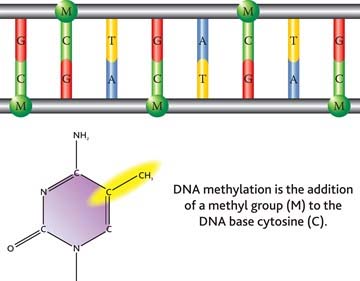
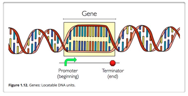
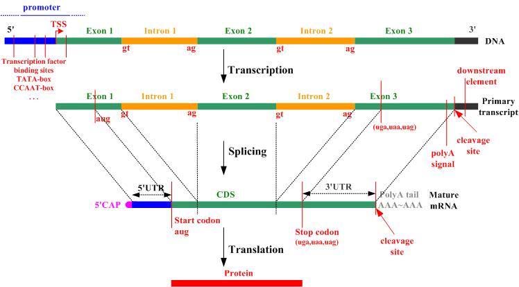
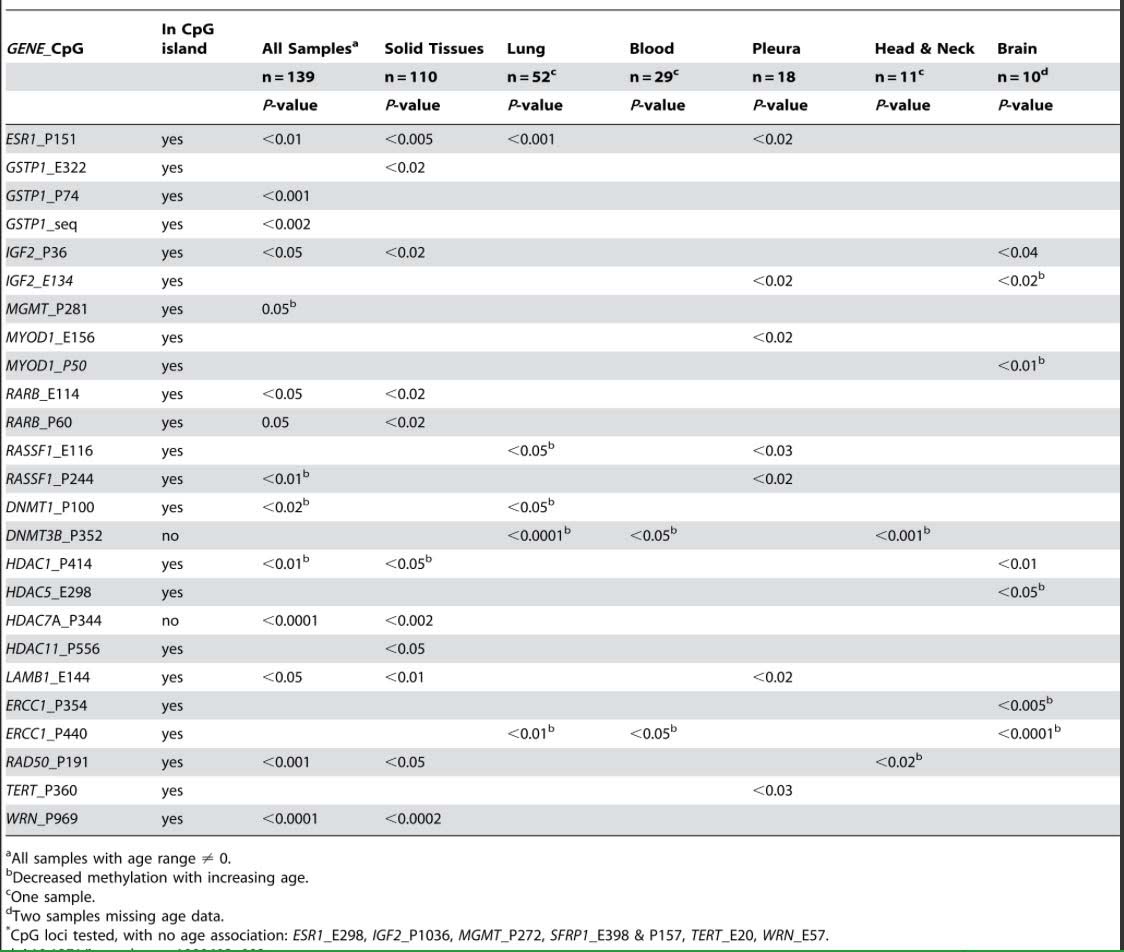
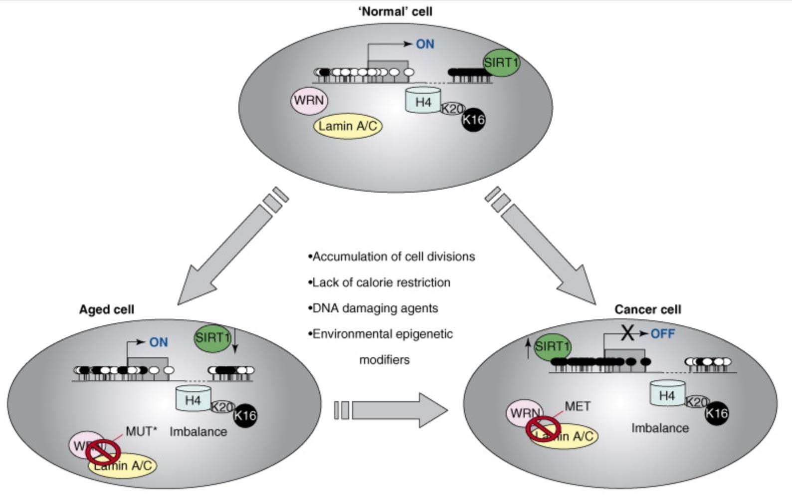
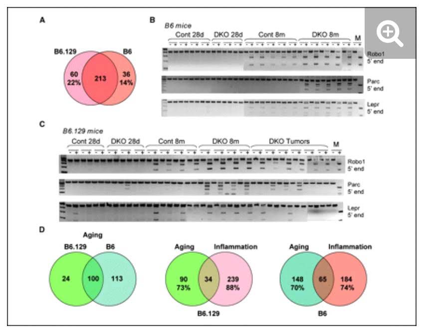
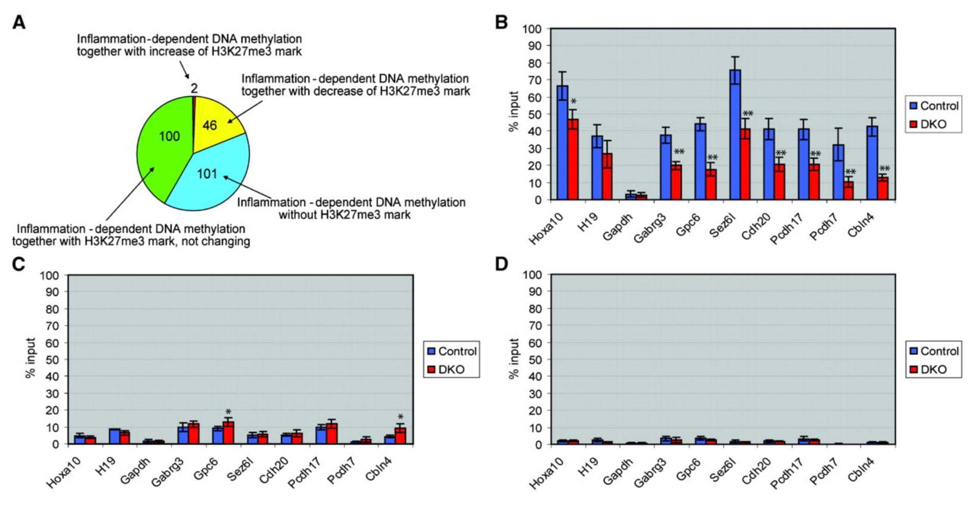
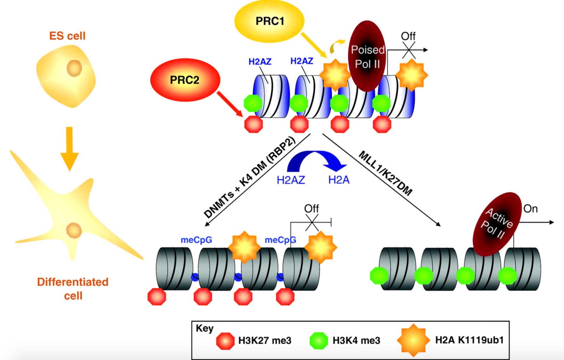
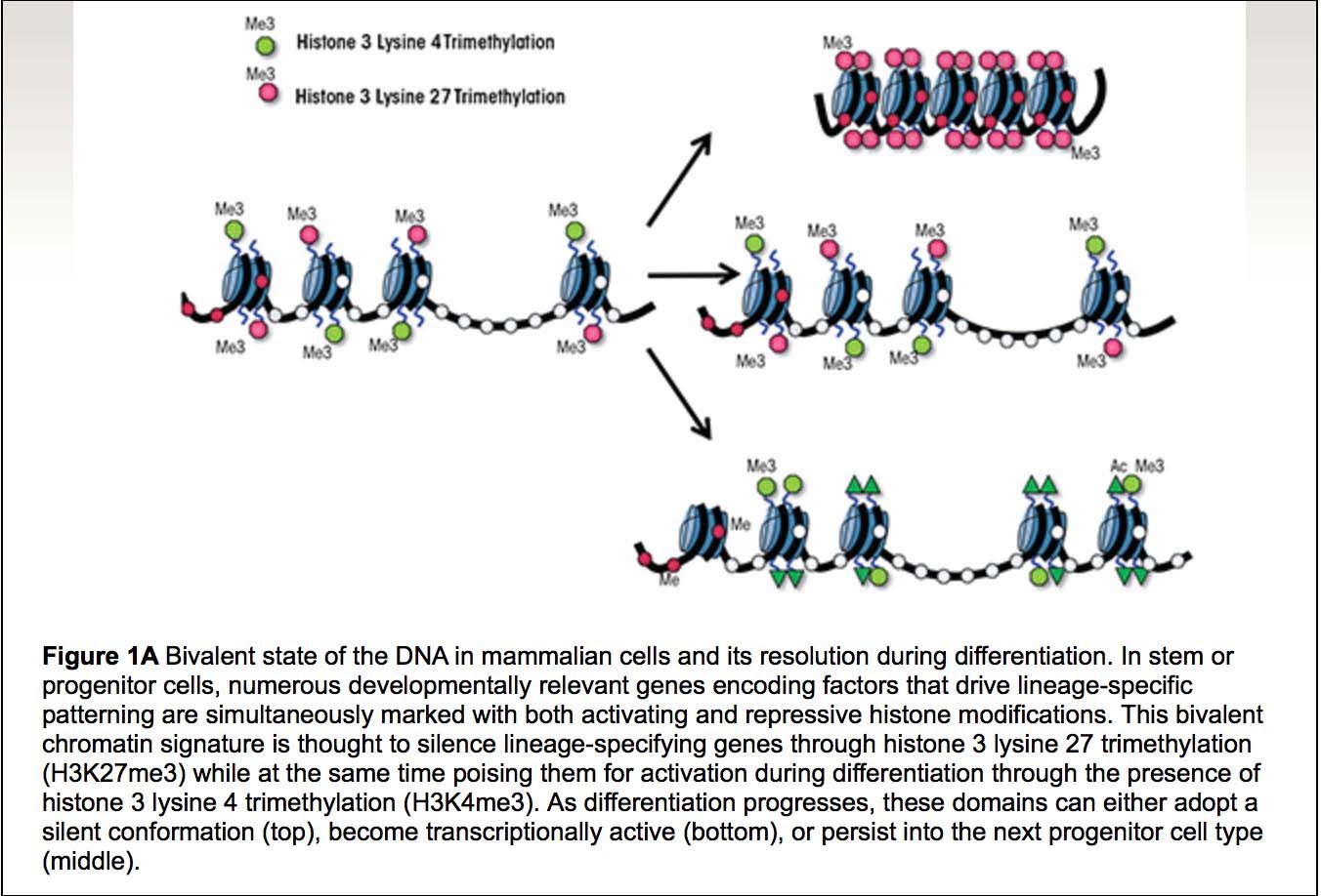
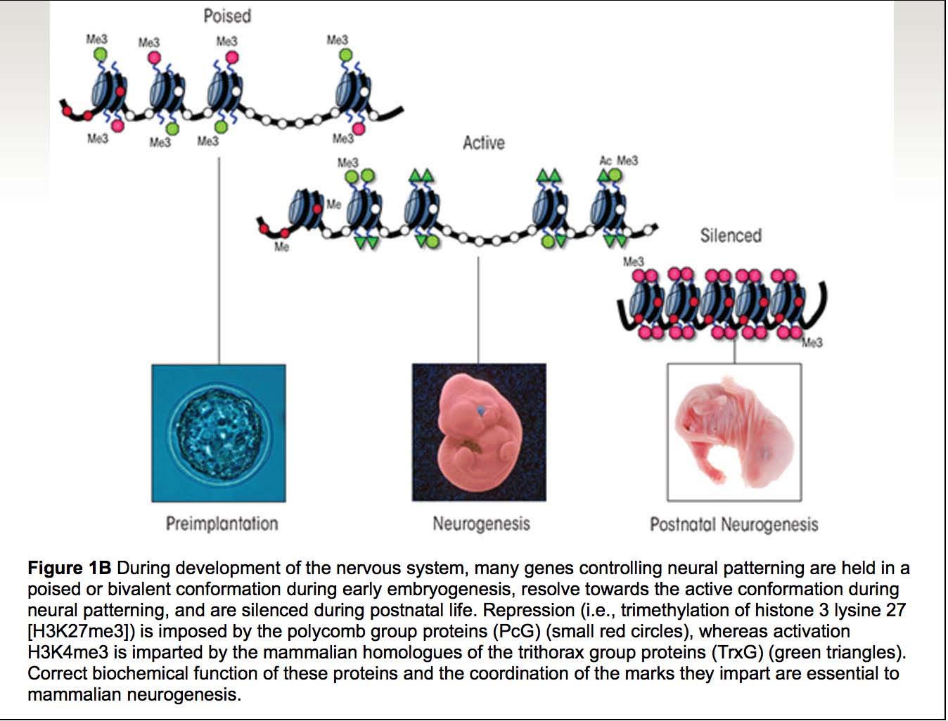
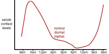
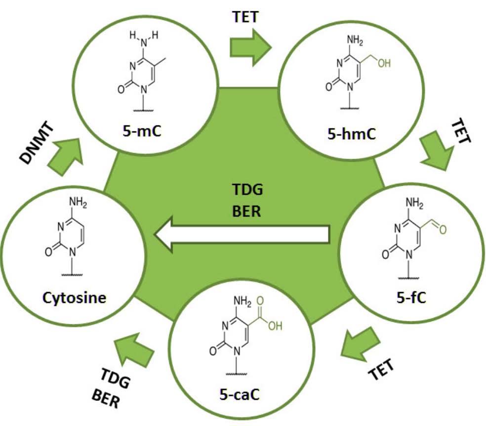
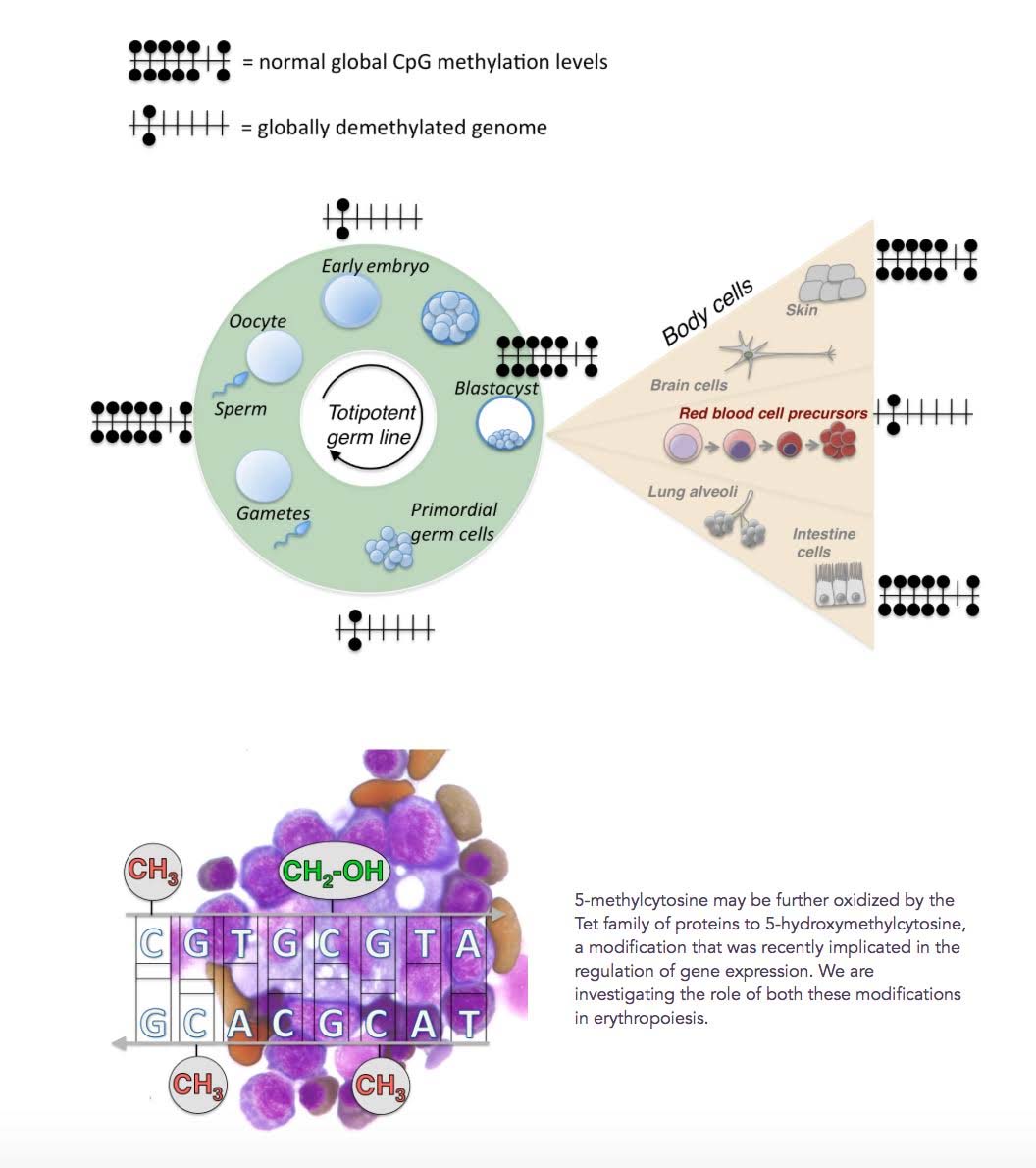
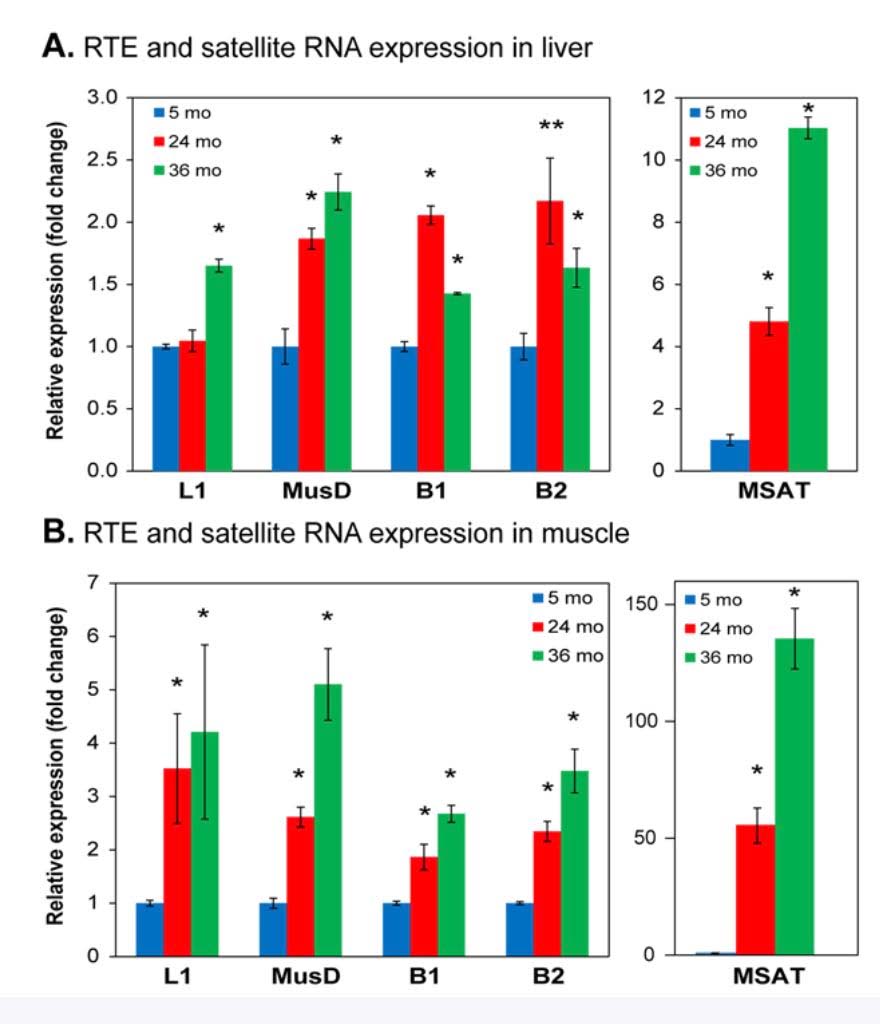
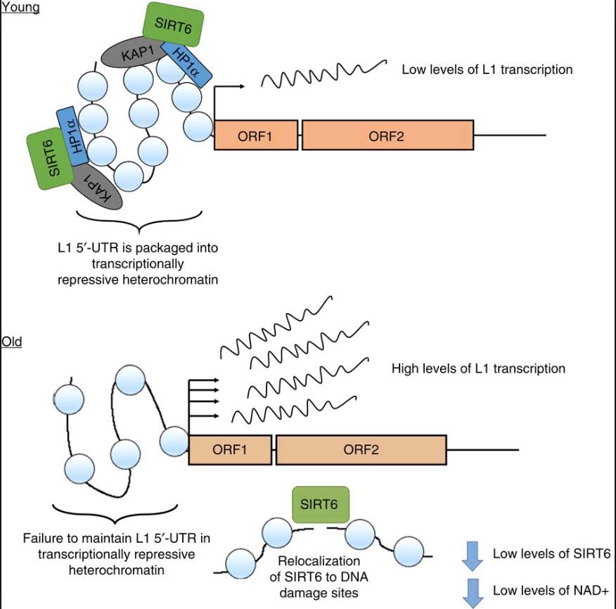
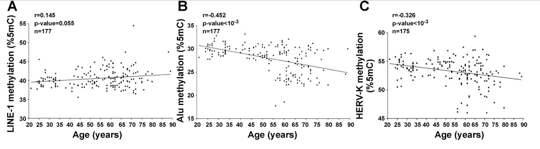
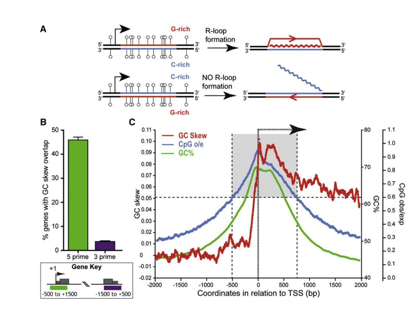
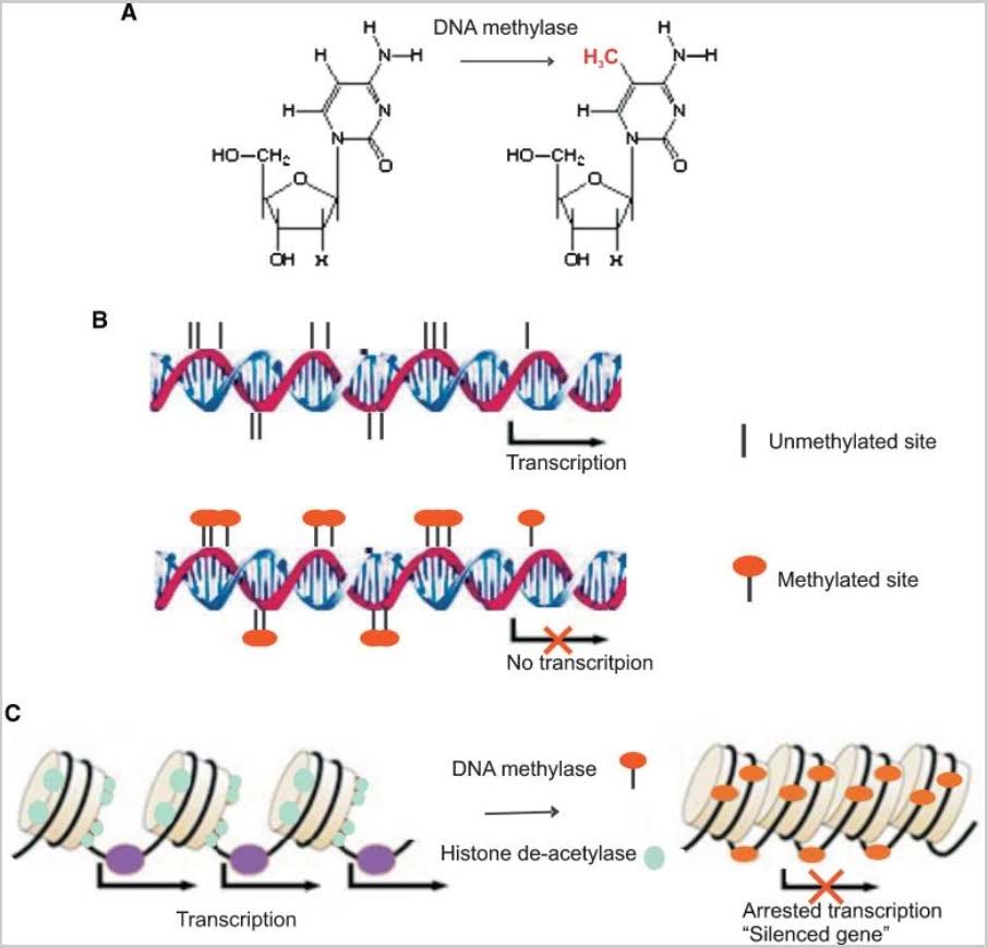
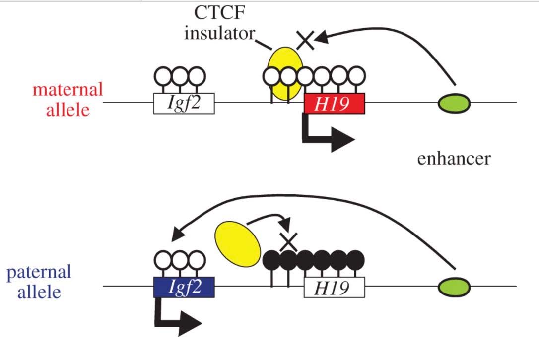
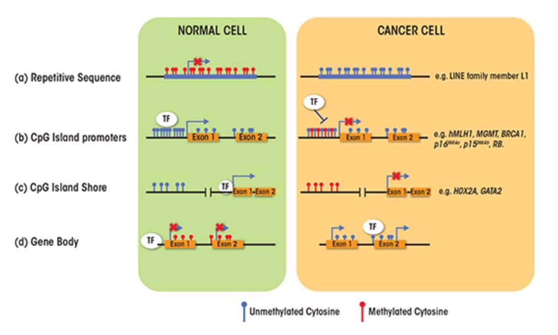
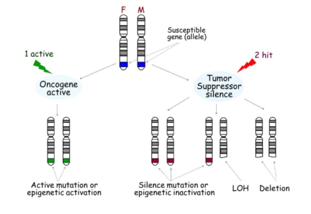
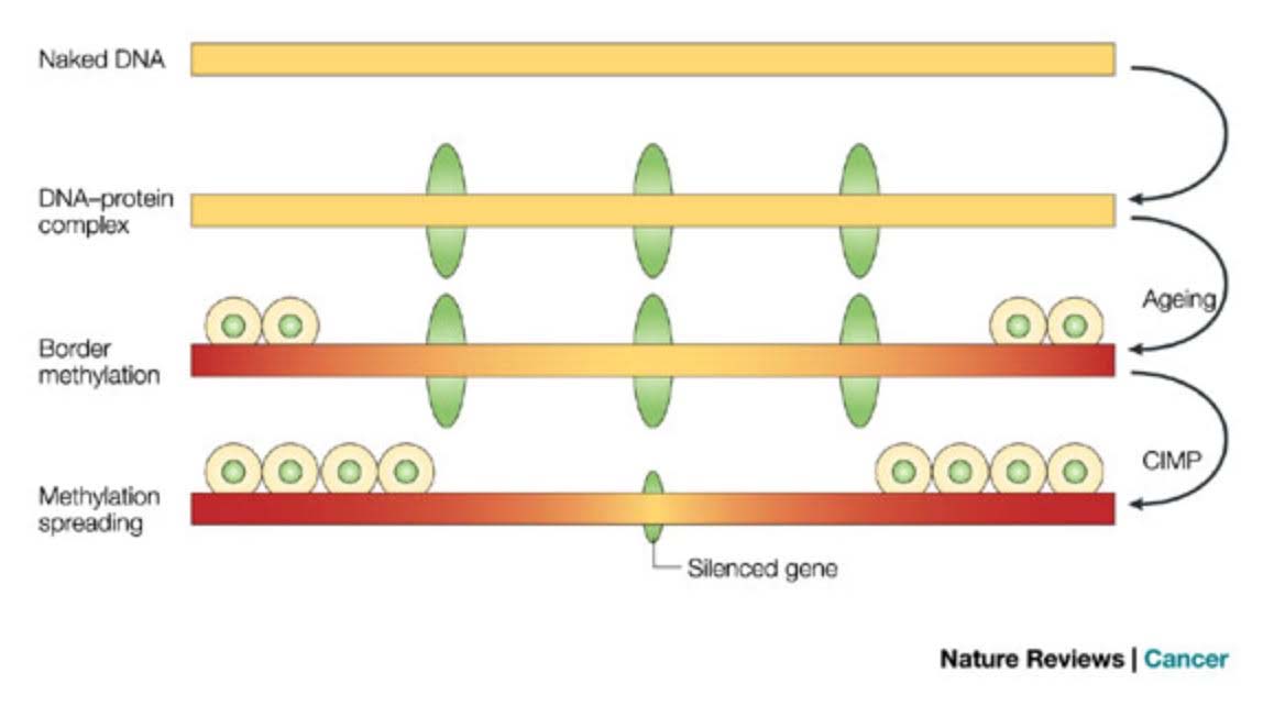
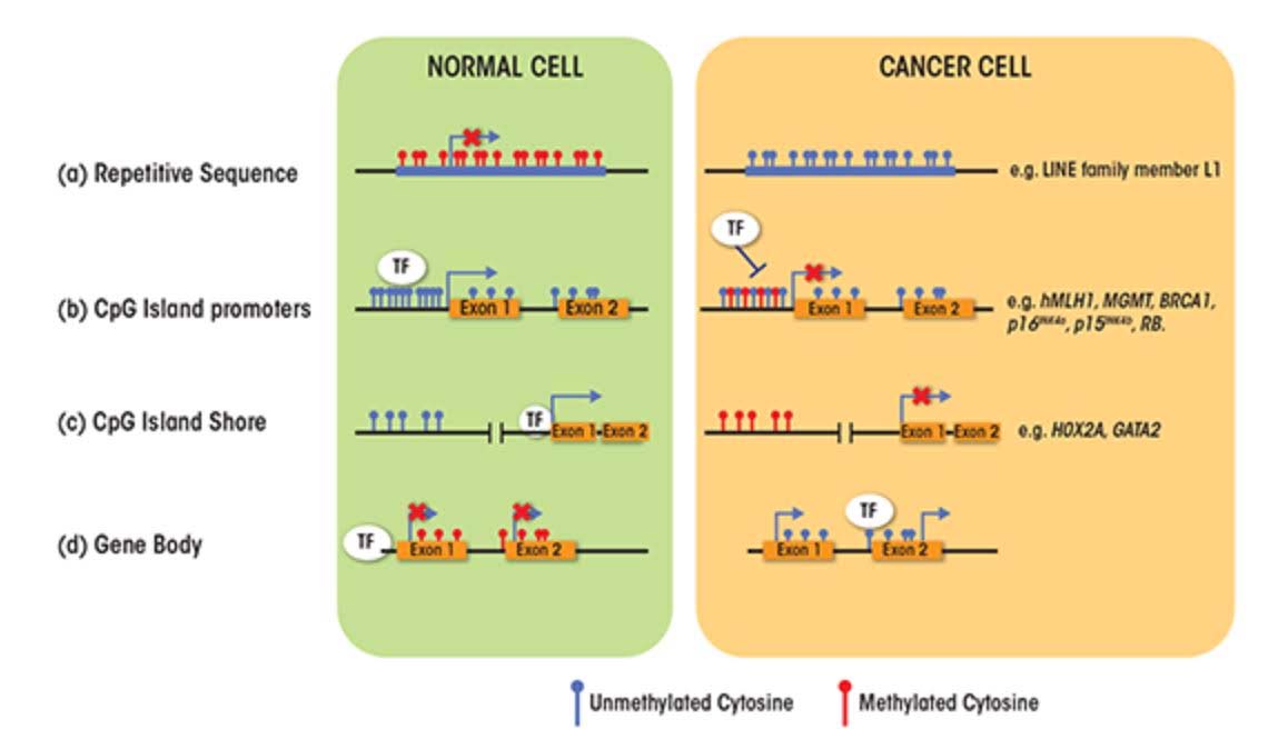
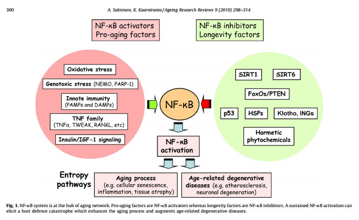
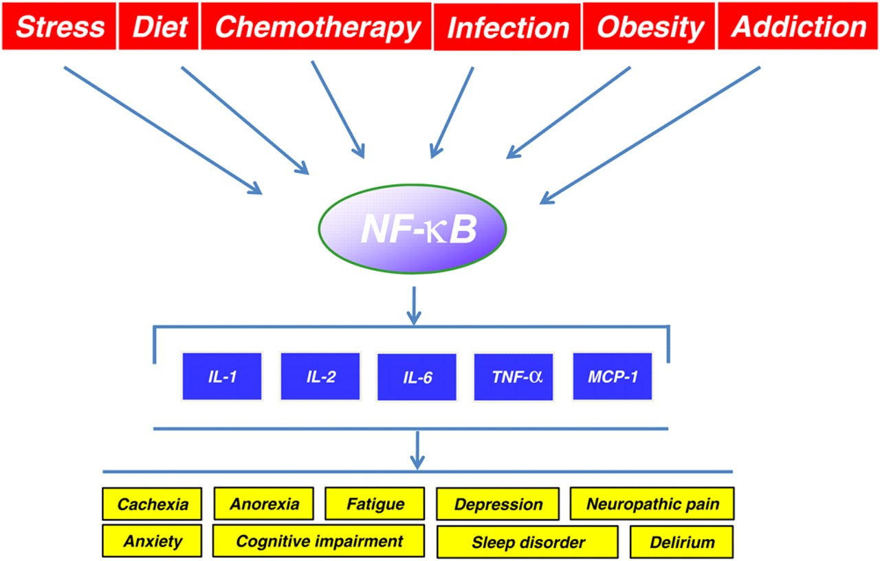
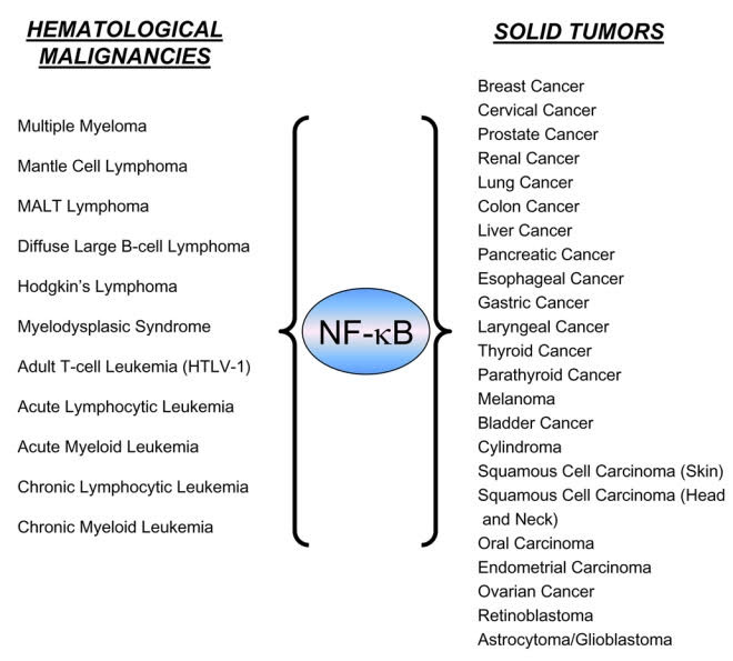
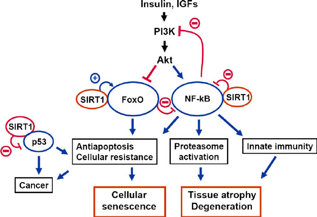
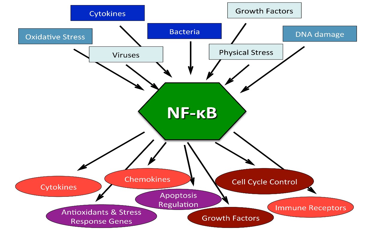
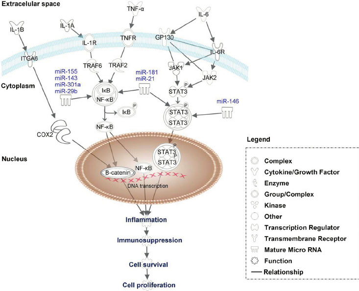
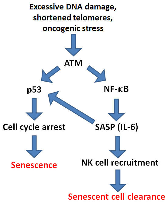
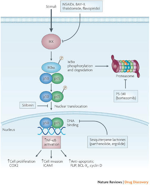
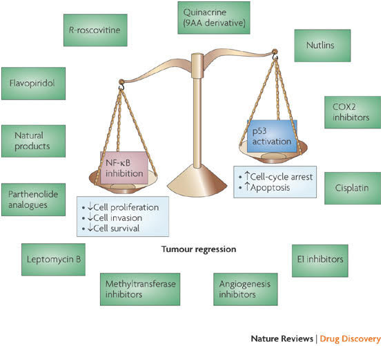
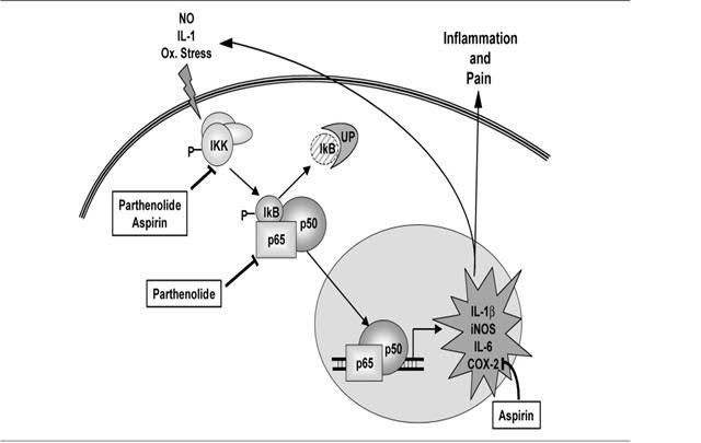
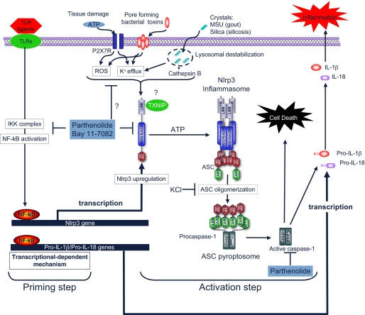
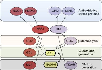
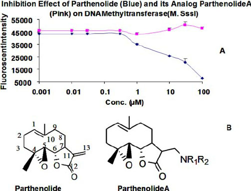
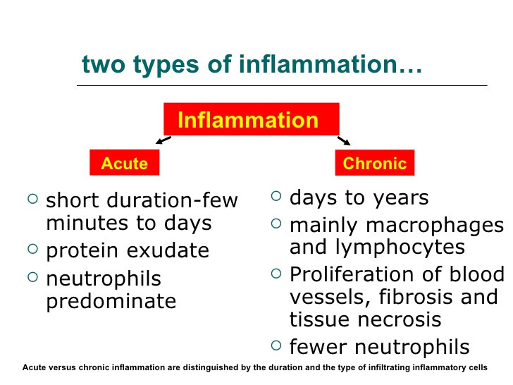
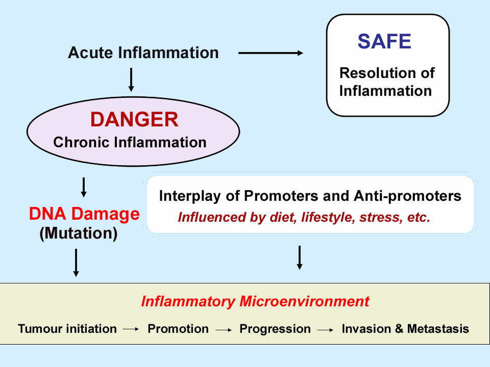
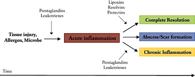
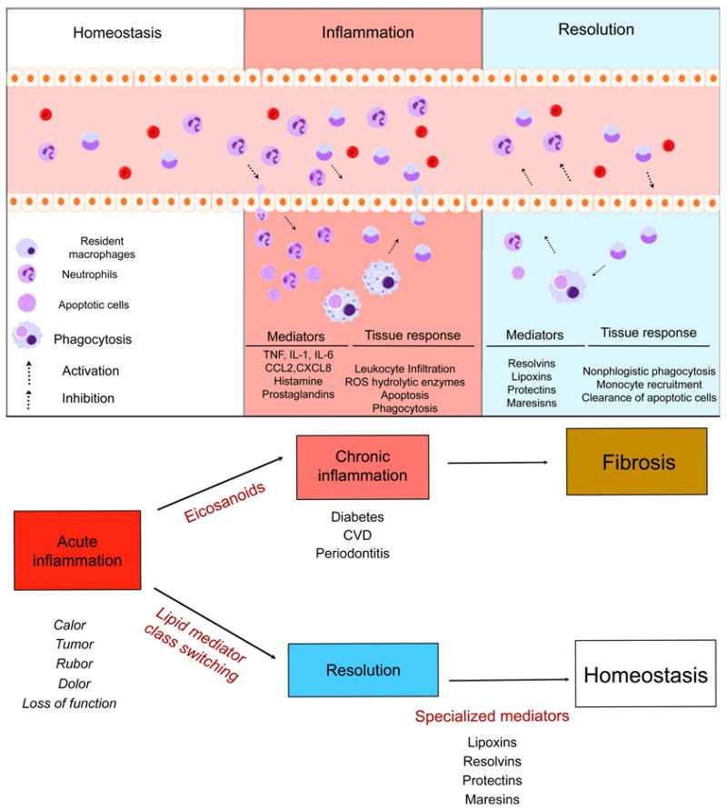
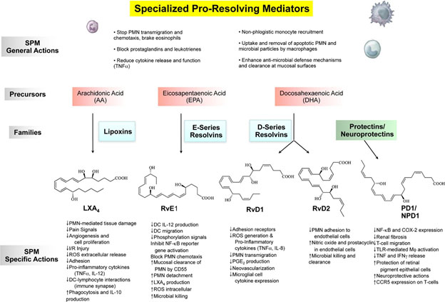
in almost all of the hundreds of thousands of papers and treatises about it.   The consequences include a general lack of agreement about how to even think about aging, and multiple disjointed and confusing perspectives and views of anti-aging interventions.

I have recently come to embrace some simple but important distinctions related to aging that provide me significant clarity in thinking about the topic and its ramifications.  This finally, after 10 years as a researcher in the area!  I share and discuss these distinctions here.  Then I discuss critical questions including the nature and causes of aging and types and levels of anti-aging interventions.

KEY DISTINCTIONS RELATED TO AGING

I start out by distinguishing between 1. Chronological aging, 2. Biological aging, 3. Diseases and degenerative processes typically associated with aging, and 4. Disease and degenerative process precursor conditions.

  1. Chronological aging (CA). This is aging as simply measured by years and days on a calendar.  There can be little argument about this.  It makes no sense to say that something causes or accelerates chronological aging or that anything slows it.  CA is by definition synched to the clock and you may no more slow it down or speed it up than you can slow down or speed up time itself.  Saying that chronological age causes something like greying hair or cancer is a statistical generalization that applies to only to some people but not to others.   Relationships of CA to other factors can only be understood as statistical distribution curves.  For example, for a given population you may specify the probability of initiation of Alzheimer’s disease as a function of age.  But you can’t say beforehand the age a given person will get it, or even for sure if he or she will ever get it.
  2. Biological aging (BA). This is the kind of aging of concern in any study where aging is looked at as cause or effect.  Sometimes it is called functional aging.  BA has been discussed in the literature, though there is no general agreement on how to measure it(ref)(ref),  When I talk about aging in the rest of this blog post it is mostly about BA.   While difficult to measure, as applied to older people, biological age can be thought of as chronological age adjusted for state of health so as to be the best predictor of mortality.  For example, my own chronological age is  nearly 88 but I estimate that my biological age is typical of that to be found in a cohort of healthy 68 year-olds who live in comparable social, lifestyle and social circumstances,   If we could agree on how to measure biological age, say be identifying accurate biomarkers for it, we would have a much better predictor of mortality than chronological age.  Because BA like CA is a whole-body factor, like for CA, relationships of BA to other factors can only be understood as statistical distribution curves.
  3. Diseases and degenerative processes typically associated with aging (DDPs), The list of these is very long including cancers, diabetes, atherosclerosis, coronary diseases, dementias, loss of balance, loss of hearing, liver and other organ failures, pneumonia, frailty, loss of muscle mass, macular degeneration, etc. People don’t die from “old age.”  They die from DDPs.   Also, many DDPs as well as therapies for them can induce system-wide modifications to the body which promote biological aging.  Cancers, radiation and chemotherapy are examples.  Some degenerative processes feed on themselves and accelerate other diseases and biological aging.  Joint inflammation can make exercise less attractive because it is painful, leading to cardiovascular diseases, sarcopenia and dementias.  Loss of balance can lead to falls which also lead to cessation of physical exercise which leads to acceleration of biological aging.
  4. DDP precursor conditions (DPCs). A large number of conditions can exist in a person which are precursors to DDPs.  I have focused on chronic inflammation. (See for example the recent blog entries in our Series on Inflammation, Part 1, Part 2, and Part 3.  (And two additional parts have been drafted and will be published soon.)  Chronic inflammation is a consequence of many diseases and degenerative processes and the cause of others.  Others of these DPCs can include hormone imbalances, abnormal body redox state, inadequate NAD/NADPH ratio, high levels of IGF-1 and insufficient levels of sirtuins.  DPCs can themselves have major influences on DDPs as well as on biological aging.
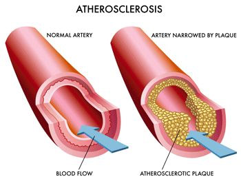
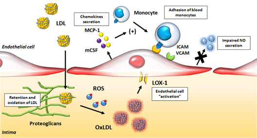
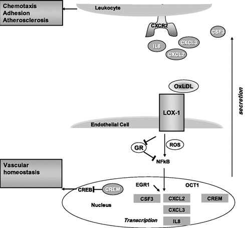
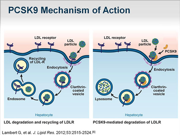
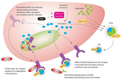
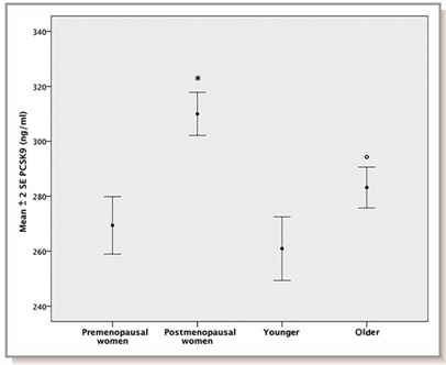
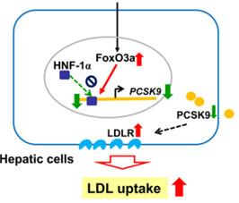
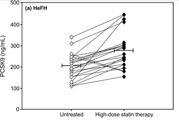
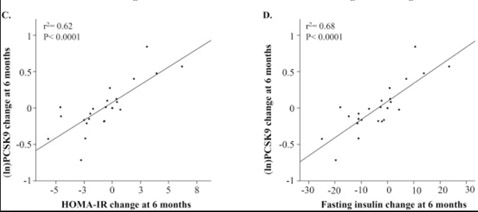
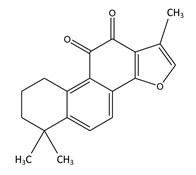
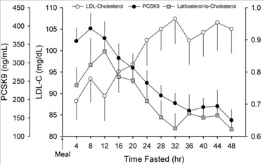
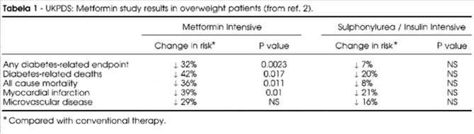
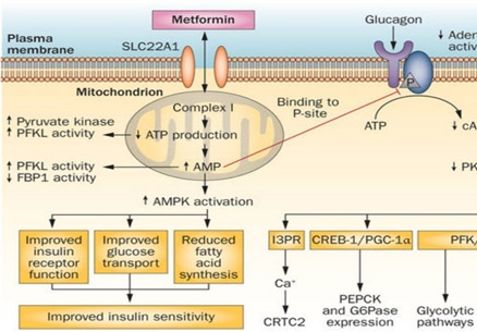
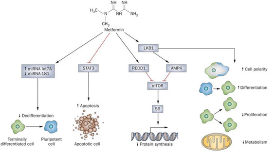
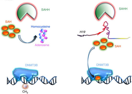
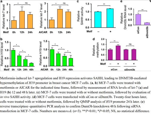
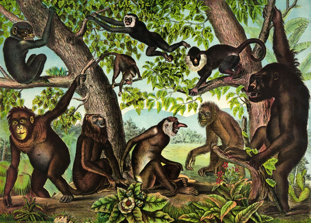
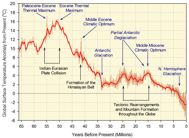
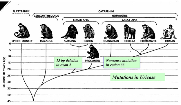
WHAT IS THE NATURE OF AGING?

  1. I believe aging (speaking primarily about biological aging) is a species specific lifelong molecular program which takes into account stochastic environmental conditions. It starts with conception and ends with death.  Aging is not the consequence of random damage nor is it simply a pseudo program, the consequence of vestigial developmental programs that fail to shut down.  There might well, however, be one master program for each species that governs both development and aging, and governs several other aspects of life for that species as well.  And vestigial pseudo progams may be part of the overall aging program, like mTOR expression failing to wind down in older people.  I refer to biological aging as a “program” because aging is highly regulated and comes to a deterministic conclusion, ie.  You die with certainty before the maximum age for your species.  Note that I am not saying that the code for this program is separable from the code for other biological programs that govern a live organism.  What I am saying is that aging behaves like a program and therefore is likely one, even if we are not completely sure how the program works.  It is clear that human embryogenesis and early development are governed by programs, ones that exercise an incredible degree of regulation.  Programmatic regulation of life is essential and continues at all ages, including providing multiple forms of homeostasis.  Even advanced aging is similarly highly regulated.  Further:
  • All living species of all kinds have maximum known lifespans, without exception, Nothing ever lives beyond the maximum age for its species.  Although stochastic variables exist in the aging program, the program itself is deterministic, surely leading to death.
  • The age-vs mortality curves for members of species do not represent those which would exist if aging was caused by random damage or any other random process. It they did we would have a tiny handful of 600+ year old people and 75 year-old house cats, 100 year old dogs and 15 year-old mice.  The same would be true if aging was the result of randomly-operating vestigial developmental programs.  More precisely, the statistical distribution for any random process results in a Poissonian distribution curve, one with an infinitely long tail.  The same is true for distributions representing combinations of random process.  The lifespan curves for all species have cut-off tails.
  • DNA methylation provides lifelong clocks that can predict BA. DNA methylation is a clear indication of one or more of the critical subroutines of aging, is one of the three major epigenetic mechanisms for gene activation and silencing ,and appears to act as a major causal factor in the overall program of aging.,   See Jim Watson’s  blog entry Aging, health and disease – view from the DNA MethylomeAlso, see this list of other entries in this blog related to DNA methylation.
  • Over history and also in recent decades there has been a steady increase in average expected human lifespan – by around 4 months for each passing years now in Western societies(ref). This increase appears to be due to constant epigenetic remodeling.  It is not known whether maximum human lifespan is experiencing a similar increase.
  • In terms of classical evolutionary biology going back to Darwin, nature cares a lot about the preservation of a species, much less about the preservation of members of that species. In fact it has long been thought that there is generally an evolutionary advantage to pruning out the old so they are not competing with the young for resources.  Both the Antagonistic Pleiotropy and The Disposable Soma therories explain aging as nature being indifferent to organisms after reproductive age.
  • To understand the nature of aging, the best place to start is by considering the most ancient of evolutionary-conserved pathways, ones at least 500 million years old going back to the pre-Cambrian period and before(ref). In the course of evolutionary history, very early on nature had to decide how to handle life, reproduction, death and the preservation of species.  Once evolution found a good solution to a problem, It tended to re-use that solution in descendant species.  Aging and death of members of a species was one of the first great problems dealt with and solved – and those ancient solutions are still built-in within us.
  • Nature cares for the preservation of life even more than for the preservation of species which can come and go.  So nature provides mechanisms for accelerating the evolution of any species when its members are too stressed out.  See the blog entry Transposable DNA elements – Part 3 TEs and and other key mechanisms of evolution: incRNAs, A to I editing, alternative splicing and exonization.   It is about how a mechanism of greatly accelerated evolution kicks in when stress on an organism gets too great.
  1. The human aging program is inexorable and act ever-more powerfully with advancing age until it kills.
    • Only one person in 250,000 lives to 105 years old
    • Only one person in five million lives to 110 years old
    • Nobody ever lives to be beyond 123 years old
  2. The program operates differently at different phases of life, though there is little data on this
    • Factors related to longevity and what kills organisms are different for different periods in their life cycles. For humans, for example, environmental and epigenetic factors seem to be the most important factors for staying alive for people in their 60s through 80s, while genetic composition is thought to be most important for centenarians,
    • The program for humans may vary somewhat by ethnicity and other factors in now-unknown ways.
    • My guess is that the aging program for humans contains one or more “cleanup” routines that sense age, probably from a DNA methylation clock, and starts to kill you around some preset age, say around 115, if nothing else has killed you by then.
  3. Different species have different aging programs. Mice live typically from 1 to 3 years depending on species and environmental conditions, small bats live to about 20 years on the average, and naked mole rats can live up to 31 years.  Yet, all three of these kinds of animals have similar weights and look a lot alike.
  4. The information repository for aging that drives the aging program is most likely epigenetic information in the nuclii of cells including DNA methylation status and state of chromatin ordering.
  5. Circadian clocks seem to play important roles in epigenetic regulation and biological aging(see this reference list).
  6. Although the overall molecular program of aging is still poorly understood, we do know important things about some key subroutines in that program for humans.
    • These subroutines relate to key molecular pathways including those associated with mTOR, IGF-1, NAD, REDOX states, and there are probably others in addittion
    • The subroutines associated with these pathways in turn affect the DDP conditions mentioned above
    • Insight is being gained on how these subroutines interact so as to affect aging is rapidly growing though still primitive.

LEVELS OF ANTI-AGING INTERVENTIONS

The above framework allows sorting out anti-aging interventions, that is:

  • Direct interventions on diseases and degenerative processes typically associated with aging (DDPs) already in place: Most of what is done in modern medicine is on this level.  You deal with a cancer or Alzheimer’s disease or a failing liver after the disease or degenerative problem is already clearly manifest and diagnosed.  If you have a medical condition that seriously threatens your life, interventions on this level sometimes can keep you alive longer – like a heart transplantation or cancer removal surgery.  However, much of what can be done on this level is not efficacious or only partially efficacious, i.e.  there is no known cure for Alzheimer’s disease or many types of cancer.  Or, the best that can be hoped for is a stabilized but still-impaired condition, such being the case when dealing with many organ or system pathologies.
  • Inteventions aimed at disease and degenerative process precursor conditions (DPCs): A few things are being done on this level in contemporary medicine, mainly in the domain of blockbuster drugs.  An example is prescription of metformin and other anti-diabetic drugs for people showing signs of insulin resistance, to delay or prevent diabetes.  Another example is prescription of statins like Lipitor to forestall cholesterol-related cardiovascular problems.  Another yet would be to take a statin when eye examination reveals druzen, a precursor condition to AMD macular degeneration.  The hope would be to prevent proliferatiom of chlorestrol-rich druzen, assuming they are causal of AMD.  To the extent that a deadly disease is prevented or delayed by an intervention on this level, life extension is provided.  I believe a lot more can be done on this level using well-researched plant-based substances.  Especially when certain herbal extracts are delivered by means that assure high bioavailability.  As avid readers of this blog know, I have recently come to favor the control of chronic inflammation via a liposomal concoction of extracts of well-researched Aryuvedic anti-inflammatory herbs.
  • Interventions aimed generally at biological aging (BA): These are interventions aimed at slowing or reversing some of the molecular and epigenetic mechanisms of aging itself.  These are “hacks” on the program of aging, targeteted to particular subroutines in the overall aging program.  They might not be aimed at any particular disease of DDP condition.  They are thought to be life-extending because they slow down or even reverse aspects of organismal aging and their application might significantly delay or forestall disease process precursor conditions.  These include several interventions known to be life-extending in animals.  They address ancient evolutionary-conserved pathways going back hundreds of millions of years to very primitive organisms.  So we think they can be efficacious in us humans as well.  They go to the root of aging to the extent we know how to do that now.  Examples are:

About the interventions and aging subroutines

Each of these interventions addresses one or more critical subroutines in the overall aging program, and this list of interventions and the subroutines that can be addressed is far from complete.  For example, it could turn out that we would want to target gene methylation with pharmacologic agents as another anti-aging intervention, or a large variety of other candidates .  I expect it might be 40-50 years before these critical subroutines are systematically understood and laid out, and fairly complete practical understanding exists of how we can effectively game them.  Meanwhile we have to go with what we know, limited though that may be.

I need point out that many of the classical anti-aging interventions that I and others have been following probably impact on major longevity pathways and therefore work by affecting aging itself (BA), as well as disease and degenerative processes associatiated with aging (DDPs).  I am thinking of matters like taking melatonin, pregnalone and DHEA to impact hormone regulation of aging, and numerous phytosubstances like curcumin and boswellia to impact on inflammation.  As a matter of fact until recently I have relied completely on natural phyto substances to extend my longevity and only now am beginning to add pharmacological agents as I grow ever-older and the challenge of staying healthy and fully vital ever-greater.  Jim Watson and I have published blog entries related to impacts of several phytosubstances on histone acetylation and deacetylation (See blog and other entries on this list).  These impacts are likely to affect epigenetic encoding of biological age, and possibly exercise profoundly impact the aging program itself.  There is very very much we don’t know yet.

It is possible that most or all of the anti-aging interventions I mentioned above are effective mainly within certain age ranges, say before age 100.  There is little or no literature on their effectiveness for the very old and centenarians.  On the other hand, with extreme aging some or all of the useful interventions of earlier years may prove to be necessary for continuing health, although not sufficient to do so without further interventions.  For example, high levels of the sirtuins SIRT1 and SIRT3 are required for DNA damage repair, and the need for effective repair is likely to continue to increase at all advanced ages.  So the need for metabolic interventions that insure high sirtuin levels is not likely to ever go away.

Reversability of aging

It has been a conservative habit to think of aging interventions as being ones “which slow or halt aging,” and rather politically incorrect for a serious scientist to even mention reversing aging.  Saying that threatens to tag oneself at worst as a quack remedy marketer, or at best someone ignorant of biology.  My personal take is that:

  • Selective aspects of the aging program can most likely be reversed, leading to significant remodeling of multiple body systems to a younger phenotype. This is consistent with encoding of aging being epigenetic, since epigenetic coding appears to be reversible.  And it is consistent with what is seen in small animals on age-extension regimens, such as mice fed with rapamycin(ref) and mice fed NAD+ boosters(ref).  It is also consistent with anecdotal reports of a few human self-experimenters, including the lab rat writing this.
  • Since the aging program is multi-faceted and employs multiple subroutines for aging which can lead to death, I very much doubt that any one or even a handful of known intervention can keep you or me alive, healthy and active beyond our species age limit of 123 years. As science progresses, I fully expect a time will come when this is no longer true.  And I am doing my best to stay alive, healthy,  and fully functional until that time and well beyond.

The post ON AGING appeared first on AGINGSCIENCES™ - Anti-Aging Firewalls™.

Inflammation Part 4 – PCSK9 inhibition

$
0
0

By James P Watson with inputs and editorial assistance by Vince Giuliano

I.  INTRODUCTION

This is the fourth in what we expect will be seven or more blog posts concerned with chronic inflammation.  It relates to a recently discovered protein PCSK9 and tells a story of how age-related loss of LDL receptors leads to increase in LDL, associated uncontrollable cardiovascular inflammation, and atherosclerotic cardiovascular disease – the main killer of older people.  It lays out why the old standard of care for atherosclerotic cardiovascular disease – the taking of statins – is slowly in the process of being augmented and replaced with a new standard of care – inhibiting PCSK9.

Part 1 of the Inflammation series is the same as Part 5 of the NAD worldThat blog entry is concerned with The pro-inflammatory effects of eNAMPT(extracellular NAMPT, nicotinamide phosphoribosyltransferase).  Part 2 of the Inflammation series relates a) the “master” pathway network of inflammation (NF-kB) to two other pathway networks clearly implicated in aging and disease processes, b) Genomic Instability (p53), and c) Oxidative stress (Nrf2)Part 3 of the Inflammation series of blog entries is concerned with the all-important resolution phase of inflammation, how acute inflammation goes away under ideal conditions instead of hunkering down to lingering and dangerous chronic inflammation.  It is concerned with recently identified substances found in fish and flaxseed oils that play important roles in resolving certain kinds of inflammation – what they can do and how they work.

Atherosclerotic cardiovascular disease is the leading cause of morbidity and mortality worldwide (Gupta, Expert Review, Dove Press).  Wannah die later rather than sooner?   Then pay attention to this.

Image source

Many National and International though leaders concerned with the disease have refocused their efforts from lowering cholesterol to lowering LDL levels (Grundy, et.al., 3rd Report of NECP). The reason for this is due in part to the discovery of PCSK9 protein, which is a circulating protein in blood that triggers the degradation of the LDL-receptor.

First, a little history about the discovery of the PCSK9 gene and why all the excitememnt about it.  PCSK9 is an abbreviation for Preprotein Convertase Subtilisin/kexin type 9, an extracellular protein that triggers the degradation of the LDL receptor, the LRP-1 receptor, and other receptors found on the surface of many cells (especially the liver).  In 2003, French researchers reported two mutations in the PCSK9 gene that caused familial autosomal dominant hypercholesterolemia (Abifadel, et.al., Nature Genetics).  In 2008, Spanish researchers found a new mutation in the promoter of the PCSK9 gene that increased gene expression of PCSK9 mRNA and plasma levels of PCSK9 (Blesa, et.al. J Clin Endocrin Met). About the same time, researchers at UT Southwestern in Dallas reported a series of PCSK9 gene nonsense mutations found in 2.6% of a large group of African Americans that reduced LDL plasma levels by 15% and reduced coronary artery disease risk by 47% (Cohen, et.al., NEJM).  In the ensuing 10 years, monoclonal antibodies that target the circulating PCSK9 protein have been developed, tested in over 25 R clinicaltrials, and FDA-approved.

What these clinical trials have shown in their “size effect” of reducing all-cause mortality (ACM) is nothing short of astounding – much more than statins ever did.  All cause mortality is reduced 55% in patients with hypercholesterolaemia.  So, we go on now to discussing how this all works.

II.  MECHANISMS RELATING LDL, ATHEROSCLEROSIS AND HEART DISEASE, LDL CELL RECEPTORS, PCSK9 AND MOST OF THE  MOLECULAR USUAL SUSPECTS OF INFLAMMATION, DISEASE AND AGING

It has long been known that there is a positive correlation between circulating LDL, atherosclerosis and heart disease.  Why is this?  The most harmful aspect of high LDL levels is most likely due to the fact that LDL is oxidized to “oxidized-LDL” (oxLDL) by free radicals (ROS) which triggers a pro-inflammatory receptor found on vascular endothelial cells called the “Lectin-like oxidized low density lipoprotein receptor 1” (LOX-1).  Here is a diagram of how this works:

Image source and reference: Role of Oxidized LDL in Atherosclerosis

Oxidized LDL lipoproteins bind to the LOX-1 receptor and are then internalized into endothelial cells, vascular smooth muscle cells, as well as monocyte/macrophages.  Inside the cell, this process triggers free radical production (ROS) and activation of NF-kB (the master switch for inflammation). NF-kB then turns on the transcription of hundreds of inflammatory genes, including cytokines (IL-8) and chemokines (CXCL2, CXCL3, CSF3), which are then secreted and trigger the CXCR2 receptor on white blood cells.  This is how the inflammatory cascade is “let loose” when your LDL-C is too high.  Here is a further diagram of how this works:

Diagram and legend source: LOX-1-dependent transcriptional regulation in response to oxidized LDL treatment of human aortic endothelial cells (2009)  “Model for LOX-1 functions in atherosclerosis and endothelial dysfunction. LOX-1 binding to OxLDL initiates reactive oxygen species (ROS) formation and an inflammatory response mediated in part by nuclear factor (NF)-κB and EGR1, leading to an upregulation and secretion of chemokines. The three CXC-chemokines (CXCL2, CXCL3, and IL-8) are all ligands for the same receptor, CXCR2, found on leukocytes. CXCR2 activation leads to chemotaxis and adhesion of leukocytes to endothelium. EGR1, CXCR2, and CSF3 are all implicated in animal models of atherosclerosis and represent novel molecular connections between LOX-1 and atherosclerosis. The transcription factor CREB is involved in maintaining vascular homeostasis. LOX-1 activation by OxLDL leads to a downregulation of CREB target genes and an upregulation of the CREB repressor CREM, thus providing a potential molecular mechanism of LOX-1-dependent endothelial dysfunction.”

In addition to LOX-1 mediated vascular inflammation,  oxLDL/LOX-1 internalization triggers endothelial/vascular smooth muscle cell dysfunction, apoptosis, cellular senescence, or osteoblastic differentiation. The osteoblastic differentiation of these cells is manifested as “vascular calcification”. If the oxLDL/LOX-1 internalization occurs in a monocyte, the macrophage is phenotypically transformed into the classic “foam cell”, which is the sine qua non of atherosclerosis.  Along with the other 4 “drivers” of atherosclerosis (Angiotensin II, pro-inflammatory cytokines, sheer stress, and advanced glycation end products) this oxLDL/LOX-1 pathway is considered to be the major molecular cause of the disease.

So, the first point in the story is this:  1. Too high LDL is bad because when oxidized it lets loose an inflammatory cascade that can lead to chemotaxis, adhesion, plugged arterial lumens, atherosclerosis and heart disease.

The next part of the story is simple: 2. under normal healthy conditions, LDL receptors on cells latch on to LDL particles and drag them into the cell where endosomes degrade the LDL and then the receptors pop back up to the surface of the cell and wait for another LDL particle to come along.  But if PCSK9 is present, the LDL receptor itself is destroyed.  The amazing clinical results mentioned above were due to inhibiting PCSK9.

The diagram below illustrates how PCSK9 works for elimination of LDL receptors and therefore reduction of capability to eliminate LDL.

Image source: Complementing the “Gold Standard”: Exploring PCSK9 MOA with Current Lipid Therapies  “When PCSK9 is present in that complex, what ultimately happens is that the LDL receptor is subsequently targeted via the endosome to the lysosome for degradation (situation depicted in right panel in diagram). Hence, we lose the LDL receptor; it does not recirculate back to the liver. If there is no PCSK9 that is bound to that complex, then once it is internalized, the cholesterol is released, the LDL particle is released from the receptor, and the receptor recirculates back to the surface, where it can then attach to more LDL cholesterol and clear more LDL cholesterol from the circulation degradation (situation depicted in left panel in diagram).”

The following diagram show how the final part of the story works: 3. anti-PCSK9 antibodies prevent PCSK9 from binding to LDL receptors so they are not destroyed in the lysosome.

DIAGRAM B  Image source

As already mentioned, although the PCSK9 inhibitor class of drugs have only been recently FDA approved, they have already shown the largest ACMR (all-cause mortality reduction) of any class of FDA-approved drugs ever approved in history.

III.  PCSK9 AND AGING

These drugs are especially important for those of us who are interested in the science of aging since it has been shown that the levels of PCSK9 protein circulating in the plasma increases with aging in rodents and in humans (Tao, et.al., J Bio Chem) (Ruscica, et.al. J Am Heart Asso).

Here is a diagram showing the relative levels of plasma PCSK9 in pre vs post menopausal women and young vs old men in a large population-based study (Diagram source: Ruscica, et.al. J Am Heart Asso).

The cause of the age-related increase in PCSK9 gene expression is due to a loss of suppression of the PCSK9 gene by the FoxO3a transcription factor.  Normally SIRT6 recruits FoxO3a to the promoter of the PCSK9 gene, but with aging, SIRT6 activity declines due to declining NAD+ levels in the cell.  As a result, there is an increase in PCSK9 gene expression with aging (Tao, et.al., J Biol Chem). Here is a diagram illustrating this:

Note that we are here extending the story line of our NAD World blog entries detailing negative impacts of declining NAD+ LEVELS.

Reference and image source :Role of Oxidized LDL in Atherosclerosis (2015)

FoxO3a and Nrf2 are considered two of the most important transcription factors that protect mammals from aging by increasing oxidative stress resistance (Li, et.al. Ox Med Cell Long). Multiple genetic variants in the Foxo3a gene have been linked in many studies world-wide with extreme longevity (Anselmi, et.al., Rejuvenation Research).  SIRT6 has also been shown to be a longevity gene in GWAS studies as well as in lab animals (Braidy, et.al., Front Cell Neuroscience).  Thus it is not surprising that aging due to declines in FoxO3a/SIRT6 activity triggerw the increase in expression of the PCSK9 gene and play a major role in vascular aging.  Moreoever, statin use causes a paraxodical INCREASE in PCSK9 levels in patients with familial hypercholesterolemia, most likely due to a compensatory homeostatic feedback mechanism (Raal, et.al., J Am Heart Asso).  The following diagram is from this study.

Another reason why PCSK9 levels increase with aging is insulin signaling. It is not surprising either that Insulin/IGF-1 signaling exacerbates this problem with age-related insulin resistance, since FoxO3a cannot enter the cell nucleus under conditions of high glucose/insulin signaling (due to the inhibition of nuclear translocation of FoxO3a by Akt).   This is why diabetes, metabolic syndrome, and insulin resistance leads to an increase in the expression of PCSK9 gene.  Here is a diagram of the relationship between insulin resistance (HOMA-IR) and insulin correlated with increases in PCSK9 expression over time

(Image source: Levenson, et.al. NMCD).

Age and diet-related increases in LDL blood levels are probably the most common clinical problem facing modern man.  Although statins reduce endogenous cholesterol synthesis, they do not directly stop the age-related increase in PCSK9 protein in the blood.  As a result, most studies have suggested that “age” is the greatest risk factor for atherosclerosis after age 50, whereas “cholesterol” is the greatest risk factor for atherosclerosis prior to age 50.  For this reason, the value of a drug that reduces the PCSK9 protein in the blood should increase with aging.  So far, this prediction has held true.  Unlike statins, where their value declines in old age (zero value by age 85), there appears to be value in reducing LDL levels even in old age, with FoxO3a and SIRT6 signaling decline.

So, in this overall story we are seeing a link-up of several “usual suspect” themes we have touched on multiple times before in this blog: insufficient expression of sirtuins and lack of NAD+, the FoxO3a transcription factor, high age-related LDL, the Insulin/IGF-1/PI3K/Akt pathway, the master trigger of inflammation NF-kB, Nrf2, reactive oxygen species, inflammatory cytokines, atherosclerosis.  With a new element which is age-related overexpression of the PCSK9 gene which kills off LDL receptors.

Cholesterol, step to the rear of the coach please

Whereas cardiologists used to consider cholesterol on this “big five” list, it is no longer considered to be as important as it once was. Instead, high blood levels of LDL is now reputed to be the #1 culprit that causes atherosclerotic disease. Since PCSK9 proteins trigger the degradation of the LDL receptor, monoclonal antibodies against PCSK9 have been successful at dropping LDL levels as low as 10 mg/dl (although no physician is recommending that you lower your LDL that much).  Getting rid of LDL reduces oxidized LDL, which dramatically reduces LOX-1 activation.  Reduced LOX-1 activation reduces endothelial cell dysfunction, apoptosis, senescence, and osteoblastic differentiation of endothelial cells (which is a major cause of vascular calcification).  It is this mechanisms that explains why PCSK9 inhibitor therapy has been shown to reduce vascular and valvular calcification (something that statins rarely do).

Despite these known (theoretical) molecular mechanisms, most cardiologists were skeptical that PCSK9 inhibitors would reduce all-cause mortality as well as statins.  However, the results of the PCSK9 inhibitor clinical trials have shattered all doubts by 10 miles!  The results on ACM reduction have been nothing short of amazing. A meta-analysis of the 24 RCTs that have been done so far (N = 10,159) show a 55% ACMR (OR = 0.45) and a 50% reduction in cardiovascular mortality (OR = 0.50). The rate of MI was also reduced by 51% (OR = 0.49). Even increases in serum creatinine kinase (CPK) was reduced (OR = 0.72). Because PCSK9 levels increase as a function of aging, it is likely that these drugs will still have beneficial effects after age 85, whereas data shows that statins probably become harmful after age 85.

F In addition to LOX-1 mediated vascular inflammation,  oxLDL/LOX-1 internalization triggers endothelial/vascular smooth muscle cell dysfunction, apoptosis, cellular senescence, or osteoblastic differentiation. The osteoblastic differentiation of these cells is manifested as “vascular calcification”. If the oxLDL/LOX-1 internalization occurs in a monocyte, the macrophage is phenotypically transformed into the classic “foam cell”, which is the sine qua non of atherosclerosis.  Along with the other 4 “drivers” of atherosclerosis (Angiotensin II, pro-inflammatory cytokines, sheer stress, and advanced glycation end products) this oxLDL/LOX-1 pathway is considered to be the major molecular cause of the disease.

DIAGRAM A above, shows how PCSK9 inhibitors work and how they are fundamentally different than statins:

ReferenceEffects of Proprotein Convertase Subtilisin/Kexin Type 9 Antibodies in Adults With Hypercholesterolemia: A Systematic Review and Meta-analysis (2015)

IV.  TURNING TO THE PERSONAL AND PRACTICAL

Costs of PCSK9 Inhibitor Therapy and Delivery methods$14,000/year in the US but cheaper overseas

This blog is designed to be a practical resource that you can use on your personal journey to improved healthspan.  It is not a substitute for your personal physician, however. For this reason, we suggest you consult with your doctor before attempting any of the interventions in this blog, including monoclonal antibodies against PCSK9.  However, your doctor may be completely unaware of this new class of drugs and your health insurance company will not pay for the drugs unless you have had an MI or a stroke AND you also have failed to control your LDL with statins (or have severe side effects from statins that cannot be treated with CoQ10).  In the US, the annual costs for PCSK9 inhibitor therapy runs about $14,000 per year so this is mow a very expensive way to try to lengthen your healthspan.  However the cost  of the same PCSK9 inhibitor drug in some other countries can be half the price found here in the US, so for those who have figured out how to do cash-based medical tourism, you can save $7,000 per year. Here are the two monoclonal antibodies against PCSK9 that are currently FDA-approved:

  • Alirocumab (Praluent) – This monoclonal antibody against the PCSK9 protein was the first to be approved by the US FDA (July, 2015). It is a twice-a-month SQ injection of 75-150 mg per dose. It has been FDA-approved for patients with heterozygous familial hypercholesterolemia or for normal patients whose cholesterol cannot be adequately controlled with diet and statins.  Patients who go on Praluent are supposed to stay on their statin, even though they may have to lower their dose to avoid statin-induced muscle pain. Side effects include nasopharyngitis (11%), injection site reactions (7%), influenza (5.7%), UTIs (4.8%), and diarrhea (4.7%).  Neurocognitive events were seen in two of the large clinical trials, but did not correlate with how low the LDL-C went.   Further studies have failed to show any neurocognitive difference between the drug and the placebo group (0.7-0.8%).  8% of patients developed anti-drug antibodies. Alirocumab costs $1200 per month or $14,350 per year in the US.  What is amazing is that PCSK9 monoclonal antibody therapy can drop your LDL-C to as low as 25 with no evidence of any major side effect.  With such dramatic reduction, vascular and valvular calcifications have been shown to be reduced.
  • Evolocumab (Repatha) – This monoclonal antibody was approved in August, 2015, shortly after Alirocumab. Like Alirocumab, it is approved for familial (genetic) hypercholesterolemia (heterozygous or homozygous) and patients who fail diet + statin therapy.  Unlike Praluent, it can be given twice-a-month dose (140 mg) or once-a-month (420 mg) SQ. Like Alirocumab, patients are supposed to remain on statins.  Most common side effects Include nasopharyngitis (11%), URI (9.3%), influenza (7.5%), back pain (6.2%), and Injection-site reactions (5.7%).  Ongoing studies are closely looking for neurocognitive side effects, but these studies are not completed.  The annual costs for Evolocumab are almost identical to those for Alirocumab.

Are PCSK9 Inhibitors Cost Effective? Answer: No

While our enthusiasm for this dramatic reduction in all-cause mortality may appear to be “exuberant”, a famous economist once said that people often suffer from “irrational exuberance”.  In the case of PCSK9 monoclonal antibodies, this is definitely true.  The “bean counters” have already figured out that these new drugs are overpriced to be cost effective (Really!….any 5th grader could tell you that!).  At $14,000 per person per year, the two PCSK9 inhibitor drugs are expected to generate $4.5 billion USD in annual sales by 2020 (Big Pharma is drowning in their saliva over this!).  If all eligible patients took these injections, it would add an approximately $150 billion annual cost to our health care system.  Only if the price of these drugs dropped to $4,536 per patient per year would the drugs start to become cost effective.  However. if you have a high LDL-C despite eating a strict cholesterol-free diet and taking your statins, taking one of these drugs could lengthen your healthspan.

ReferenceAre PCSK9 meds worth the cost? Only if Amgen, Sanofi and Regeneron slash prices by two-thirds: JAMA

Alternative to PSCK9 Inhibitor TherapyReduce Insulin signaling with diet, SIRT6 activation, fasting, and the Salvia extract, Tanshinone IIA

For those who do not have $14,000 per year to “burn”, there is a much simpler way of “shutting off” PCSK9.  As mentioned in the beginning of this section, the “longevity gene” FoxO3a functions as a suppressor of PCSK9 gene expression when the FoxO3a transcription factor can enter the cell nucleus. Unfortunately to do this, you must reduce insulin signaling, since insulin prevents FoxO3a from entering the cell nucleus (due to the Insulin/IGF-1/PI3K/Akt pathway).  Once you reduce insulin signaling, FoxO3a can enter the cell nucleus and be recruited by SIRT6.

Unfortunately, with aging, SIRT6 cannot be activated due to declining levels of NAD+ within the cell.  This can be ameliorated (at least in theory) by restoring NAD+ levels within the cell with NAD+ precursors like NR or NMN, or possibly with NAD+ given IV.  (you can look at our blog postings on the NAD World that describe the mechanisms involved.)   Specifically, however there is a fascinating phytochemical that has been isolated from the Salvia miltiorrhiza Bunge plant called Tanshinone IIA. This compound is the most pharmacologically bioactive compound found in the Salvia plan and has anti-inflammation, anti-cancer, anti-LDL-cholesterol, neuroprotective, and hypolipidemic properties.  Only recently was the molecular mechanism of the Tanshinone IIA compound elucidated by a team of researchers from Taiwan.  Below is a molecular diagram of the molecule and the reference for how it works.

There are other molecular mechanisms that regulate the PCSK9 gene besides the SIRT6-mediated FoxO3a suppression, however.  This includes binding sites for several transcription factors in the promoter region of the PCSK9 gene (SREBP-1/2, HNF1A, farnesoid X receptor, PPAR-gamma, liver X receptor, and histone nuclear factor P).  Fasting has also been shown to be a very powerful way of reducing plasma levels of PCSK9, reducing PCSK9 levels by as much as 58%, compared to fed conditions.  This is an even greater effect than the monoclonal antibodies!

Image and legend source  Effects of fasting on PCSK9 levels   “Trends in plasma LDL-c, PCSK9, and lathosterol-to-cholesterol ratio during a 48 h fast. Plasma levels of LDL-c increased modestly over the initial 32 h of the two-day fast (P = 0.004) and then stabilized. Plasma PCSK9 levels declined steadily after 8 h of fasting and reached a nadir by 36 h (P = 0.024). The decline in plasma PCSK9 occurred in tandem with a decrease in the lathosterol-to-cholesterol ratio (P = 0.091). Data (mean ± SEM) were derived from 18 healthy subjects (described in Table 1) who underwent an observed 48 h fast. PCSK9, proprotein convertase, subtilisin/kexin type 9; LDL-c, low-density lipoprotein cholesterol.”

References:

Conclusion: The story of the strange, extracellular protein called PCSK9 has now become the focal point of a $4.5 billion/year gravy train for Big Pharma companies, due to the dramatic effect that monoclonal antibodies have of clearing this “bad” protein out of the blood.  There is no debate that the use of these monoclonal antibodies has a greater effect on reducing all-cause mortality than any other drug, diet, supplement, or lifestyle intervention at this time, with an ACM reduction of 55%.  However until the cost of these PCSK9 monoclonal antibodies drops below $4,536 per person per year, they are not cost effective.  Since the increased expression of the PCSK9 gene is due in part to the Insulin/IGF-1 signaling pathway, which prevents FoxO3a transcription factor from migrating into the cell nucleus, the most cost effective way to reduce PCSK9 expression is to reduce your insulin levels.  This can easily be done with diet and exercise.  Moreover, a phytochemical from the Salvia plant called Tanshinone IIA, and SIRT6 activation with NAD+/NR/NMN may also be ways to prevent the increase in PCSK9 gene expression which are NOT due to insulin.  The loss of SIRT6 activity due to the decline in NAD+ levels within the nucleus is a likely contributor to the age-related increase in PCSK9.  Nevertheless, because oxidized LDL is such a powerful “driver” of atherosclerosis, a dramatic reduction in PCSK9 plasma protein levels should be a major goal for lengthening lifespan.  Despite its unpopularity, fasting has been scientifically shown to be the cheapest and most effective way to reduce PCSK9. With a 48-hour fast, PCSK9 levels can be lowered by 58%, which is even more than the monoclonal antibodies do!

The post Inflammation Part 4 – PCSK9 inhibition appeared first on AGINGSCIENCES™ - Anti-Aging Firewalls™.

Part 2 of interventions that reduce all cause mortality (ACM) – Metformin

$
0
0

by James P Watson and Vince Giuliano

There is a community of self-experimenters who are taking the drug Metformin, not because they are diabetic or prediabetic, the FDA-approved reasons for prescribing the drug, but rather because they believe it probably has an impact in promoting general health and retarding aging(ref). In fact, it is probably the pharmaceutical most used for this purpose. The purpose of this blog entry is to discuss Metformin as it has been shown to reduce all cause mortality in various studies, and discuss its understood mechanisms of operation.

This blog entry is Part 2 of interventions that reduce all cause mortality.  The first blog entry in this series, which was also Part 4 of the inflammation series. Was concerned with PCSK9 inhibition.

What Is Metformin?

Metformin is a traditional and inexpensive drug that is the first line of treatment for people with Type 2 Diabetes or prediabetic conditions. It is a drug that has been extensively studied, both with respect to the impacts of using it and as to the molecular mechanisms of its operations.

Image source  Metformin “originates from the French lilac or goat’s rue (Galega officinalis), a plant used in folk medicine for several centuries.[114]

“Metformin has been used for over 40 years as an effective glucose-lowering agent in type 2 DM. Typically it reduces both basal and post-prandial hyperglycaemia by about 25-30% on over 90% of type 2 DM patients when given either alone or in combination with other therapies —”(ref)

Clinical trials of Metformin

Clinical trials.gov lists 1989 clinical trials mentioning Metformin but only three clinical trials mentioning Metformin and longevity. All three of these are placebo controlled. One is called the Metformin in Longevity Study (MILES)sponsored by the Albert Einstein College of Medicine is currently listed as active but not recruiting and is the only study where aging is the unique endpoint condition. A second study is concerned with Metformin and Longevity Genes In Pre-Diabetics where the conditions being tested for our insulin resistance aging and inflammation. This study is listed as completed. A third study is concerned with Metformin And Longevity as related to with patients with prostate cancer.  Its current status is unknown.

The TAME trial

The American Federation of Aging Research is sponsoring a clinical trial of the impact of consuming Metformin on a general population on aging, TAME standing for Targeting Aging With Metformin. A number of prominent aging researchers, particularly Nir Barzilai are backing the trial and it has received widespread mainstream media publicity. We do not know the relationship between the TAME trial and the MILES trial mentioned above.

The TAME trial has been approved by the FDA and will seek to follow two populations, one of 1500 people who will receive metformin, and an equal size control population who will receive a placebo. The trial will look at whether Metformin delays the onset of aging–related diseases or disease precursor conditions such as cancer, cardiovascular diseases and Alzheimer’s disease, and of course ACM. This study will include people 70 to 80 years old in locations across the United States. They will be followed for 5 to 7 years. Participants will be included who may have or be at risk for certain diseases such as cancer, heart disease, or cognitive impairment. The study will not include Type 2 diabetics because it is intended to measure the anti-aging effects of Metformin apart from its known capability to significantly reduce ACM in diabetics..

The TAME trial was reportedly first discussed with the FDA in 2014. To our knowledge, however, now in 2017 the trial is yet to be funded. If the TAME study is funded and when its results are known many years from now, we should have trustworthy statistics related to the longevity impact of normal healthy people taking Metformin. At present, the best surrogate measure for the longevity impact of consuming Metformin is ACM. Unfortunately, the clinical trials that produce the statistics were based on populations of diabetic people. Knowing the molecular mechanisms involved in Metformin’s activities. we assume that there is also a longevity impact on people in the general population, but we do not know what that impact is,

Metformin – 24-36% reduction in All Cause Mortality, 40-57% reduction in cancer mortality

Metformin used to be in 1st place on our list of drugs for all-cause mortality reduction, but we knocked it down several notches as a result of reviewing the recent data on fish oils, PCSK9 inhibitors (see our recent blog entry on them}, testosterone, and SGLT2 inhibitors.

One of the best studies of Metformin vs other more conventional therapies for type II diabetes was done in Britain and was called the United Kingdom Prospective Diabetes Study (UKPDS). This was a 20-year prospective study of over 5,000 patients treated with either Metformin or other conventional therapy (sulphonylurea or insulin).  Here is the data on how much Metformin reduced diabetes-related deaths, all-cause mortality, myocardial infarction, and microvascular diabetic disease.

Reference and table source: Metformin and The United Kingdom Prospective Diabetes Study: A Commentary (2000)

This ACM reduction is quite astounding! Not all studies show such a dramatic effect, however.  A reference below is a meta-analysis that casts doubt on the value of metformin in reducing all-cause mortality, even in diabetics.  However, the initial results of the UKPDS study were so dramatic and a long term follow-up article showed that the reductions in cardiovascular mortality (15%) and call-cause mortality (13%) in the UKPDS trial persisted for 18 years, although the “size effect” of the reductions was smaller.

Will dramatic beneficial effects of Metformin be seen in nondiabetics who are put on metformin for “anti-aging” purposes or will we see side effect that outweigh the benefits?  Only when Dr. Nir Bazali’s TAME trial is funded and the many years from now when the study is completed will we have an answer to that question.  In diabetics, Metformin monotherapy vs Sulfonylurea therapy shows a 58% higher all-cause mortality in those on sulfonylureas. In older men with prostate CA and diabetes, Metformin users had a 24% ACMR in the 1st 6 months of use and a reduction in prostate cancer-specific mortality of 24% as well (HR = 0.76).  However, this effect waned over time.

Metformin may reduce death by several causes. Meta-analysis of papers on cancer and Metformin show a 57% reduction in cancer incidence and cancer mortality in diabetic patients on Metformin vs other diabetic drugs (OR = 0.43)(Landman, et.al., Diabetes Care).  Thus the ACMR probably is due to more than just diabetes deaths. Insulin therapy, on the other hand, dramatically increases the risk of cancer in diabetics by 69% to 363%! (No surprise!).

How does Metformin work?

Here are some diagrams that help illustrate that:

Reference and image source: Metformin—mode of action and clinical implications for diabetes and cancer (2014)

Anti-diabetic Molecular Mechanisms of Metformin

The above diagram illustrates how Metformin enters the cell by active transport systems, such as the SLC22A1 transporter.  Once it is inside the cell, it migrates to complex I in the mitochondria where it inhibits ATP synthesis. As a result, the cell thinks it is in “energy bankruptcy”, effectively activating all of the caloric restriction pathways. The most direct CR pathway activated by Complex I inhibition and ATP synthesis blockade is AMPK.  AMPK is the enzyme that you activate when you exercise.  ATP depletion and the resultant increase in AMP levels drives AMPK to improve insulin receptors on the cell surface, improve glucose transport, and reduce fatty acid synthesis.  This results in improved insulin sensitivity.  AMPK also inhibits glucagon signaling, however. This results in a reduction in all of the downstream effects of PKA enzyme activity, which include glycolytic pathways and gluconeogenic pathways.  However the above diagram fails to illustrate many additional molecular mechanisms of Metformin. These include cancer chemoprevention pathways such as the ones diagramed below:

Cancer Chemopreventative Molecular Mechanisms of Metformin

Reference and image source: Beyond aspirin—cancer prevention with statins, metformin and bisphosphonates (2013)

The 4 Major “Anti-Cancer” Molecular Mechanisms of MetforminmicroRNA, STAT3, mTOR, AMPK

In the diagram above, the illustrators try to show how Metformin reduces cancer risk by 30-50%.

The 4 major mechanisms for this include 1.  increases in miRNA Let7A and decreases in miRNA-181, which lead to reduced “dedifferentiation” of terminally differentiated cells into pluripotent “cancer stem cells”; 2.  the inhibition of the STAT3 pathway, which is involved with apoptosis and cellular senescence; 3.the activation of REDD1 and 4.  LKB1, which activates AMPK and inhibits mTOR.  This results in decreased protein synthesis and increased autophagy. Let-7 is a key microRNA that is activated by KSRP, which is a downstream target of AMPK.  LKB1 also increases cell polarity, cell differentiation, and reduces cell proliferation and metabolism.  All of these mechanisms may play a role in the dramatic reduction in cancer risk seen with Metformin use.  However there may be yet-more mechanisms of how Metformin works, other than the anti-diabetic effects and these anti-cancer effects.  Many believe there is possibly a more direct way that Metformin slows down epigenetic aging involving DNA methylation.  This mechanism is described in the next section.

References:

Reappraisal of Metformin Efficacy in the Treatment of Type 2 Diabetes: A Meta-Analysis of Randomised Controlled Trials ( 2012)

10-Year Follow-up of Intensive Glucose Control in Type 2 Diabetes (2008)

Metformin and The United Kingdom Prospective Diabetes Study: A Commentary (2000)

Effect of intensive blood-glucose control with metformin on complications in overweight patients with type 2 diabetes (UKPDS 34). UK Prospective Diabetes Study (UKPDS) Group (1998)

Metformin reduced diabetes-related end points and all-cause mortality in overweight patients with type 2 diabetes (1998)

Here is a list of publications that discuss the use of Metformin along with statins and other pharmaceutical off-label for the prevention or treatment of cancers.

Metformin Alters Genome-wide DNA MethylationMetformin can slow epigenetic aging

Until recently, all attention on Metformin was focused on its effects on glucose metabolism, mitochondrial complex I inhibition, AMPK activation, and the 4 anti-cancer mechanisms (Let-7, miR-181, STAT3, REDD1/mTOR, LKB1/AMPK).  However just recently this year, an entirely new molecular mechanism was proposed by Zhong and colleagues from Yale University.  They showed in a very elegant study that Metformin induces genome-wide changes in DNA methylation by modulating the activity of S-adenosylhomocysteine hydrolase (SAHH), the enzyme that degrades S-adenosylhomocysteine (SAH).  As it turns out, SAH is a molecular intermediate in the methionine cycle and is the byproduct of DNA methyltransferase transferring a methyl group from methionine to DNA cytosines.  If the methionine cycle slows down due to homocysteine build-up or if SAH levels build up for other reasons, DNMT3B is directly inhibited by feedback inhibition by SAH.  DNMT3B is one of the major de novo DNA methylation enzymes and the only one of the 3 DNMTs that increases with aging. Here is a diagram of the effect of SAH on DNMT3B.

Image source: H19 lncRNA alters DNA methylation genome wide by regulating S-adenosylhomocysteine hydrolase (2015)

SAHH is the only enzyme in human cells that can degrade SAH.  Unfortunately, the SAHH enzyme is inhibited by a long noncoding RNA called “H19”. As it turns out, H19 is normally degraded by a microRNA called Let-7.  Metformin upregulates Let-7 by an AMPK/KSRP-mediated molecular mechanism (already described in the previous section).  As a consequence, Let-7 can trigger the degradation of H19 which then allows SAHH to get rid of the SAH build-up.  The net effect is that DNMT3B is no longer inhibited and can resume normal genome-wide DNA methylation. Here is a diagram and the reference for this study:

This pathway too is thought to contribute to the anti-cancer as well as anti-aging properties of  Metformin.

Image and legend source: Metformin alters DNA methylation genome-wide via the H19/SAHH axis (2017)

Additional references:   

Regulation of tumor cell migration and invasion by the H19/let-7 axis is antagonized by metformin-induced DNA methylation (2015)

S-adenosylhomocysteine hydrolase downregulation contributes to tumorigenesis (2008)

Dosage, Side Effects, Cost, and Cost Efficacy of Metformin Therapy

one of the purposes of this blog is to provide a practical resource that you can personally use on your journey to improved healthspan.  It is not meant to be a substitute for your own personal physician.  Vince and I also do not recommend that you “play doctor” and treat yourself.  While we both advocate self-experimentation, we cannot recommend this as a substitute for a licensed health care provider who can be much more objective about our health that we can. With that in mind, here is some possibly useful information about Metformin, dosage, side effects, cost, cost efficacy, and alternatives.

Formulations – Metformin comes in regular forms in doses of 500 mg, 750 mg,850 mg, and 1,000 mg. It also comes in an extended release form in doses of 500mg and 1,000 mg that last much longer.

Daily dosage – For diabetes, Metformin doses are typical starting doses are 500 mg twice a day or 850 once a day, then increased as needed to lower HgA1c. For the extended release forms, typical starting doses are 500mg or 1,000 mg once per day and increased slowly, based on patient’s HgA1c.

Side effects – Lactic acidosis is a rare but serious complication of Metformin therapy that can kill you.  It occurs most often in renal failure patients, in dehydrated states, with excessive alcohol intake, and in sepsis. It most commonly occurs with plasma levels of metformin are greater than 5 mcg/ml.

Reference: Metformin dosage guide from Drugs.com

Cost – Metformin is generic, so the cost is very reasonable. The cash price for 14 tablets of 500 mg Metformin is about $10, although much cheaper prices can be found online, especially from Canada pharmacies.

Cost effectiveness – Metformin is very cost effective, since it is generic and has been generic for a long time.  When compared to lifestyle intervention for treating T2D, Metformin is either as cost effective as lifestyle programs or more cost effective, provided that the person complies with lifestyle modification.

Reference: The 10-Year Cost-Effectiveness of Lifestyle Intervention or Metformin for Diabetes Prevention  (2012)

Alternatives – For those who prefer a natural product over Metformin, there is a good natural product that has a very similar mechanism of action called Berberine.  Berberine inhibits gluconeogenesis, inhibits mitochondrial complex I, activates AMPK, and inhibits mTOR.  It is unclear whether if exercises impacts through the other channels of operation of metformin described above, such as related to cancer, methylation or longevity.  It does appear, however, that this alternative has one other mechanism of action that the drugstore may not even know about – that is the activation of PPAR-gamma. For those who prefer an AMPK-promoting synthetic compound, the synthetic compound called AICAR has shown similar results. AICAR is very effective as an “exercise mimetic”, if such a thing exists.

Conclusions on Metformin 

More and more molecular mechanisms are being discovered that may ultimately explain the many pleotropic effects of Metformin.  The anti-diabetic effects are mostly mediated by mitochondrial inhibition of Complex I in the electron transport chain.  This leads to cellular ATP depletion, which then activates AMPK.  AMPK activation induces a host of downstream effects that improve insulin sensitivity and inhibit glucagon signaling.  The anti-cancer effects of metformin may be more diverse and include miRNA-181, Let-7, STAT3, REDD4/mTOR, and LKB1/AMPK.  However there is a brand new, more recently discovered molecular mechanism called the Let-7/H19/SAHH/DNMT3B pathway.  This probably explains the longevity effects of Metformin, which cannot be fully explained by the molecule’s effect on AMPK, mTOR, and glucose metabolism genes. Metformin is cheap, cost effective, with few side effects except for lactic acidosis. It is usually the first line of therapy for type 2 diabetes mellitus.

——————————————

We expect to publish several more blog entries in this series on interventions that reduce all cause mortality. The next one, currently in the works, will probably be the Fish Oil Story

MEDICAL DISCLAIMER

FROM TIME TO TIME, THIS BLOG DISCUSSES DISEASE PROCESSES.  THE INTENTION OF THOSE DISCUSSIONS IS TO CONVEY CURRENT RESEARCH FINDINGS AND OPINIONS, NOT TO GIVE MEDICAL ADVICE.  THE INFORMATION IN POSTS IN THIS BLOG IS NOT A SUBSTITUTE FOR A LICENSED PHYSICIAN’S MEDICAL ADVICE. IF ANY ADVICE, OPINIONS, OR INSTRUCTIONS HEREIN CONFLICT WITH THAT OF A TREATING LICENSED PHYSICIAN, DEFER TO THE OPINION OF THE PHYSICIAN. THIS INFORMATION IS INTENDED FOR PEOPLE IN GOOD HEALTH.  IT IS THE READER’S RESPONSIBILITY TO KNOW HIS OR HER MEDICAL HISTORY AND ENSURE THAT ACTIONS OR SUPPLEMENTS HE OR SHE TAKES DO NOT CREATE AN ADVERSE REACTION.

The post Part 2 of interventions that reduce all cause mortality (ACM) – Metformin appeared first on AGINGSCIENCES™ - Anti-Aging Firewalls™.


TALES OF NAD+ POWERPOINT PRESENTATION

$
0
0

By Vince Giuliano

Here is the PowerPoint presentation I made at the 3 rd Annual NAD Summit Conference on January 27 at 2 PM in San Diego. TALES OF NAD+  Just double click on the following link to download it.

Giuliano Lecture on NAD2-4-18

The presentation consist of relating a number of stories, tales related to the enzyme NAD+.  There are three sets tales I talk about, respectfully FUNDAMENTAL NAD STORIES, tales about THE NAD- CHAMBER OF HORRORS, things that can go wrong in your body if you do not have enough NAD+. And NAD+ PAC-MEN, processes that can eat up your NAD+ while you are not looking.   Jim Watson contributed a lot to making this presentation.          


These are all stories that are implicit in our published series of blog entries on the NAD world summarized below. Hopefully, they are easier to fathom then as origionally presented in our complex and encyclical blog entries themselves.

The NAD- CHAMBER OF HORRORS tales are about some of the main things that can go horribly wrong if you don’t have enough NAD+ in your body or your NAD+/NADH ratio goes screwy. These include but are not limited to:

  • Inadequate production of sirtuins: SIRT1, SIRT6 and SIRT7
  • PARP starvation and compromised DNA repair; genomic instability
  • Inadequate production of key mitochrondial proteins, mitochondrial dysfunction and death,
  • Extensive mitochondria-originated ROS flooding
  • Metabolic reprogramming to Warburg metabolism
  • Misfolded proteins don’t get cleaned up
    Compromised stress resistance
  • Histones don’t get adequately deacetylated
  • Reduced antioxidant defenses and oxidative damage to proteins
  • Deacetylated and inactivated tumor suppressor proteins
  • Microtubule railways hijacked, inflammasomes activated, and chronic destructive inflammation
  • Cell senescence
  • Impaired autophagy
  • Endoplasmic reticulum stress

Also, these horror tales include ones about the about downstream consequences of the above factors such as:

  • Genesis and persistence of most diseases of aging including cancers, atherosclerosis, diabetes and dementias
  • Low energy, tiredness, difficulty focusing
  • Hypertension, increased susceptibility to sunburn and skin cancer
  • Makes you fat and stupid
  • Poor sleep
  • Accelerated aging
  • Many many other unwanted consequences and forms of suffering

The NAD+ PAC-MEN tales are about processes that can eat up your NAD+ while you are not looking, including

  • PARPs (1 and 2)
  • SIRTS (1-7)
  • CD 38
  • NQ01 gene not turned on
  • DBC1
  • CD157, ART1, ART2, ART3, and ART4; ADP-ribose glycohydrolases
  • Warburg metabolism
  • High fat diet
  • Aging
  • At least 45 additional factors regulate NAD+ levels and SIRT1 levels which are closely related.

I also comment on some things that can be done about a few of the Horror and Pac-Men factors identified.  Some of the subjects touched upon include the NAD salvage cycle, futile redox cycling, role of BET proteins, impact of and upon circadian gene regulation, NQO1-mediated PGC-1α protection, NQ01 and P53 stabilization , activation of NQO1 by beta-lapachone,  and some of the other ways to turn on the NQ01 gene.

Finally, I comment on the possible reason why IV infusions of NAD may be beneficial when levels of NAD+ are so depleted that oral supplements may not be capable of  overcoming the effects of a deleterious positive feedback cycle,

Resistance is not necessarily futile!


If you would like more extensive background, you can review the series of blog entries on the NAD world by Jim Watson and I:

The  Part 1 blog entry in the NAD World series provided an overview treatment of the NAD World and its nuances: to identify the major molecular entities involved, their roles, health and longevity ramifications, the reasons for the current excitement, and to begin to clarify what is actually known and what the remaining uncertainties are. 

The Part 2 blog entry in the NAD World series concentrates on the reasons for focusing on NAD+, particularly with respect to interventions that are seriously likely to lead to longer healthier lives.  It discusses molecular processes in the NAD salvage cycle that are responsible for the health-inducing and life-extending properties of calorie restriction, and further discuss the key roles of Sirtuins, SIRT1, SIRT6 and SIRT7 in particular.

The Part 3 blog entry in the NAD World series identifies 30 Major Factors that Control SIRT1 Expression, SIRT1 Activity, and SIRT1-mediated Aging, the NAD+/NADH ratio and what affects it.

The Part 4 blog entry in the NAD World series is concerned with the NQ01 gene, the Warburg effect, SIRT 1 and inflammation, and possible interventions.

The Part 5 blog entry in the NAD World series is concerned with the conflicting roles of  extra-cellular NAMPT, which behaves as both an enzyme and a cytokine, how it is implicated in inducing many disease processes as well as having some positive health impacts.

We expect there will be more such blog entries, in fact we are considering a Part 6 entry that will include:  How Google is getting into NAD+ businesss, why Google’s strategy will fail, why NR/NMN/NAD+ will not stop aging.


The post TALES OF NAD+ POWERPOINT PRESENTATION appeared first on AGINGSCIENCES™ - Anti-Aging Firewalls™.

THE DOUBLE GENE KNOCKOUT BEHIND OUR OBESITY EPIDEMIC

$
0
0

By James P Watson with assistance and added commentary by Vince Giuliano

This is a story of how we got here to our obesity and metabolic disease epidemic.  Goes a bit back – around 30-40 million years – with another important Chapter that started about 45 years ago.  It is a story about climate change, what happened during global cooling, about industrialized agriculture, and about dietary changes wayback then and now.  And about how the gooey contents of thousands of grimy black rail tank cars seen everywhere in Iowa and the Midwest is making us sick because of what happened millions of years ago in icy Europe.

Once we were Great Apes

    Image source  Eocene Primates – Library of Congress vintage illustration

We still are Great Apes in fact, though we don’t remember most things we have gone through in our long and challenging primate past.  This story starts with two specific loss-of-function gene mutations that occurred long ago during our primate evolution, mutations that have huge implications today for dietary choices. These two gene mutations play a major causal role in the current world-wide epidemic of obesity, metabolic syndrome (central truncal obesity, hypertriglyceridemia, low HDL, high BO, and elevated FBS), type 2 diabetes, cardiovascular disease, and accelerated aging.

No genotyping are needed for these two gene mutations that are mostly “primate specific.”  These two mutations occurred during the late Eocene and mid-Miocene periods of primate evolution, during periods of global cooling and accelerated primate evolution. Together, the two gene mutations mean that we are all “double knock-outs” for very important genes that were essential for our survival as primate predecessors.  The problem now, however, is that these knock-outs  lead to high fructose-induced visceral fat build-up, which is the cause of metabolic syndrome, obesity, and type 2 diabetes (the 3 major disease scourges of our times).

The basic argument is pretty simple.  It is that during these ancient epochs of global cooling, our simian ancestors underwent two forever loss-of function gene mutations, one mutation (30-40 million years ago) wiping out our ability to synthesize Vitamin C, the other mutation (10-15 million years ago) undermining our ability to degrade uric acid.  Back then, these mutations together enabled us to use fructose to accumulate layers of fat which helped us to stay alive during the long cold winters, a time when you could not go to a trendy Fusion restaurant, or pick up healthy ingredients from a Whole Foods, or order a pizza from a local shop.  Nowadays fructose seems to be everywhere.  High fructose corn syrup is stuff carried in grimy black railroad tank cars and is universally included in thousands of manufactured food and drink products.  And the same layers of visceral fat give us the not-so-nice things we mentioned earlier – the current world-wide epidemic of obesity, metabolic syndrome (central truncal obesity, hypertriglyceridemia, low HDL, high BO, and elevated FBS), type 2 diabetes, cardiovascular disease, and accelerated aging.

So, those two gene mutations – 50 and 15 million years ago – together with universal use of high fructose corn syrup in manufactured foods – starting about 45 years ago – have combined to give us a major health epidemic.

The good news is that there are some very specific dietary and lifestyle interventions that can successfully prevent 100% of this modern day, 3-disease scourge.   However, to really understand why these specific dietary changes makes such a difference in metabolic syndrome, obesity, and T2D, it is crucial to understand these two gene mutations.  These gene mutations are the “crux” of the modern day scourge – visceral fat. To understand how we reached this condition, we can start by considering climate change that occurred during primate evolution – the global cooling periods of the Eocene and Miocene epochs.

Image source  Periods of global cooling

Humans are “Double Knock-outs”

The GLO gene loss – 30-40 million years ago

“THE BEST IDEA IS TO GET FAT USING FRUCTOSE”

Humans lost the ability to synthesize Vitamin C due to the deletion of multiple Exons in the L-gulono lactone oxidase (GLO) gene. In non-primate mammals, Vitamin C may be the single most important non-enzyme antioxidant. Denham Harmon’s “Free Radical Theory if Aging” would predict that a mutation in such a key antioxidant gene would not survive the “survival of the fittest” world of evolution. Denham Harmon’s theory would predict all animals with this mutation would become extinct. Instead, this mutation has survived 40 million years along the “evolutionary road kill highway”.  Today, all primates carry this mutation except for prosimians, a feat that defies Hardy-Weinberg equilibriums in gene alleles.  Today the GLO gene is so damaged that it is called a pseudogene and has not been transcribed to create a protein in 30 million years!  The old explanations for why all primates have this loss-of-function mutation (except prosimians) does not “hold water”. For instance, Linus Pauling suggested that this loss-of-function mutation occurred because our ancestors were eating fruit and getting enough Vitamin C this way.  But this does not explain why the mutation has reached nearly 100% penetrance!  For such widespread expression of a mutant gene, there has to be what the evolutionary biologists call “positive selection pressure”. The abundance of fruit is not a “selection pressure”, especially in cold weather when there were no grocery stores open in the winter that carried fruit in the primate food section, 30 million years ago! LOL!

The major advantage that GLO knock-out primates may have had was their ability to accumulate fat. Fat may have helped these GLO gene mutant primates survive cold weather. Low Vitamin C levels trigger fat build-up by at least two molecular mechanisms:

  1. Low Vit C reduces Uric acid secretion (Uric acid increases NADPH oxidase activity inside the cell, triggering oxidative stress and insulin resistance)
  2. Vit C partially blocks the effects of fructose-mediated induction of metabolic syndrome. Low Vit C levels do the opposite – promote fructose-mediated metabolic syndrome

In the late Eocene period, when the GLO gene mutation flourished, global cooling occurred due to declines in atmospheric CO2. This resulted in cold winters and seasonal food shortages. Richard Johnson and colleagues at the U of Colorado have hypothesized that the primates carrying the GLO gene knock-out were able to build up fat stores better than the primates carrying functional copies of the GLO gene.  This is a much more plausible explanation for why this knock-out allele reaches nearly 100% penetrance in Eocene primates.

The Urate Oxidase (uricase) gene loss – a multistage loss at 15 million and 9.8 million years ago

BETTER YET, USE URIC ACID TO HELP YOU GET FAT CONSUMING FRUCTOSE

Almost all primates carry another loss-of-function mutation in another antioxidant gene.

Again, Denham Harmon’s “Free radical theory of aging” would predict that any mammal carrying such a gene mutation would become extinct! Instead, the knock-outs survived and the functional gene primates died off! (Sorry Mr. Harmon, but you should have studied evolution before you made up your crazy theory!) This 2nd anti-oxidant gene knock-out occurred much more recently – 10-15 million years ago, when another period of global cooling occurred. This time another ice age occurred during the Miocene period of evolution. From comparative genetic studies of various primates, It appears that mutations started showing up in the promoter region of the urate oxidase gene. This effectively down-regulated expression of the gene long before it became nonfunctional. Then multiple mutations started occurring in the protein coding portions of the urate oxidase gene (I.e. in the 2nd and 5th exons). Gibbons developed a nonsense mutation in codon 33 and a 13-bp deletion in the ORF. Chimps developed 3 deleterious mutations. In great apes, the urate oxidase gene mutations occurred approximately 15 million years ago and in lesser apes, the uricase gene mutations occurred 9.8 million years ago. Since humans are genetically more similar to the great apes, we share the same mutation in codon 33.

Image source

It is thought that this series of uricase mutations primarily happened in Europe where the winters were long and cold and fresh fruit nonexistent.  Since there has always been year-round fruit in parts of Africa, it seems doubtful that the mutations happened only there. Later, according to the Back to Africa hypothesis, hominoid migrations back to Africa resulted in the mutations being carried back there.

Outside of the cell uric acid is considered an anti -oxidant, but inside the cell, there is evidence that uric acid promotes oxidative stress by multiple mechanisms. First of all, uric acid causes mitochondrial dysfunction, increasing the efflux of mitochondrial free radicals. Uric acid also activates NADPH oxidase, which is a plasma membrane bound producer of free radicals. Thus uric acid causing oxidative stress via mitochondrial and plasma membrane sources of free radicals. Why would a loss-of-function gene mutation like this survive if it increased oxidative stress? Again, the old theories fail to explain the ubiquitous loss of urate oxidase gene function in the large majority of primates. Unlike the GLO gene mutations (which all appear to come from a very old common ancestor), many different urate oxidase gene mutations occurred independently, suggesting again that some natural “selection pressure” existed between 9.8 and 15 million years ago. What was it? Again, cold climates encroached on primate habitats, giving mutants with fat-gaining abilities a survival advantage over their lean counterparts. As it turns out, uric acid is actually a great molecule to promote fat build-up, by increasing the absorption of fructose in the intestine and the liver.  Uric acid increases gene expression of Glut5 and Glut2, which are the two major transporters if fuctose.

So, the metabolism of fructose causes much oxidative stress in cells (compared to glucose), a quite “good thing” 10-15 million years ago.  Body fat accumulation gave us a distinct survival advantage back then when global cooling was occurring for the 2nd time. More body fat in periods of food scarcity may not be the only advantages conveyed by these mutations.   “We suggest that both mutations may have provided a survival advantage to early primates by helping maintain blood pressure during periods of dietary change (including low salt) and environmental stress(ref) (ref).”

Enter the age of HFCS (last 45 years) –

 “WHOOPS  GETTING FAT USING FRUCTOSE IS NOW TURNING OUT TO BE A VERY BAD IDEA

The sugar fructose is at the epicenter of the current perfect storm of the metabolic disease epidemics we are experienced today.  First, because of the two ancient gene mutations described above which make our metabolisms extremely susceptible to wreckage by fructose, and second because of the massive industrial scale usage of high fructose corn syrup (HFCS) as the preferred sweetner in practically all manufactured food and drink products.  As an industrial food product, HFCS is ideal in several respects.  First of all, it is extremely cheap because of the industrial scale of corn production subsidized by tens of billions of dollars of federal subsidies.  It is produced, shipped and used on mega-scales.  A single train of HFCS cars like that in the video here can ship a million gallons of it.  It keeps well and blends into canned soups, packaged meals, cookies, cakes, breads, ice cream, canned fruits, salad dressings, breakfast cereals, sweetened yogurts, cheese products, cough drops, jams, jellys, candy bars, snack packs, ketchup and barbecue sauce, coffee creamers, – you name it.  Thousands or tens of thousands of food products, including “nutrition bars” and “natural juice cocktails.” Adding HFCS can delay food product expiration dates and must people think fructose is from fruit and therefore must be good.  And it is used in gigantic  CAFOs — Concentrated Animal Feeding Operations.

This Youtube video of a corn syrup train gives an idea of the scale of high fructose corn syrup in our society:

“High fructose corn syrup” used to evoke an image in me (Vince) of a wholesome and good product, made out of fresh juice squeezed by hand from healthy corn kernels by Aztec maidens.  Perhaps, a cousin of strawberry or orange juice, definitely related to real fruit.

   A corn maiden image

Very nice, but far from what is.  HFCS is not chemically the same as anything found in nature and is manufactured in large industrial facilities that look a lot like oil refineries.  The process is complicated, involving large vats of murky fermenting liquid with Aspergillus fungus, liquid chromatography and multiple steps of chemical tweaking.

HFCS manufacturing plant in Cedar Rapids Iowa Image source, one of 14+ such plants in the corn belt.

Food and drink products with HFCS are sweeter and cheaper than products made with cane sugar.  Manufacturers of soft drinks have been able to increase bottle size dramatically without charging much more – from 8 to 20 ounces.  And machines that inexpensively dispense immense soda drinks are in all fast food restaurants, pizza shops and convenience stores.  While there has been little visable added price cost to consumer, the true costs have been massive, to the consumer and to our health system.  The costs show up as increased obesity, metabolic syndrome, diabetes, and chronic disease. So, today, fructose intake has sky rocketed, with the annual consumption of sugar increasing from 4 lbs/year to 130-200 lbs per year from 1700 AD to 2015 AD. Fructose (compared to glucose) is a major cause of metabolic syndrome, obesity, and type 2 diabetes.  And tooth decay.

If humans did not carry these two loss of function gene mutations, the high fructose corn syrup and high sugar intake that characterizes the Western Diet might not have produced the current epidemic of obesity, metabolic syndrome, and type 2 diabetes. However with this 30-50 fold increase in total sugar and an even greater increase in fructose, our “double knock-out genotype” that protected us from cold winters and famine now has become a true Genetic Scourge.

The health news in the last decade has not been kind to HFCS.  HFCS can promote abnormal heart growth, increasing the risk for heart failure. According to a 2015 study(ref).  HFCS has been linked to opioid addiction (ref).  Further, one study in 2015 concluded that “Nearly half of all commercial high-fructose corn syrup (HFCS) used in processed foods like soda pop, ketchup and candy is tainted with toxic mercury, according to a peer-reviewed study published in the journal Environmental Health. Among 20 samples of commercial HFCS tested for the heavy metal, nine tested positive, says the Institute for Agriculture and Trade Policy (IATP), the consumer group that spearheaded the study. — According to The Washington Post (WP), the average American consumes roughly 12 tablespoons of HFCS daily, while teenagers and other “high consumers” are believed to consume up to 80 percent more, or roughly 22 tablespoons, of the substance every single day. Based on the findings of the study, this means that mercury is being ingested at levels never before seen, presenting serious health risks that could cause permanent health damage.”

HFCS seems mainly to be a US and Canadian problem.  Europe countries like UK don’t have this problem because they do not authorize the use of this ingredient. It is banned in the UK by a production quota.  In Canada, favorable treatment of HFCS under NAFTA rules may have contributed significantly to obesity.  “The study, covering 1985-2000, found that lower tariffs on HFCS were associated with an increase of about 41.6 kcal in caloric sweeteners supplied and likely consumed per person per day in Canada. This increase in the supply of HFCS correlates with a large rise in obesity rates, from 5.6% in 1985 to 14.8% in 1998, as well as increases in diabetes. Even seemingly small increases in caloric intake can contribute to weight gain, with small daily increases adding up over time(ref).”

The age of HFCS may be topping off as we write this.  From Wikipedia: “In August 2016 in a move to please consumers with health concerns, McDonald’s announced they would be replacing all HFCS in their buns with sucrose (table sugar) and would cut out preservatives and other artificial additives from their menu items.[73]Marion Gross, senior vice president of McDonald’s stated, “We know that they [consumers] don’t feel good about high-fructose corn syrup so we’re giving them what they’re looking for instead.”[73] Over the early 21st century, other companies such as YoplaitGatorade, and Hershey’s also phased out HFCS, replacing it with conventional sugar because consumers perceived sugar to be ‘healthier’.[74][75] Companies such as PepsiCo and Heinz have also released products that use sugar in lieu of HFCS, although they still sell HFCS-sweetened products.[71][74]

If you are concerned with deleterious gene mutations, start (and possibly end) here

So, if you want to do genotyping to determine a personalized dietary plan, I suggest gene sequencing your GLO gene, located on the short arm of chromosome 8; and gene sequencing your urate oxidase gene, located on chromosome 1.  I can tell you now what you will find – these genes don’t work anymore!  What does this mean? It means you are prone to Western Diet-induced obesity, metabolic syndrome, hypertriglyceridemia, low HDL, High BP, beer belly, coronary artery disease, Stroke, and type II diabetes!

These two gene mutations are 1000 times more important than all of those SNPs that the nutrigenomics companies are trying to peddle on ignorant customers! First and foremost, accept your identity as a “double knock-out”! Your ancestors have carried these gene mutations for millions of years! Don’t sweat the single point mutations that don’t even come close to the significance of the GLO and uricase mutations! And there is a “primate specific diet” that you should be on if you genotype your primate genes.

What you can do with diet

The “personalized primate diet” that you should be on as a result of your genetic analysis should include the following:

  1. Exogenous Vitamin C supplementation.  This of course is what many of us do.  Unfortunately this may not work very well because of the poor GI absorption of Vit C.  Gene therapy with a messenger  RNA encoding for the GLO mRNA would probably be a better way to go, since  many mammals endogenously synthesize up to 14 grams of Vit C per day.  Another way to go could be to take liposomal Vitamin C.
  2. Avoid “added fructose” like a plague! Fructose is a major cause of hyperuricemia, via a mechanism described in Richard Johnson’s paper below. The mechanism involves transient ATP depletion, and AMP deamination by AMO .deaminase, which supplies more purines to the Xanthine/Uric acid pathway.  Avoid manufactured foods, carry a magnifying glass and read the labels in supermarkets and don’t buy anything with high fructose corn syrup listed as an ingredient. Fructose naturally present in fresh fruits and vegetables is not a problem.
  3. Reduce animal protein and increase plant-derived proteins. Animal proteins have a lot more hypoxanthine and xanthine in the protein, increasing uric acid
  4. Reduce alcohol drinks that tend to promote a “beer belly.” In general, this means “beer” (that is where beer bellies come from). The discussion part of Richard Johnson’s paper explains how beer increases uric acid.
  5. Consider taking allopurinol, a xanthine oxidase inhibitor medication used specifically to control uric acid levels. It only costs pennies a day and I would recommend a goal of achieving uric acid levels that mirror those found in mammals without the uricase mutation.

References: 

Theodore E. Woodward Award: The Evolution of Obesity: Insights from the Mid- Miocene  2015

All humans are double knockouts. Humans lack the ability to synthesize vitamin C due to a mutation in L-gulono-lactone oxidase that occurred during the late Eocene, and humans have higher serum uric acid levels due to a mutation in uricase that occurred in the mid Miocene. In this paper we review the hypothesis that these mutations have in common the induction of oxidative stress that may have had prosurvival effects to enhance the effects of fructose to increase fat stores. Fructose was the primary nutrient in fruit which was the main staple of early primates, but this food likely became less available during the global cooling that occurred at the time of these mutations. However, in today’s society, the intake of fructose, primarily in the form of added sugars, has skyrocketed, while the intake of natural fruits high in vitamin C has fallen. We suggest that it is the interaction of these genetic changes with diet that is responsible for the obesity epidemic today. Hence, we propose that Neel’s thrifty gene hypothesis is supported by these new insights into the mechanisms regulating fructose metabolism.”

Added Fructose 2015

“Data from animal experiments and human studies implicate added sugars (eg, sucrose and high-fructose corn syrup) in the development of diabetes mellitus and related metabolic derangements that raise cardiovascular (CV) risk. Added fructose in particular (eg, as a constituent of added sucrose or as the main component of high-fructose sweeteners) may pose the greatest problem for incident diabetes, diabetes-related metabolic abnormalities, and CV risk. Conversely, whole foods that contain fructose (eg, fruits and vegetables) pose no problem for health and are likely protective against diabetes and adverse CV outcomes. Several dietary guidelines appropriately recommend consuming whole foods over foods with added sugars, but some (eg, recommendations from the American Diabetes Association) do not recommend restricting fructose-containing added sugars to any specific level. Other guidelines (such as from the Institute of Medicine) allow up to 25% of calories as fructose-containing added sugars. Intake of added fructose at such high levels would undoubtedly worsen rates of diabetes and its complications. There is no need for added fructose or any added sugars in the diet; reducing intake to 5% of total calories (the level now suggested by the World Health Organization) has been shown to improve glucose tolerance in humans and decrease the prevalence of diabetes and the metabolic derangements that often precede and accompany it. Reducing the intake of added sugars could translate to reduced diabetes-related morbidity and premature mortality for populations.”

Uric acid-dependent inhibition of AMP kinase induces hepatic glucose production in diabetes and starvation: evolutionary implications of the uricase loss in hominids 2015

“Reduced AMP kinase (AMPK) activity has been shown to play a key deleterious role in increased hepatic gluconeogenesis in diabetes, but the mechanism whereby this occurs remains unclear. In this article, we document that another AMP-dependent enzyme, AMP deaminase (AMPD) is activated in the liver of diabetic mice, which parallels with a significant reduction in AMPK activity and a significant increase in intracellular glucose accumulation in human HepG2 cells. AMPD activation is induced by a reduction in intracellular phosphate levels, which is characteristic of insulin resistance and diabetic states. Increased gluconeogenesis is mediated by reduced TORC2 phosphorylation at Ser171 by AMPK in these cells, as well as by the up-regulation of the rate-limiting enzymes PEPCK and G6Pc. The mechanism whereby AMPD controls AMPK activation depends on the production of a specific AMP downstream metabolite through AMPD, uric acid. In this regard, humans have higher uric acid levels than most mammals due to a mutation in uricase, the enzyme involved in uric acid degradation in most mammals, that developed during a period of famine in Europe 1.5 × 10⁷ yr ago. Here, working with resurrected ancestral uricases obtained from early hominids, we show that their expression on HepG2 cells is enough to blunt gluconeogenesis in parallel with an up-regulation of AMPK activity. These studies identify a key role AMPD and uric acid in mediating hepatic gluconeogenesis in the diabetic state, via a mechanism involving AMPK down-regulation and overexpression of PEPCK and G6Pc. The uricase mutation in the Miocene likely provided a survival advantage to help maintain glucose levels under conditions of near starvation, but today likely has a role in the pathogenesis of diabetes.—Cicerchi, C., Li, N., Kratzer, J., Garcia, G., Roncal-Jimenez, C. A., Tanabe, K., Hunter, B., Rivard, C. J., Sautin, Y. Y., Gaucher, E. A., Johnson, R. J., Lanaspa, M. A. Uric acid-dependent inhibition of AMP kinase induces hepatic glucose production in diabetes and starvation: Evolutionary implications of the uricase loss in hominids.”

Uric Acid: A Danger Signal From the RNA World That May Have a Role in the Epidemic of Obesity, Metabolic Syndrome, and Cardiorenal Disease: Evolutionary Considerations  2011

“All human beings are uricase knockouts; we lost the uricase gene as a result of a mutation that occurred in the mid-Miocene epoch approximately 15 million years ago. The consequence of being a uricase knockout is that we have higher serum uric acid levels that are less regulatable and can be readily influenced by diet. This increases our risk for gout and kidney stones, but there is also increasing evidence that uric acid increases our risk for hypertension, kidney disease, obesity, and diabetes. This raises the question of why this mutation occurred. In this article we review current hypotheses. We suggest that uric acid is a danger and survival signal carried over from the RNA world. The mutation of uricase that occurred during the food shortage and global cooling that occurred in the Miocene epoch resulted in a survival advantage for early primates, particularly in Europe. Today, the loss of uricase functions as a thrifty gene, increasing our risk for obesity and cardiorenal disease.”

Redefining metabolic syndrome as a fat storage condition based on studies of comparative physiology 2013

 “Objective: The metabolic syndrome refers to a constellation of signs including abdominal obesity, elevated serum triglycerides, low HDL-cholesterol, elevated blood pressure, and insulin resistance. Today approximately one third of the adult population has the metabolic syndrome. While there is little doubt that the signs constituting the metabolic syndrome frequently cluster, much controversy exists over the definition, pathogenesis, or clinical utility.  Design and Methods: Here we present evidence from the field of comparative physiology that the metabolic syndrome is similar to the biological process that animals engage to store fat in preparation for periods of food shortage.”

Fructose, uricase, and the Back-to-Africa hypothesis  2010

“In the Middle Miocene (approximately 17 to 12 Ma) at least two radiations of fossil apes from East Africa into Eurasia occurred, and, while controversial, some paleoanthropological studies suggest that one of the Eurasian lineages may have returned to Africa to evolve into humans and the African apes. Here, we present a novel argument supporting this hypothesis. Specifically, the global cooling that occurred in the middle Miocene rendered hominoids living in Europe at risk for starvation as seasonal climate change resulted in less availability of fruits during the winter months. During this time, a mutation in uricase occurred in early hominids that resulted in a rise in serum uric acid. Uric acid has been found to potentiate the effect of fructose to increase fat stores, suggesting that the mutation provided a survival advantage. Such a survival advantage would have been less likely to occur in Africa, where the continued presence of tropical rainforests would have been more likely to provide food throughout the year. Furthermore, Miocene apes in Europe were in protected sites where geographic isolation could have allowed the uricase mutation to be rapidly expressed in the entire population. While speculative, we suggest that the uricase mutation supports an extra-Africa origin of humans.”


The Planetary Biology of Ascorbate and Uric acid and their Relationship with the Epidemic of Obesity and Cardiovascular Disease  2009 

Humans have relatively low plasma ascorbate levels and high serum uric acid levels compared to most mammals due to the presence of genetic mutations in L-gulonolactone oxidase and uricase, respectively. We review the major hypotheses for why these mutations may have occurred. In particular, we suggest that both mutations may have provided a survival advantage to early primates by helping maintain blood pressure during periods of dietary change and environmental stress. We further propose that these mutations have the inadvertent disadvantage of increasing our risk for hypertension and cardiovascular disease in today’s society characterized by Western diet and increasing physical inactivity. Finally, we suggest that a “planetary biology” approach in which genetic changes are analyzed in relation to their biologic action and historical context may provide the ideal approach towards understanding the biology of the past, present and future.”

Humans have relatively low plasma ascorbate levels and high serum uric acid levels compared to most mammals due to the presence of genetic mutations in L-gulonolactone oxidase and uricase, respectively. We review the major hypotheses for why these mutations may have occurred. In particular, we suggest that both mutations may have provided a survival advantage to early primates by helping maintain blood pressure during periods of dietary change and environmental stress. We further propose that these mutations have the inadvertent disadvantage of increasing our risk for hypertension and cardiovascular disease in today’s society characterized by Western diet and increasing physical inactivity. Finally, we suggest that a “planetary biology” approach in which genetic changes are analyzed in relation to their biologic action and historical context may provide the ideal approach towards understanding the biology of the past, present and future.


Effect of Plasma Uric Acid on Antioxidant Capacity, Oxidative Stress, and Insulin Sensitivity in Obese Subjects  2014

Uric acid, evolution and primitive cultures 2004  “Hypertension is epidemic and currently affects 25% of the world’s population and is a major cause of stroke, congestive heart failure, and end-stage renal disease. Interestingly, there is evidence that the increased frequency of hypertension is a recent event in human history and correlates with dietary changes associated with Westernization. In this article, we review the evidence that links uric acid to the cause and epidemiology of hypertension. Specifically, we review the evidence that the mutation of uricase that occurred in the Miocene that resulted in a higher serum uric acid in humans compared with most other mammals may have occurred as a means to increase blood pressure in early hominoids in response to a low-sodium and low-purine diet. We then review the evidence that the epidemic of hypertension that evolved with Westernization was associated with an increase in the intake of red meat with a marked increase in serum uric acid levels. Indeed, gout and hyperuricemia should be considered a part of the obesity, type 2 diabetes, and hypertension epidemic that is occurring worldwide. Although other mechanisms certainly contribute to the pathogenesis of hypertension, the possibility that serum uric acid level may have a major role is suggested by these studies.”

Lessons from comparative physiology: could uric acid represent a physiologic alarm signal gone awry in western society? 2008 

Uric acid has historically been viewed as a purine metabolic waste product excreted by the kidney and gut that is relatively unimportant other than its penchant to crystallize in joints to cause the disease gout. In recent years, however, there has been the realization that uric acid is not biologically inert but may have a wide range of actions, including being both a pro- and anti-oxidant, a neurostimulant, and an inducer of inflammation and activator of the innate immune response. In this paper, we present the hypothesis that uric acid has a key role in the foraging response associated with starvation and fasting. We further suggest that there is a complex interplay between fructose, uric acid and vitamin C, with fructose and uric acid stimulating the foraging response and vitamin C countering this response. Finally, we suggest that the mutations in ascorbate synthesis and uricase that characterized early primate evolution were likely in response to the need to stimulate the foraging “survival” response and might have inadvertently had a role in accelerating the development of bipedal locomotion and intellectual development. Unfortunately, due to marked changes in the diet, resulting in dramatic increases in fructose- and purine-rich foods, these identical genotypic changes may be largely responsible for the epidemic of obesity, diabetes and cardiovascular disease in today’s society.”

Soluble Uric Acid Activates the NLRP3 Inflammasome 2017

“Uric acid is a damage-associated molecular pattern (DAMP), released from ischemic tissues and dying cells which, when crystalized, is able to activate the NLRP3 inflammasome. Soluble uric acid (sUA) is found in high concentrations in the serum of great apes, and even higher in some diseases, before the appearance of crystals. In the present study, we sought to investigate whether uric acid, in the soluble form, could also activate the NLRP3 inflammasome and induce the production of IL-1β. We monitored ROS, mitochondrial area and respiratory parameters from macrophages following sUA stimulus. We observed that sUA is released in a hypoxic environment and is able to induce IL-1β release. This process is followed by production of mitochondrial ROS, ASC speck formation and caspase-1 activation. Nlrp3−/−macrophages presented a protected redox state, increased maximum and reserve oxygen consumption ratio (OCR) and higher VDAC protein levels when compared to WT and Myd88−/− cells. Using a disease model characterized by increased sUA levels, we observed a correlation between sUA, inflammasome activation and fibrosis. These findings suggest sUA activates the NLRP3 inflammasome. We propose that future therapeutic strategies for renal fibrosis should include strategies that block sUA or inhibit its recognition by phagocytes. — Uric acid, the product of purine catabolism, is a damage-associated molecular pattern (DAMP) released from ischemic tissues and dying cells1,2. Once crystalized, uric acid activates the immune system3,4, since it acts as a pro-oxidant molecule that reduces nitric oxide availability1, increases the production of reactive oxygen species (ROS), stimulates chemotaxis and also activates NF-κB and MAPK pathways1. Uric acid crystals also induce the release of proinflammatory cytokines such as IL-1β4,5, a signaling molecule secreted when the formation of a high molecular weight complex named “inflammasome” is activated6.  — Crystals trigger inflammasome activation4,7,8,9 mainly through frustrated phagocytosis, a process characterized either by aberrant actin polymerization10 or by lysosomal damage11, in which lysosome contents leak into the cytosol. Lysosomal proteases, once in the cytosol, in addition to acting as DAMPs12, are able to digest vital proteins and affect other organelles, such as the mitochondria. In addition, uric acid crystals can directly engage cellular membranes without the involvement of any known cellular receptor. Recent evidences demonstrate that serum uric acid levels are also associated with inflammatory effects, such as in gout-related disease and preeclampsia13,14. However, it is still unknown if soluble uric acid (sUA) activates the inflammasome complex and lead to IL-1β production. Primates lost the uricase gene and the direct consequence is that they have higher serum uric acid levels than other animals15. The mutation in the uricase gene that occurred during food scarcity and global cooling resulted in a survival advantage at that time16. Today, however, it is associated with hypertension, kidney disease, obesity and diabetes17. Accumulating evidence has revealed a positive relationship between serum uric acid levels and cardiovascular mortality in patients with chronic kidney disease18,19. Despite an unknown mechanism, local accumulation of uric acid may activate the inflammasome complex, leading to inflammation and fibrosis20.”

The post THE DOUBLE GENE KNOCKOUT BEHIND OUR OBESITY EPIDEMIC appeared first on AGINGSCIENCES™ - Anti-Aging Firewalls™.

2018 meeting of the International Dose-Response Society

$
0
0

By Vince Giuliano

As in previous years, I am posting this note regarding the forthcoming 2018 annual meeting of the International Dose-response Society.  It will be held as usual on the Campus of the University of Massachusetts in Amherst MA on April 17-18.  The program appears to be particularly interesting this year, and I plan to attend.

As regular readers of this blog know, my opinion is that non-linear responses at very low doses to a broad variety of stimuli is a fundamental characteristic of all biological entities at each of their multiple levels of organization In various writings I have repeatedly pointed out how this property, broadly known as hormesis, is fundamental to biology and understanding of development and aging.  It is likely to be a fundamental pillar of any emerging Grand Unified Theory of Biology. Non-linear responses to dangerous stresses, for example, trigger evolution by  an identifiable mechanism, namely transposable DNA elements (ref).  Some of the articles Jim Watson and I have produced on this hormesis phenomenon are listed here.     Suffice it to say that the International Dose-response Society is the central professional group concerned with hormesis, and the 2018 program looks at some of the highly practical and exciting applications of it and further groundbreaking research in this field.

Conference Program

The theme of the 2018 program continues to be PRECONDITIONING IN BIOLOGY AND MEDICINE – MECHANISMS AND TRANSLATIONAL RESEARCH,  similar to that of last year’s program.

The announcement website for the 2018 meeting including registration information can be found here.  You can download a PDF for the detailed program from that site.

From last year’s preliminary conference program: “Low levels/doses of numerous stressors (e.g., exercise, intermittent fasting, hypoxia, heat, cold, radiation, electricity, toxins, chemicals/drugs) are known to stimulate a wide range of preconditioning/adaptive responses that may profoundly affect the success of medical interventions for a vast spectrum of disorders. Stressors that trigger adaptive responses also offer ways to enhance healthy aging, improve human performance, and prevent damage in tissues exposed afterward to injurious levels of stressors, including severe psychological stress. Leading researchers will present numerous examples of the adaptive response and show how understanding molecular mechanisms(s), optimizing dosimetry and selecting the appropriate stressors will be important in enabling scientific and technological advances that can translate into future benefits for society.”

A little personal anecdote.  As a child from about age 3 to 16  my mother  draged me from shoe store to shoe store in Detroit, looking for shoes that best fit my feet and that were most economical.  She would never buy me a pair of shoes until we visited at least 3 or 4 stores.  At each store I would have to try on multiple pairs of shoes.  And for  each pair I would have to stick my feet in a buzzing fluroscope x-ray machine and keep them there while my mother and the shoe salesman looked at the flickering green image of  my feet and foot bones.  They  had long discussions about the fit.  All the time  I kept getting x ray exposure on my feet and lower body while the interminable fit discussions dragged on.  Those machines were not lined with lead, and sprayed x-rays everywhere including on my sexual organs.  I am talking about the 1930s and early 1940s.  This was before the concern about radiation that happened after the Hiroshima first atomic bomb explosion.  As a teeneager I got the Smyth Report and read all about the atomic bomb and the horrible dangers of radfiation.

  • For most of my life after that, until I learned about hormesis , I thought that the immense amount of childhood exposure to  radiation had doubtlessly damaged my DNA, shortened my life, and possibly made it impossile for me to have healthy children.  (Turns out I am very healthy at 88 and have had numerous very healthy children and grandchildren.)
  • Finally when I learned about non-linear dose response curves for radiation in the last ten years, my take on that childhood radiation exposure completely flipped.  I read a ton of research studies of the impacts of radiation that indicate that within a “hormetic” low dose of exposure, the radiation is actually health-inducing.  I now think that is what happened to me as a result of shoe shopping and am glad for my mother’s diligance.

There are papers in this 2018 conference related to this radiation hormesis effect.  And the feeling is intimate and welcoming.   If you see me at this conference, please say hi.


The post 2018 meeting of the International Dose-Response Society appeared first on AGINGSCIENCES™ - Anti-Aging Firewalls™.

Inflammation Part 5: Inflammasomes – science of and disease implications

$
0
0

By James P. Watson and Vince Giuliano

Inflammasomes are multimeric self-assembling protein complexes within the cytosol of mammalian cells, pattern-recognizing components of the innate immune system.  They can be thought of as finely tuned alarm, triggering and amplifying systems that are responsive to cellular stresses and infections.  When activated they can promote the maturation and release of pro-inflammatory cytokines and danger signals as well as pyroptosis, a rapid, pro-inflammatory form of cell death.

Inflammasomes are among the newest (discovered in 2002), most complex, and least understood entities encountered related to inflammation,.  They are very interesting because a) their actions can explain phenomena that were previously completely enigmatic such as sterile inflammation and the actions of beta amloid and tau tangles in Alzheimer’s disease. b) Inflammasomes play important roles in a number of chronic disease processes. And c) practical actions can be taken to control their activation and consequently limit runaway chronic inflammation.  It appears that, certain plant based substances, for example reseveratrol, can be effective in this regard.

This is Part 5 of what will likely be a nine-part series of blog entries on inflammation.  Being a central aspect of every degenerative disease of old age, chronic inflammation can be thought of as the Great Executioner, the most central machinery of most people’s ultimate illnesses and deaths. This blog entry is concerned with the basic science of inflammasomes and how they relate to a number of disease processes. There is much to be said also about how practically to limit or prevent the formation or expression of inflammasomes, or otherwise impact them. We are planning to discuss them in a Part 6 of this series on inflammation which will be on Substances That Can Limit or Prevent The Formation or Expression of Inflammasomes.

Parts 1,2, 3 and 4 of this series are already published.  Part 1 of the series is the same as Part 5 of the NAD worldThat blog entry is concerned with The pro-inflammatory effects of eNAMPT(extracellular NAMPT, nicotinamide phosphoribosyltransferase).  Part 2 relates 1) the “master” pathway network of inflammation (NF-kB) to two other pathway networks clearly implicated in aging and disease processes, 2) Genomic Instability (p53), and 3) Oxidative stress (Nrf2).  Part 3  is concerned with the all-important resolution phase of inflammation, how acute inflammation goes away under ideal conditions instead of hunkering down to lingering and dangerous chronic inflammation. Part 4  of the inflammation series, is concerned with  PCSK9 inhibition – Also that blog entry is Part 1 of a series on interventions that reduce all cause mortality (ACM).

We expect Part 7 The Making of a Dietary Supplement is the story of a dietary supplement that was developed and tested by my colleagues and I.  This supplement is made of synergistic combination of traditional anti-inflammatory herbs and offers unusual bioavailability because of its liposomal nature. Specifically, the blog entry is about the scientific basis for the supplement.

An anticipated Part 8 will be concerned with the frequent decoupling of transcription and translation in protein synthesis, the code for what goes on at the Internal Ribosome Entry Sites” (or IRES).  And it will be concerned with implications of decoupling for both generation of inflammation and its consequences, particularly insulin resistance.   An expected Part 9 will be concerned about several traditional herbal remedies for inflammation and how their effectiveness and bioavability can be multiplied by judicious combination and by current nano-delivery techniques as ancient Chinese and Ayurvedic medicine joins up with high-tech in the emerging present.

Some bottom-line messages of this blog entry

As seen by Vince

  1. Inflammasomes have existed in mammalian species for hundreds of millions of years. They are key parts of our inane immune defense system.  They play key roles in activating inflammation and assuring its continuance as long as it is needed, and protecting us against a large variety of assaults that would otherwise be deadly, including viral assaults and toxins.
  2. But inflammasomes have been recognized in science only since 2002 so there is much still to be learned about them. And they are hardly recognized or acknowledged yet for their potential importance in medical practice.
  3. Inflammasomes are necessary and useful, and they are also part of the programmed pruning apparatus that kills off older people. They are also closely associated with the initiation and progression of many diseases.
  4. What we know about inflammasomes is consistent with most if not all the previous knowledge we have accumulated about inflammation and inflammatory processes, and what can be done about them.
  5. Yet, embracing inflammasomes has already extended our understanding in very basic ways. They help explain chronic inflammation, age-related inflammation, and inflammatory diseases for which there are no inflammatory pathogens, like Alzheimer’s and Parkinson’s Diseases.  And they provide new more-satisfying explanations for why some time-honored approaches to controlling inflammation work and others do not.
  6. Reviewing what we know now about inflammasomes, we can say their story is a major and essential chunk of the inflammation story, a chunk that we must grapple with if we are to understand chronic inflammation and best bring it under effective control.
  7. Understanding inflammasomes may be as important a step for dealing with chronic inflammation as understanding microbes once was for dealing with infectious diseases.
  8. The good news is that inflammasomes can be hacked. Old-friend phytosubstances like resveratrol, quercetin and curcumin can inhibit cytokine release from them (the activation phase), and a number of others can inhibit their formation in the first place (the priming phase). We expect to deal with such hacks in our expected Part 6 blog entry.

OVERVIEW ON INFLAMMASOMES

An inflammasome is a cytosolic high molecular weight protein complex that can detect and be activated by any of a number of signal sources that could initiate a possible problem for the cell, including microbial toxins, live bacteria, xeno-compounds, viruses, fungi, mycobacteria, or Protozoa exposure.  These activators are also known as Pathogen-Associated Molecular Patterns (PAMPs) and Danger-Associated Molecular Patterns (DAMPs).  A number of different inflammasomes have been identified including a number named because of the NLR protein they contain, including NLRP1, and the most-studied one NLRP3.  Others include the IPAF (NLRC4) inflammasome.   Not all inflammasomes contain a member of the NLR family, such as the recently-discovered AIM2 (absent in melanoma) inflammasome.  The primary purpose of the Inflammasome is to trigger the activation of the mature form of Caspace-1, IL-1 and IL-18, potent inflammatory cytokines that are secreted by the cell after activation by the Inflammasome.  Another purpose can be to accelerate cell death via pyroptosis.  The Inflammasome components are mostly proteins encoded by NF-kB transcription factor regulated genes. However the JAK-STAT pathway also plays a role.


Image source and further introduction to inflammasomes

A good review article on inflammasomes is  NLRP3 inflammasome and its inhibitors: a review.  “Inflammasomes are newly recognized, vital players in innate immunity. The best characterized is the NLRP3 inflammasome, so-called because the NLRP3 protein in the complex belongs to the family of nucleotide-binding and oligomerization domain-like receptors (NLRs) and is also known as “pyrin domain-containing protein 3”. The NLRP3 inflammasome is associated with onset and progression of various diseases, including metabolic disorders, multiple sclerosis, inflammatory bowel disease, cryopyrin-associated periodic fever syndrome, as well as other auto-immune and auto-inflammatory diseases. Several NLRP3 inflammasome inhibitors have been discovered.  There is so great chemical diversity among 2nd-phase inflammasome activators, that it is thought that they may induce a common cellular stress response instead of binding directly to the NLRP3 inflammasome.”

A very interesting property of inflammasomes is that the formation self-assembly phase (priming phase) is decoupled from the activation phase, both of which happen in response to different stimuli.  Once formed, inflammasomes may lurk for long periods and be responsible for persistent seemingly-uncaused (“sterile”) chronic inflammation.  This is the kind of inflammation associated with advanced aging and the chronic diseases of aging that kill older people.

The assembly phase is described in the article Structural mechanisms of inflammasome assembly. (2015)   ”Inflammasomes are supramolecular signaling complexes that activate a subset of caspases known as the inflammatory caspases, an example of which is caspase 1. Upon stimulation by microbial and damage-associated signals, inflammasomes assemble to elicit the first line of host defense via the proteolytic maturation of cytokines interleukin-1β and interleukin-18, and by induction of pyroptotic cell death.  Inflammasome assembly requires activation of an upstream sensor, a downstream effector and, in most cases, an adaptor molecule such as apoptosis-associate speck-like protein containing a caspase recruitment domain (ASC). Depending on whether ASC is required, inflammasomes can be categorized into ASC-dependent and ASC-independent inflammasomes. — Canonical inflammasomes assemble upon the activation of two classes of sensor molecules: NOD-like receptors (NLRs) and AIM2-like receptors (ALRs). The human genome encodes 22 NLRs [3], but only NLRP1 [4], NLRP3 [5, 6], NLRP6 [7], NLRP7 [8], NLRP12 [9] and the NAIP/NLRC4 complex [10, 11] have been reported to assemble into their respective inflammasomes. These essential sensor proteins are composed of an effector domain such as the caspase recruitment domain (CARD), Pyrin domain (PYD) or baculoviral IAP repeat domain for downstream interactions, a nucleotide-binding and oligomerization domain NACHT, and a leucine-rich repeats domain (Fig. 1).”

This diagram offers another view of inflammasome assembly:


Image and legend from Inflammasomes: mechanism of assembly, regulation and signalling  (2016)  Formation of The NAIP–NLRC4 inflammasome. “Binding of their specific ligand activates mouse and human NLR family, apoptosis inhibitory proteins (NAIPs) and allows them to interact with and activate NLRC4 (Refs 21,22,23). This interaction results in the generation of a new nucleating surface, which in turn allows NLRC4 molecules to recruit and further activate NLRC4 molecules in a domino-like reaction68, 69. The completed NAIP–NLRC4 complex is a multi-subunit disk-like structure that contains 9–11 molecules of NLRC4, but only one NAIP molecule68, 69. Following receptor complex assembly, the caspase recruitment domains (CARDs) of NLRC4 cluster and recruit bridging ASC molecules to initiate ASC polymerization. A NAIP–NLRC4 complex can also directly initiate caspase 1 activation in the absence of ASC (not shown), although ASC recruitment seems to be the default situation. b | The absent in melanoma 2 (AIM2) inflammasome. DNA is bound in regular intervals by the HIN domain of AIM2, resulting in the clustering of the AIM2 pyrin domains (PYDs)75, 76. This allows the PYDs of AIM2 to recruit ASC and to initiate ASC polymerization into filaments. ASC filaments aggregate to form a macromolecular assembly that is known as the ASC speck. The CARD of ASC, which is exposed on the surface of the ASC filaments recruits pro-caspase 1 and in turn initiates filaments that are formed by the CARD of caspase 1 and that are necessary for proximity-induced activation of the caspase79, 80. LRR, leucine-rich repeat; NOD, nucleotide-binding oligomerization domain.”

A central message portrayed in the following diagram is that the roles of the different inflammmasomes are to protect us from assaults that could otherwise be deadly:  lethal toxins in the case of NLRP1. PAMPS and DAMPS in the case of NLRP3, Salmonella in the case of NLRC4, F novicida in the case of AIM2, and some toxins in the case of Pyrin (as examples).

Some canonical inflammasomes and factors influencing their activation.

Image and legend source: 2016 review The cell biology of inflammasomes: Mechanisms of inflammasome activation and regulationCanonical inflammasomes NLRP1, NLRP3, NLRC4, AIM2, and pyrin. Ligands and upstream mediators involved in inflammasome activation. The NLRP1b inflammasome responds to lethal factor produced by B. anthracis and assembles an inflammasome by recruiting caspase-1 through its CARD domain or through ASC as an adaptor. The NLRP3 inflammasome responds to intracellular damage induced by pathogenic or sterile insults. The NLRC4 inflammasome assembles in response to recognition of bacterial flagellin or components of the type III secretion system via NAIPs and can form complexes with or without recruiting ASC. AIM2 inflammasome senses double-stranded DNA through its HIN200 domain. Pyrin responds to Rho modification induced by bacterial toxins. Inflammasome activation leads to caspase-1 activation that in turn cleaves its downstream effectors: the newly identified pyroptosis executioner gasdermin D and pro-form of cytokines IL-1β and IL-18. DAMP, danger associated molecular pattern; GBP, guanylate binding protein; PAMP, pathogen associated molecular pattern; T3SS, type III secretion systems.”

See also Inflammasomes, the cardinal pathology mediators are activated by pathogens, allergens and mutagens: A critical review with focus on NLRP3. (2017).

What moves around within a cell upon NLRP3 inflammasome activation is very interesting: “Resting NLRP3 localizes to endoplasmic reticulum structures, whereas on inflammasome activation both NLRP3 and its adaptor ASC redistribute to the perinuclear space where they co-localize with endoplasmic reticulum and mitochondria organelle clusters.(ref).”  Also, Mitochondrial dysfunction is considered crucial for NLRP3 inflammasome activation, an important point that is elaborated below.

Post-translational regulation of Inflammasomes

Because inflammasomes can create so much inflammatory havoc when activated, they are subject to multiple forms of post-translational regulation.

Image source:  Post-translational regulation of Inflammasomes   (2016)

NF-kB is important for NLRP 3 inflammasome priming

We discussed NF-kB often identified as the master regulator and initiator of inflammation, in our Part 2 blog entry for this series on inflammation. Is interesting to note that this role of NF-kB is reinforced rather than reduced when we bring inflammasomes into the inflammation picture.

RefInitiation and perpetuation of NLRP3 inflammasome activation and assembly.  (2015)   “ The NLRP3 (NOD-like receptor family, pyrin domain containing 3) inflammasome is a multiprotein complex that orchestrates innate immune responses to infection and cell stress through activation of caspase-1 and maturation of inflammatory cytokines pro-interleukin-1β (pro-IL-1β) and pro-IL-18. Activation of the inflammasome during infection can be protective, but unregulated NLRP3 inflammasome activation in response to non-pathogenic endogenous or exogenous stimuli can lead to unintended pathology. NLRP3 associates with mitochondria and mitochondrial molecules, and activation of the NLRP3 inflammasome in response to diverse stimuli requires cation flux, mitochondrial Ca(2+) uptake, and mitochondrial reactive oxygen species accumulation. It remains uncertain whether NLRP3 surveys mitochondrial integrity and senses mitochondrial damage, or whether mitochondria simply serve as a physical platform for inflammasome assembly. The structure of the active, caspase-1-processing NLRP3 inflammasome also requires further clarification, but recent studies describing the prion-like properties of ASC have advanced the understanding of how inflammasome assembly and caspase-1 activation occur while raising new questions regarding the propagation and resolution of NLRP3 inflammasome activation. Here, we review the mechanisms and pathways regulating NLRP3 inflammasome activation, discuss emerging concepts in NLRP3 complex organization, and expose the knowledge gaps hindering a comprehensive understanding of NLRP3 activation.)” The following diagram is from this publication:


“Priming the NLRP3 inflammasome for activation  NLRP3 inflammasome priming is accomplished by NFκB-activating receptors including Toll-like receptors, interleukin-1 receptor, tumor necrosis factor receptor, and the cytosolic PRR NOD2. Nuclear translocation of NFκB leads to increased synthesis of NLRP3 and inflammasome-dependent cytokine pro-IL-1β. Priming also induces post-translational modifications to inflammasome components including NLRP3 deubiqutination and ASC ubiquitination and phosphorylation.”

The very practical message is therefore: existence of inflammasomes or not, NFκB plays a critical role in the initiation of inflammation, and therefore practical control of the expression of NFκB via the dietary intake of anti-inflammatory herbs may be used to control chronic inflammation. Soon to come in this blog, a new entry On The Making Of A Dietary Supplement.

Nrf2 and inflammasomes

Above, we mentioned that it appears that the approaches known for limiting chronic inflammation also work to limit the formation or expression of inflammasomes. An example is the expression of Nrf2. The 2915 publication The Crosstalk between Nrf2 and Inflammasomes illustrates this observation.  ““The Nrf2 (nuclear factor E2-related factor or nuclear factor (erythroid-derived 2)-like 2) transcription factor is a key player in cytoprotection and activated in stress conditions caused by reactive oxygen species (ROS) or electrophiles. Inflammasomes represent central regulators of inflammation. Upon detection of various stress factors, assembly of the inflamasome protein complex results in activation and secretion of proinflammatory cytokines. In addition, inflammasome activation causes pyroptosis, a lytic form of cell death, which supports inflammation. There is growing evidence of a crosstalk between the Nrf2 and inflammasome pathways at different levels. For example, Nrf2 activating compounds inhibit inflammasomes and consequently inflammation. This review summarizes what is known about the complex and predominantly antagonistic relationship of both stress-activated pathways.”

Here is a diagram from the publication that relates Nrf2, NF-kB, autophagy and NLRP3 inflammasome activation:

About inflammasomes and pyroptosis

By activating caspase pathways, inflammasomes can induce lyctic cell death.

Ref: The intersection of cell death and inflammasome activation. ( 2016) “Inflammasomes sense cellular danger to activate the cysteine-aspartic protease caspase-1, which processes precursor interleukin-1β (IL-1β) and IL-18 into their mature bioactive fragments. In addition, activated caspase-1 or the related inflammatory caspase, caspase-11, can cleave gasdermin D to induce a lytic cell death, termed pyroptosis. The intertwining of IL-1β activation and cell death is further highlighted by research showing that the extrinsic apoptotic caspase, caspase-8, may, like caspase-1, directly process IL-1β, activate the NLRP3 inflammasome itself, or bind to inflammasome complexes to induce apoptotic cell death. Similarly, RIPK3- and MLKL-dependent necroptotic signaling can activate the NLRP3 inflammasome to drive IL-1β inflammatory responses in vivo. Here, we review the mechanisms by which cell death signaling activates inflammasomes to initiate IL-1β-driven inflammation, and highlight the clinical relevance of these findings to heritable autoinflammatory diseases. We also discuss whether the act of cell death can be separated from IL-1β secretion and evaluate studies suggesting that several cell death regulatory proteins can directly interact with, and modulate the function of, inflammasome and IL-1β containing protein complexes.”

Additional references:

Inflammasomes as polyvalent cell death platforms. ( 2016)

Caspase-11: the driving factor for noncanonical inflammasomes. ( 2013)

A brain in flame; do inflammasomes and pyroptosis influence stroke pathology? (2017)

Pyroptotic cell death defends against intracellular pathogens  (2015)

INFLAMMASOMES AND MITOCHONDRIA 

We have already made a few mentions of the role mitochondria in NLRP3 inflammasome activation, such as the role of ROS produce by damaged mitochondria. However, mitochondrial misbehavior and NLRP3 inflammasome activation appeared to be tightly coupled processes. Here are a few documents that identify the mechanisms. We have italicized what we think to be particularly relevant points:

Mitochondria and the NLRP3 inflammasome: physiological and pathological relevance (2016)“ —  Emerging evidence suggests that mitochondrial events are associated with NLRP3 activation in disease conditions. Mitochondrial dysfunction acts upstream of NLRP3 activation by providing reactive oxygen species (ROS) to trigger NLRP3 oligomerization or by inducing α-tubulin acetylation to relocate mitochondria to the proximity of NLRP3. In addition, mitochondria work as a platform for inflammasome assembly. Mitochondrial events may also lie downstream of NLRP3 activation. While the molecular mechanisms of mitochondrial dysfunction associated with NLRP3 activation are still unclear, they may involve the perturbation of mitochondria by K+ efflux and subsequent intracellular disequilibrium. Thus, mitochondria and NLRP3 machinery appear to be closely interwoven at multiple levels.”

RefA role for mitochondria in NLRP3 inflammasome activation   (‎2011)   “Previous studies suggested that NLRP3 inflammasome activity is negatively regulated by autophagy and positively regulated by reactive oxygen species (ROS) derived from an uncharacterized organelle. Here we show that mitophagy/autophagy blockade leads to the accumulation of damaged, ROS-generating mitochondria, and this in turn activates the NLRP3 inflammasome. Resting NLRP3 localizes to endoplasmic reticulum structures, whereas on inflammasome activation both NLRP3 and its adaptor ASC redistribute to the perinuclear space where they co-localize with endoplasmic reticulum and mitochondria organelle clusters. Notably, both ROS generation and inflammasome activation are suppressed when mitochondrial activity is dysregulated by inhibition of the voltage-dependent anion channel. This indicates that NLRP3 inflammasome senses mitochondrial dysfunction and may explain the frequent association of mitochondrial damage with inflammatory diseases.”

This diagram shows two dynamics that mitochondria and inflammasomes share in the development of inflammatory diseases:

Image and legend source “NLRP3 forms an inflammasome with its adaptor ASC, and its excessive activation can cause inflammatory diseases. However, little is known about the mechanisms that control assembly of the inflammasome complex. Here we show that microtubules mediated assembly of the NLRP3 inflammasome. Inducers of the NLRP3 inflammasome caused aberrant mitochondrial homeostasis to diminish the concentration of the coenzyme NAD+, which in turn inactivated the NAD+-dependent α-tubulin deacetylase sirtuin 2; this resulted in the accumulation of acetylated α-tubulin. Acetylated α-tubulin mediated the dynein-dependent transport of mitochondria and subsequent apposition of ASC on mitochondria to NLRP3 on the endoplasmic reticulum. Therefore, in addition to direct activation of NLRP3, the creation of optimal sites for signal transduction by microtubules is required for activation of the entire NLRP3 inflammasome.”

The pathway identified as Signal 1 shows mitochondrial damage resulting in reduction of NAD+ and SIRT2, accumulation of acetylated alpha-tublin, and involvement of microtubules and mitochondrial transport — topics covered in depth in other blog entries.  The possibility for therapeutic interventions seems very rich, including upgrading the expression of NAD+ and steps to minimize mitochondrial damage. What a incredibly rich web of interacting pathways! I have a feeling the collectively we are beginning to get on top of them though it may take a long time for this to be complete. (Vince comment).,

We thought we understood inflammation pretty well before we knew about inflammasomes, but this last comment is indicative of the additional insight into inflammation that inflammasomes give us.  A simple practical consequence may be that an excellent way to prevent spurious assembly or activation of NLRP3 inflammasomes may be to keep mitochondria healthy. Actually, it’s a bit more complex than that and circular. As pointed out in the next article quoted, inflammasomes in turn can result in damaged mitochondria.

RefCaspase-1–dependent mitochondrial damage and block of mitophagy (2014)  “Inflammasomes are intracellular sensors that couple detection of pathogens and cellular stress to activation of Caspase-1, and consequent IL-1β and IL-18 maturation and pyroptotic cell death. Here, we show that the absent in melanoma 2 (AIM2) and nucleotide-binding oligomerization domain-like receptor pyrin domain-containing protein 3 (NLRP3) inflammasomes trigger Caspase-1–dependent mitochondrial damage. Caspase-1 activates multiple pathways to precipitate mitochondrial disassembly, resulting in mitochondrial reactive oxygen species (ROS) production, dissipation of mitochondrial membrane potential, mitochondrial permeabilization, and fragmentation of the mitochondrial network. Moreover, Caspase-1 inhibits mitophagy to amplify mitochondrial damage, mediated in part by cleavage of the key mitophagy regulator Parkin. In the absence of Parkin activity, increased mitochondrial damage augments pyroptosis, as indicated by enhanced plasma membrane permeabilization and release of danger-associated molecular patterns (DAMPs). Therefore, like other initiator caspases, Caspase-1 activation by inflammasomes results in mitochondrial damage.” There we have it: mitochondrial damage and NLRP3 inflammasome activation feed upon each other.

Ref. Mitochondria: diversity in the regulation of the NLRP3 inflammasome (2015)  Ref. Bottom-line summary: 1  “Mitochondrial dysfunction induces specific activation of the NLRP3 inflammasome.  2. Mitochondria recruit NLRP3 and aid the assembly of the NLRP3 inflammasome. 3. Autophagy/mitophagy clears damaged mitochondria and induces inflammasome activation. 4. Mitochondria and NLRP3 dysfunction promotes metabolic diseases.”

Ref. Role of mitochondrial dysfunction in NLRP3 inflammasome  (2015)  “Mitochondrial dysfunction is considered crucial for NLRP3 inflammasome activation partly through its release of mitochondrial toxic products such as mitochondrial ROS (mROS) and mitochondrial DNA (mtDNA).  While previous studies have shown that classical NLRP3-activating stimulations lead to mROS generation and mtDNA release, it remains poorly understood whether and how mitochondrial damage-derived factors may contribute to NLRP3 inflammasome activation. Here, we demonstrate that impairment of the mitochondrial electron transport chain by rotenone licenses NLRP3 inflammasome activation only upon costimulation with ATP, but not with nigericin or alum. Rotenone-induced priming of NLRP3 in the presence of ATP triggered the formation of speck-like NLRP3 or ASC aggregates and the association of NLRP3 with ASC, resulting in NLRP3-dependent caspase-1 activation. Mechanistically, rotenone confers a priming signal for NLRP3 inflammasome activation only in the context of aberrant high-grade, but not low-grade, mROS production and mitochondrial hyperpolarization. By contrast, rotenone/ATP-mediated mtDNA release and mitochondrial depolarization are likely to be merely an indication of mitochondrial damage rather than triggering factors for NLRP3 inflammasome activation. Our results provide a molecular insight into the selective contribution made by mitochondrial dysfunction to the NLRP3 inflammasome pathway.”

Activation of the NLRP3 inflammasome can lead to mitochondrial damage

Ref. Inflammasome activation leads to Caspase-1–dependent mitochondrial damage and block of mitophagy (2014)  “Sensors of the innate immune system trigger the induction of inflammatory responses upon infection and cellular stress. One such sensor is the inflammasome pathway, which is best described for its role in the production of the inflammatory cytokines IL-1β and IL-18. Other effector functions of the inflammasome pathway remain less well defined, and here we show that activation of this pathway leads to induction of mitochondrial damage and dismantling of the organelle. We link such mitochondrial damage to another inflammasome-regulated process called pyroptosis, which is a proinflammatory form of cell death. In summary, we characterize the role of mitochondrial damage during activation of the inflammasome pathway, of relevance to host defense and other physiological and pathophysiological settings. – Inflammasomes are intracellular sensors that couple detection of pathogens and cellular stress to activation of Caspase-1, and consequent IL-1β and IL-18 maturation and pyroptotic cell death. Here, we show that the absent in melanoma 2 (AIM2) and nucleotide-binding oligomerization domain-like receptor pyrin domain-containing protein 3 (NLRP3) inflammasomes trigger Caspase-1–dependent mitochondrial damage. Caspase-1 activates multiple pathways to precipitate mitochondrial disassembly, resulting in mitochondrial reactive oxygen species (ROS) production, dissipation of mitochondrial membrane potential, mitochondrial permeabilization, and fragmentation of the mitochondrial network. Moreover, Caspase-1 inhibits mitophagy to amplify mitochondrial damage, mediated in part by cleavage of the key mitophagy regulator Parkin. In the absence of Parkin activity, increased mitochondrial damage augments pyroptosis, as indicated by enhanced plasma membrane permeabilization and release of danger-associated molecular patterns (DAMPs). Therefore, like other initiator caspases, Caspase-1 activation by inflammasomes results in mitochondrial damage.”

Ref. Mitochondrial function is required for extracellular ATP-induced NLRP3 inflammasome activation   ‎2017 “Accumulating evidence suggests that mitochondria are common mediators of NLRP3 inflammasomeactivation induced by a wide range of inflammatory stimuli — “.

Mitochondrial dysfunction, inflammasomes, chronic inflammation and aging

The processes outlined just above are extremely relevant to a central topic in this blog series, chronic inflammation. The 2012 publication Mitochondrial dysfunction and oxidative stress activate inflammasomes: Impact on the aging process and age-related diseases summarizes the situation: “Oxidative stress and low-grade inflammation are the hallmarks of the aging process and are even more enhanced in many age-related degenerative diseases. Mitochondrial dysfunction and oxidative stress can provoke and potentiate inflammatory responses, but the mechanism has remained elusive. Recent studies indicate that oxidative stress can induce the assembly of multiprotein inflammatory complexes called the inflammasomes. Nod-like receptor protein 3 (NLRP3) is the major immune sensor for cellular stress signals, e.g., reactive oxygen species, ceramides, and cathepsin B. NLRP3 activation triggers the caspase-1-mediated maturation of the precursors of IL-1β and IL-18 cytokines. During aging, the autophagic clearance of mitochondria declines and dysfunctional mitochondria provoke chronic oxidative stress, which disturbs the cellular redox balance. Moreover, increased NF-κB signaling observed during aging could potentiate the expression of NLRP3 and cytokine proforms enhancing the priming of NLRP3 inflammasomes. Recent studies have demonstrated that NLRP3 activation is associated with several age-related diseases, e.g., the metabolic syndrome. We will review here the emerging field of inflammasomes in the appearance of the proinflammatory phenotype during the aging process and in age-related diseases.”A number of additional publications dealing with these points including the following references:

Mitochondria play a central role in NLRP3 inflammasome activation  ‎(2014)

Mitochondria: diversity in the regulation of the NLRP3 inflammasome  (‎2015)

Inflammasome Activation by Mitochondrial Oxidative Stress in Macrophages Leads to the Development of Angiotensin. II–Induced Aortic Aneurysm (2015)

Mitophagy: a balance regulator of NLRP3 inflammasome activation (2016)

Mitochondrial control of the NLRP3 inflammasome

INFLASOMES AND DISEASE PROCESSESA great many publications have focused on the roles of inflammasome’s in various disease processes. Typical examples of these follow.1. Inflammasome and AtherosclerosisThe NLRP3 Inflammasome subtype links cholesterol crystals to the formation of atherosclerotic “vasculitis.”Ref. NLRP3 inflammasomes are required for atherogenesis and activated by cholesterol crystals (2010)  “The inflammatory nature of atherosclerosis is well established but the agent(s) that incite inflammation in the artery wall remain largely unknown. Germ-free animals are susceptible to atherosclerosis, suggesting that endogenous substances initiate the inflammation1. Mature atherosclerotic lesions contain macroscopic deposits of cholesterol crystals in the necrotic core, but their appearance late in atherogenesis had been thought to disqualify them as primary inflammatory stimuli. However, using a new microscopic technique, we revealed that minute cholesterol crystals are present in early diet-induced atherosclerotic lesions and that their appearance in mice coincides with the first appearance of inflammatory cells. Other crystalline substances can induce inflammation by stimulating the caspase-1-activating NLRP3 (NALP3 or cryopyrin) inflammasome2,3, which results in cleavage and secretion of interleukin (IL)-1 family cytokines. Here we show that cholesterol crystals activate the NLRP3 inflammasome in phagocytes in vitro in a process that involves phagolysosomal damage. Similarly, when injected intraperitoneally, cholesterol crystals induce acute inflammation, which is impaired in mice deficient in components of the NLRP3 inflammasome, cathepsin B, cathepsin L or IL-1 molecules. Moreover, when mice deficient in low-density lipoprotein receptor (LDLR) were bone-marrow transplanted with NLRP3-deficient, ASC (also known as PYCARD)-deficient or IL-1α/β-deficient bone marrow and fed on a high-cholesterol diet, they had markedly decreased early atherosclerosis and inflammasome-dependent IL-18 levels. Minimally modified LDL can lead to cholesterol crystallization concomitant with NLRP3 inflammasome priming and activation in macrophages. Although there is the possibility that oxidized LDL activates the NLRP3 inflammasome in vivo, our results demonstrate that crystalline cholesterol acts as an endogenous danger signal and its deposition in arteries or elsewhere is an early cause rather than a late consequence of inflammation. These findings provide new insights into the pathogenesis of atherosclerosis and indicate new potential molecular targets for the therapy of this disease.”2. Inflammasomes, Insulin Resistance, and Type II Diabetes

The NLRP3 Inflammasome links obesity, free fatty acids, and ceremides to obesity-induced inflammation and insulin resistance.   This explains many features of metabolic syndrome and T2DM not well explained by protein glycation.

Ref. The NLRP3 inflammasome instigates obesity-induced inflammation and insulin resistance  (2011)  “The emergence of chronic inflammation during obesity in the absence of overt infection or well-defined autoimmune processes is a puzzling phenomenon. The Nod-like receptor (NLR) family of innate immune cell sensors, such as the nucleotide-binding domain, leucine-rich–containing family, pyrin domain–containing-3 (Nlrp3, but also known as Nalp3 or cryopyrin) inflammasome are implicated in recognizing certain nonmicrobial originated ‘danger signals’ leading to caspase-1 activation and subsequent interleukin-1β (IL-1β) and IL-18 secretion. We show that calorie restriction and exercise-mediated weight loss in obese individuals with type 2 diabetes is associated with a reduction in adipose tissue expression of Nlrp3 as well as with decreased inflammation and improved insulin sensitivity. We further found that the Nlrp3 inflammasome senses lipotoxicity-associated increases in intracellular ceramide to induce caspase-1 cleavage in macrophages and adipose tissue. Ablation of Nlrp3 in mice prevents obesity-induced inflammasome activation in fat depots and liver as well as enhances insulin signaling. Furthermore, elimination of Nlrp3 in obese mice reduces IL-18 and adipose tissue interferon-γ (IFN-γ) expression, increases naive T cell numbers and reduces effector T cell numbers in adipose tissue. Collectively, these data establish that the Nlrp3 inflammasome senses obesity-associated danger signals and contributes to obesity-induced inflammation and insulin resistance.”

3.  The NLRP3 inflammasome and neuroinflammation

it appears that Inflammasomes play important roles in practically every disease condition the involves neuroinflammation, including autoimmune diseases. A small sampling from the literature follows:

Ref. NLRP3 Inflammasome in Neurological Diseases, from Functions to Therapies (2017)

“Neuroinflammation has been identified as a causative factor of multiple neurological diseases. The nucleotide-binding oligomerization domain-, leucine-rich repeat- and pyrin domain-containing 3 (NLRP3) inflammasome, a subcellular multiprotein complex that is abundantly expressed in the central nervous system (CNS), can sense and be activated by a wide range of exogenous and endogenous stimuli such as microbes, aggregated and misfolded proteins, and adenosine triphosphate, which results in activation of caspase-1. Activated caspase-1 subsequently leads to the processing of interleukin-1β (IL-1β) and interleukin-18 (IL-18) pro-inflammatory cytokines and mediates rapid cell death. IL-1β and IL-18 drive inflammatory responses through diverse downstream signaling pathways, leading to neuronal damage. Thus, the NLRP3 inflammasome is considered a key contributor to the development of neuroinflammation. In this review article, we briefly discuss the structure and activation the NLRP3 inflammasome and address the involvement of the NLRP3 inflammasome in several neurological disorders, such as brain infection, acute brain injury and neurodegenerative diseases. In addition, we review a series of promising therapeutic approaches that target the NLRP3 inflammasome signaling including anti-IL-1 therapy, small molecule NLRP3 inhibitors and other compounds, however, these approaches are still experimental in neurological diseases. At present, it is plausible to generate cell-specific conditional NLRP3 knockout (KO) mice via the Cre system to investigate the role of the NLRP3 inflammasome, which may be instrumental in the development of novel pharmacologic investigations for neuroinflammation-associated diseases.”

See also references:

Alcohol-induced IL-1β in the brain is mediated by NLRP3/ASC inflammasome activation that amplifies neuroinflammation. (2013)

The inflammasome sensor, NLRP3, regulates CNS inflammation and demyelination via caspase-1 and interleukin-18. (2010)

Microglial NLRP3 inflammasome activation mediates IL-1β-related inflammation in prefrontal cortex of depressive rats. (2014)

4.  The NLRP3 inflammasome and age-related macular degeneration (AMD)

There is a large number of published articles on this topic.  Activation of the NLRP3 inflammasome, along with complement activation and oxidative damage, have been implicated in retinal pigment epithelium (RPE) pathology in age-related macular degeneration (AMD) Further, there is speculation that AMD may be controlled by plant-based dietary substances that can regulate inflammasome formation and activation. You can start with this review article:

NLRP3 inflammasome: activation and regulation in age-related macular degeneration. (2015)

Age-related macular degeneration (AMD) is the leading cause of legal blindness in the elderly in industrialized countries. AMD is a multifactorial disease influenced by both genetic and environmental risk factors. Progression of AMD is characterized by an increase in the number and size of drusen, extracellular Ref:  deposits, which accumulate between the retinal pigment epithelium (RPE) and Bruch’s membrane (BM) in outer retina. The major pathways associated with its pathogenesis include oxidative stress and inflammation in the early stages of AMD. Little is known About the interactions among these mechanisms that drive the transition from early to late stages of AMD, such as geographic atrophy (GA) or choroidal neovascularization (CNV). As part of the innate immune system, inflammasome activation has been identified in RPE cells and proposed to be a causal factor for RPE dysfunction and degeneration. Here, we will first review the classic model of inflammasome activation, then discuss the potentials of AMD-related factors to activate the inflammasome in both nonocular immune cells and RPE cells, and finally introduce several novel mechanisms for regulating the inflammasome activity.”

See also references:

VEGF-A and the NLRP3 Inflammasome in Age-Related Macular Degeneration  Marneros AG, et al. Adv Exp Med Biol. 2016

NLRP3 inflammasome blockade inhibits VEGF-A-induced age-related macular degeneration. Marneros AG, et al. Cell Rep. 2013

NLRP3 inflammasome activation in retinal pigment epithelial cells by lysosomal destabilization: implications for age-related macular degeneration.Tseng WA, et al. Invest Ophthalmol Vis Sci. 2013

NLRP3 inflammasome: activation and regulation in age-related macular degeneration.  Gao J, et al. Mediators Inflamm. 2015

Complement Component C5a Primes Retinal Pigment Epithelial Cells for Inflammasome Activation by Lipofuscin-mediated Photooxidative Damage.  Brandstetter C, et al. J Biol Chem. 2015

NLRP3 Upregulation in Retinal Pigment Epithelium in Age-Related Macular Degeneration.  Wang Y, et al. Int J Mol Sci. 2016

A2E induces IL-1ß production in retinal pigment epithelial cells via the NLRP3 inflammasome.  Anderson OA, et al. PLoS One. 2013

Light induces NLRP3 inflammasome activation in retinal pigment epithelial cells via lipofuscin-mediated photooxidative damage.  Brandstetter C, et al. J Mol Med (Berl). 2015

Increased VEGF-A promotes multiple distinct aging diseases of the eye through shared pathomechanisms.  Marneros AG, et al. EMBO Mol Med. 2016

Amyloid β induces NLRP3 inflammasome activation in retinal pigment epithelial cells via NADPH oxidase- and mitochondria-dependent ROS production.  Wang K, et al. J Biochem Mol Toxicol. 2017

Key messages appear to be: “• Visible light irradiation of lipofuscin-loaded RPE cells activates inflammasome. • Inflammasome activation results from lysosomal permeabilization and enzyme leakage. • Inflammasome activation induces secretion of inflammatory cytokines by RPE cells. • Photooxidative damage by visible light as new mechanism of inflammasome activation. • Novel link between hallmark pathogenetic features of retinal degenerative diseases.(ref).”

5.  Central nervous system bacterial infection

We can easily fall into thinking of inflammasomes as evil assemblages which lock in chronic inflammation. This can lead us to forget their evolutionary role of protecting us against numerous potentially deadly assaults. One such assault, for example, can be Staphylococcus aureus (S. aureus) infection in the central nervous system (CNS), which can be fatal.

Ref. Critical role for the AIM2 inflammasome during acute CNS bacterial infection. (2014)   “Interleukin-1β (IL-1β) is essential for eliciting protective immunity during the acute phase of Staphylococcus aureus (S. aureus) infection in the central nervous system (CNS). We previously demonstrated that microglial IL-1β production in response to live S. aureus is mediated through the Nod-like receptor protein 3 (NLRP3) inflammasome, including the adapter protein ASC (apoptosis-associated speck-like protein containing a caspase-1 recruitment domain), and pro-caspase 1. Here, we utilized NLRP3, ASC, and caspase 1/11 knockout (KO) mice to demonstrate the functional significance of inflammasome activity during CNS S. aureus infection. ASC and caspase 1/11 KO animals were exquisitely sensitive, with approximately 50% of mice succumbing to infection within 24 h. Unexpectedly, the survival of NLRP3 KO mice was similar to wild-type animals, suggesting the involvement of an alternative upstream sensor, which was later identified as absent in melanoma 2 (AIM2) based on the similar disease patterns between AIM2 and ASC KO mice. Besides IL-1β, other key inflammatory mediators, including IL-6, CXCL1, CXCL10, and CCL2 were significantly reduced in the CNS of AIM2 and ASC KO mice, implicating autocrine/paracrine actions of IL-1β, as these mediators do not require inflammasome processing for secretion. These studies demonstrate a novel role for the AIM2 inflammasome as a critical molecular platform for regulating IL-1β release and survival during acute CNS S. aureus infection.

A similar protective pathway is activated by the Myxoma virus:

Ref. Myxoma virus lacking the pyrin-like protein M013 is sensed in human myeloid cells by both NLRP3 and multiple Toll-like receptors, which independently activate the inflammasome and NF-κB innate response pathways. ( 2011)  “The myxoma virus (MYXV)-encoded pyrin domain-containing protein M013 coregulates inflammatory responses mediated by both the inflammasome and the NF-κB pathways. Infection of human THP-1 monocytic cells with a MYXV construct deleted for the M013 gene (vMyxM013-KO), but not the parental MYXV, activates both the inflammasome and NF-κB pathways and induces a spectrum of proinflammatory cytokines and chemokines, like interleukin-1β (IL-1β), tumor necrosis factor (TNF), IL-6, and monocyte chemoattractant protein 1. Here, we report that vMyxM013-KO virus-mediated activation of inflammasomes and secretion of IL-1β are dependent on the adaptor protein ASC, caspase-1, and NLRP3 receptor. However, vMyxM013-KO virus-mediated activation of NF-κB signaling, which induces TNF secretion, was independent of ASC, caspase-1, and either the NLRP3 or AIM2 inflammasome receptors. We also report that early synthesis of pro-IL-1β in response to vMyxM013-KO infection is dependent upon the components of the inflammasome complex. Activation of the NLRP3 inflammasome and secretion of IL-1β was also dependent on the release of cathepsin B and production of reactive oxygen species (ROS). By using small interfering RNA screening, we further demonstrated that, among the RIG-I-like receptors (RLRs) and Toll-like receptors (TLRs), only TLR2, TLR6, TLR7, and TLR9 contribute to the NF-κB-dependent secretion of TNF and the inflammasome-dependent secretion of IL-1β in response to vMyxM013-KO virus infection. Additionally, we demonstrate that early triggering of the mitogen-activated protein kinase pathway by vMyxM013-KO virus infection of THP-1 cells plays a critical common upstream role in the coordinate induction of both NF-κB and inflammasome pathways. We conclude that an additional cellular sensor(s)/receptor(s) in addition to the known RLRs/TLRs plays a role in the M013 knockout virus-induced activation of NF-κB pathway signaling, but the activation of inflammasomes entirely depends on sensing by the NLRP3 receptor in response to vMyxM013-KO infection of human myeloid cells.”

See also references:

The inflammasome sensor, NLRP3, regulates CNS inflammation and demyelination via caspase-1 and interleukin-18. (2010)  “These results suggest that NLRP3 plays an important role in a model of multiple sclerosis by exacerbating CNS inflammation, and this is partly mediated by caspase-1 and IL-18”

Inflammasome activation in multiple sclerosis and experimental autoimmune encephalomyelitis (EAE). (2017)  “Experiments with the mouse model of MS, experimental autoimmune encephalomyelitis (EAE), specifically describe the NLRP3 inflammasome as critical and necessary to disease development.”

Ref. Critical role for The AIM2 inflammasome during acute CNS bacterial infection. (2014)

6.  Inflammasome’s in Alzheimer’s disease

They play important roles and may offer therapeutic pathways.

Ref.   The role of inflammasome in Alzheimer’s disease (2016)

“Highlights: Inflammasomes involved in IL-1β and IL-18 maturation contribute to AD progression, Environmental factors, i.e. fatty acids, can trigger the activation of inflammasome., Modulating inflammasome activation could be a potential strategy for treating AD.  Alzheimer’s disease (AD) is a chronic, progressive and irreversible neurodegenerative disease with clinical characteristics of memory loss, dementia and cognitive impairment. Although the pathophysiologic mechanism is not fully understood, inflammation has been shown to play a critical role in the pathogenesis of AD. Inflammation in the central nervous system (CNS) is characterized by the activation of glial cells and release of proinflammatory cytokines and chemokines. Accumulating evidence demonstrates that inflammasomes, which cleave precursors of interleukin-1β (IL-1β) and IL-18 to generate their active forms, play an important role in the inflammatory response in the CNS and in AD pathogenesis. Therefore, modulating inflammasome complex assembly and activation could be a potential strategy for suppressing inflammation in the CNS. This review aims to provide insight into the role of inflammasomes in the CNS, with respect to the pathogenesis of AD, and may provide possible clues for devising novel therapeutic strategies.”

Graphical abstract for the above:


See also references:

Inflammasome activation and innate immunity in Alzheimer’s disease. (2017) .

Inflammasomes as therapeutic targets for Alzheimer’s disease.  (2017)

The NLRP3 and NLRP1 inflammasomes are activated in Alzheimer’s disease. (2016)

Amyloid-beta oligomers set fire to inflammasomes and induce Alzheimer’s pathology. ( 2008)

7.  Inflammasomes and multiple sclerosis

RefInflammasome activation in multiple sclerosis and experimental autoimmune encephalomyelitis (EAE). (2017)    “The aptly named inflammasomes are powerful signaling complexes that sense inflammatory signals under a myriad of conditions, including those from infections and endogenous sources. The inflammasomes promote inflammation by maturation and release of the pro-inflammatory cytokines, IL-1β and IL-18. Several inflammasomes have been identified so far, but this review focuses mainly on the NLRP3 inflammasome. By still ill-defined activation mechanisms, a sensor molecule, NLRP3 (NACHT, LRR and PYD domains-containing protein 3), responds to danger signals and rapidly recruits ASC (apoptosis-associated speck-like protein containing a CARD) and pro-caspase-1 to form a large oligomeric signaling platform-the inflammasome. Involvement of the NLRP3 inflammasome in infections, metabolic disorders, autoinflammation, and autoimmunity, underscores its position as a central player in sensing microbial and damage signals and coordinating pro-inflammatory immune responses. Indeed, evidence in patients with multiple sclerosis (MS) suggests inflammasome activation occurs during disease. Experiments with the mouse model of MS, experimental autoimmune encephalomyelitis (EAE), specifically describe the NLRP3 inflammasome as critical and necessary to disease development. This review discusses recent studies in EAE and MS which describe associations of inflammasome activation with promotion of T cell pathogenicity, infiltration of cells into the central nervous system (CNS) and direct neurodegeneration during EAE and MS.”

RefThe role of interferon-β in the treatment of multiple sclerosis and experimental autoimmune encephalomyelitis – in the perspective of inflammasomes.  (2013) Review.

 8. Additional publications relating to mechanisms of inflamasomes and of The relationship of inflammasomes to disease processes

We mostly exclude publications that are primarily focused on negative regulation of inflammasomes here, but plan to list many of them in the Part 6 blog entry.

Research progress on the NLRP3 inflammasome and its role in the central nervous system.  2013 

[Inflammatory bowel diseases and inflammasome].  2011

The NLRP3 inflammasome in kidney disease and autoimmunity. 2016

NLRP3 inflammasome and host protection against bacterial infection.  2013

NLRP3 inflammasome: from a danger signal sensor to a regulatory node of oxidative stress and inflammatory diseases.  2015

 Interferons and inflammasomes: Cooperation and counterregulation in disease.  2016

.Negative regulation of NLRP3 inflammasome signaling.

The inflammasome: an emerging therapeutic oncotarget for cancer prevention.  2016

Negative regulators and their mechanisms in NLRP3 inflammasome activation and signaling. 2017

The inflammasome and lupus: another innate immune mechanism contributing to disease pathogenesis?. 2014

Activation and regulation of the inflammasomes. 2013 Review.

NLRP3 inflammasome-driven pathways in depression: Clinical and preclinical findings.  2017

The nucleic acid-sensing inflammasomes..

The NLRP3 inflammasome is up-regulated in cardiac fibroblasts and mediates myocardial ischaemia-reperfusion injury.2013

Increased Expression of the NOD-like Receptor Family, Pyrin Domain Containing 3 Inflammasome in Dermatomyositis and Polymyositis is a Potential Contributor to Their Pathogenesis.

Hypercapnia induces IL-1β overproduction via activation of NLRP3 inflammasome: implication in cognitive impairment in hypoxemic adult rats.   2018

The purinergic 2X7 receptor participates in renal inflammation and injury induced by high-fat diet: possible role of NLRP3 inflammasome activation.  . 2013

NLRP3 inflammasome signaling is activated by low-level lysosome disruption but inhibited by extensive lysosome disruption: roles for K+ efflux and Ca2+ influx.  2016

Regulation and Function of the Nucleotide Binding Domain Leucine-Rich Repeat-Containing Receptor, Pyrin Domain-Containing-3 Inflammasome in Lung Disease.

Epidermal keratinocytes sense dsRNA via the NLRP3 inflammasome, mediating interleukin (IL)-1β and IL-18 release.

The nucleotide-binding domain and leucine-rich repeat protein-3 inflammasome is not activated in airway smooth muscle upon toll-like receptor-2 ligation. . 2013

P2X7R/cryopyrin inflammasome axis inhibition reduces neuroinflammation after SAH.  2013

Alcohol-induced IL-1β in the brain is mediated by NLRP3/ASC inflammasome activation that amplifies neuroinflammation.. 2013

The inflammasome sensor, NLRP3, regulates CNS inflammation and demyelination via caspase-1 and interleukin-18.   2010

Myxoma virus lacking the pyrin-like protein M013 is sensed in human myeloid cells by both NLRP3 and multiple Toll-like receptors, which independently activate the inflammasome and NF-κB innate response pathways. 2011 .

NLRP3 Inflammasome Is Expressed and Functional in Mouse Brain Microglia but Not in Astrocytes.. 2015

P2X7R blockade prevents NLRP3 inflammasome activation and brain injury in a rat model of intracerebral hemorrhage: involvement of peroxynitrite.  2015

Increased Expression of the NOD-like Receptor Family, Pyrin Domain Containing 3 Inflammasome in Dermatomyositis and Polymyositis is a Potential Contributor to Their Pathogenesis.  2016

Hypercapnia induces IL-1β overproduction via activation of NLRP3 inflammasome: implication in cognitive impairment in hypoxemic adult rats.  2018

The NLRP3 inflammasome is up-regulated in cardiac fibroblasts and mediates myocardial ischaemia-reperfusion injury.  2013

Microglial NLRP3 inflammasome activation mediates IL-1β-related inflammation in prefrontal cortex of depressive rats.  2014

The purinergic 2X7 receptor participates in renal inflammation and injury induced by high-fat diet: possible role of NLRP3 inflammasome activation.  2013.

NLRP3 inflammasome signaling is activated by low-level lysosome disruption but inhibited by extensive lysosome disruption: roles for K+ efflux and Ca2+ influx.   2016  

Epidermal keratinocytes sense dsRNA via the NLRP3 inflammasome, mediating interleukin (IL)-1β and IL-18 release.  2017

Initiation and perpetuation of NLRP3 inflammasome activation and assembly.  2015

The nucleotide-binding domain and leucine-rich repeat protein-3 inflammasome is not activated in airway smooth muscle upon toll-like receptor-2 ligation.  2013

P2X7R/cryopyrin inflammasome axis inhibition reduces neuroinflammation after SAH.. 2013

——————————————————————————

Again, we expect that Part 6 of this series on inflammation will be on Substances That Can Limit Or Prevent The Formation Or Expression Of Inflammasomes.

—————————————————————————————


James Watson
Colleagues

Show details

The post Inflammation Part 5: Inflammasomes – science of and disease implications appeared first on AGINGSCIENCES™ - Anti-Aging Firewalls™.

AGING, CELL AND TISSUE REPAIR, RENEWAL AND REGENERATION, INFLAMMATION AND THE SASP

$
0
0

By Vince Giuliano

INTRODUCTION

Many of us who have studied aging have long believed that, sooner or later we would discover keys to a long and healthy life via biological repair, renewal and regenerative processes. Based on what we have been learning very recently, the real possibility is sooner.  These keys include DNA repair, effective mobilization of stem cells, and the possibility of restoring cells to earlier epigenetic states. We know now that seriously damaged organs can be renovated.    
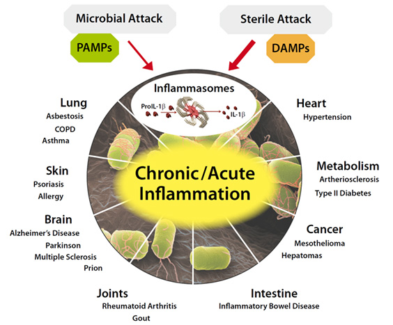
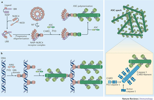
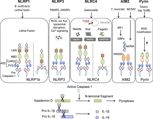
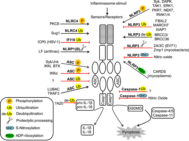
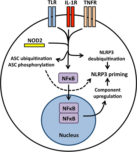
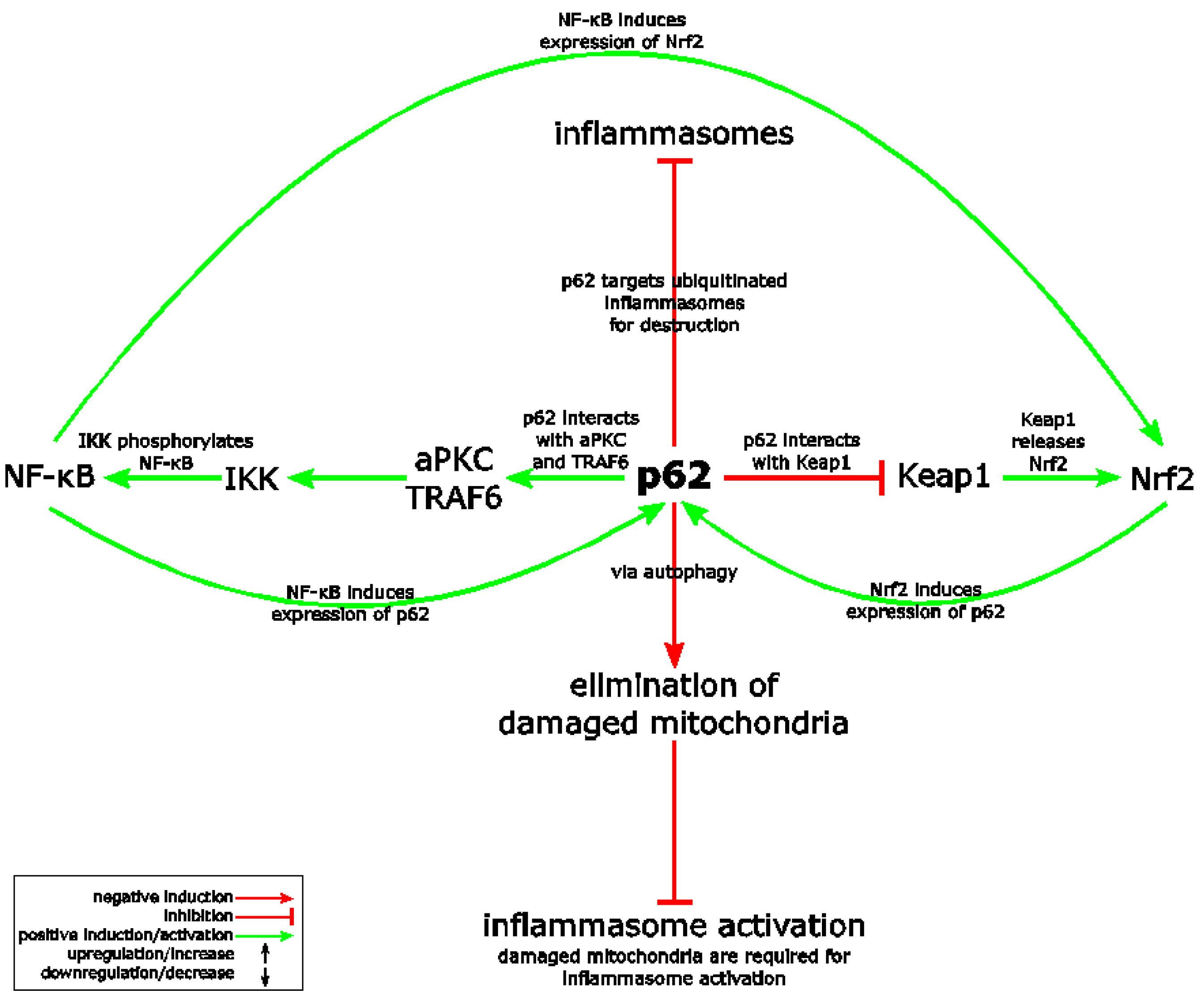
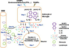
Further, deep understanding of these processes and effective interventions related to them may allow us to significantly extend our lifespans and health spans.  We have also been deeply concerned with understanding certain processes of aging which we have regarded to be deleterious and worth fighting, including inflammation and the SASP, what happens when old senescent cells do not go away but instead become bad citizens. Now we know they are not all bad, and indeed may be harnessed in the interest of health and longevity.’

We are entering an era where organ repair, renewal and regeneration has been demonstrated in laboratory fish and small animals, where we are understanding the molecular mechanisms through which these can take place, and we know how to trigger them.  We know a substance that can repair and renew seriously damaged hearts in zebrafish and mice, and grow new fins on zebrafish.  We know how a serious injury can trigger de-differentiation of neighboring cells to earlier epigenetic stem-cell like states, where these cells can proceed to regenerate complex organs. This blog entry reports on these exciting developments.

The basic message in a nutshell

When we are young and peppy, cell and organ regeneration and renewal go on as background processes in our body all the time. Here is how it works: molecular distress signals such as associated with injuries, inflammation, or cell senescence, trigger the partial regression of normal mature body cells back to earlier less differentiated epigenetic states.  This is a process of de-differentiation, one that moves cells backwards in their developmental trajectory towards becoming stem cell, but that stops short of going all the way.  Then these de-differentiated cells re-differentiate so as to create healthy new cells.  And renew organs, as needed.  If the injury is to cells that are supposedly permanent and do not reproduce, such as neurons in the brain, no problem. In this case neighboring microglial de-differentiate and then re-differentiate changing their identity to becoming neurons. The de-differentiation takes place with the body using the same factors that Yamanaka and other researchers have used to de-differentiate cells in-vitro removed from the body:  Oct3/4, Sox2, Klf4, and c-Myc, commonly referred to as OSKM.  All this happens on the local level when needed, whether within the heart, the brain, the liver or elsewhere. As we grow older, however, typical changes occur that effectively slows down or stops this renewal process. These include certain epigenetic changes associated with aging, and states of chronic inflammation.  These inhibiting factors appear to be only partially understood as of now. Despite this, the very good news appears to be that to a large extent we may be able to control and reverse the adverse epigenetic changes and neutralize chronic inflammation. We already know the interventions that do this, and they are very simple and broadly available. We don’t need additional scientific breakthroughs; we don’t need extremely expensive new drugs or high-technology treatments. This may be the blockbuster news with regard to antiaging approaches that we have been waiting for for so many years. Here, now.

FIRST, ABOUT AGING

A good argument can be made that aging is the result of decline and loss of tissue homeostasis, that is loss of capability tissues and organs to maintain and repair themselves. An excellent article published in December 2016 is In Vivo Amelioration of Age-Associated Hallmarks by Partial Reprogramming.    “Embryonic development occurs as a unidirectional progression from a single-cell zygote to an adult organism. During embryogenesis and early stages of life, cells undergo a spatiotemporally orchestrated differentiation process, leading to the generation of all of the cell types that comprise an adult organism. These events take place within a stable environment that minimizes molecular and cellular damage. As an organism ages, however, there is a continuous and progressive decline in the mechanisms responsible for minimizing cellular damage.”  In this and many other blog entries, I suggest that there may be some simple and powerful things we can do prevent and possibly reverse that decline.

Of high relevance to the current blog entry is the following paragraph relating background from the same publication: “The last decade of scientific research has dramatically improved our understanding of the aging process (Johnson et al., 2013Kenyon, 2010Riera et al., 2016). The notion that cells undergo a unidirectional differentiation process during development was proved wrong by the experimental demonstration that a terminally differentiated cell can be reprogrammed into a pluripotent embryonic-like state (Gurdon, 1962Takahashi and Yamanaka, 2006). Cellular reprogramming to pluripotency by forced expression of the Yamanaka factors (Oct4, Sox2, Klf4, and c-Myc [OSKM]) occurs through the global remodeling of epigenetic marks (Buganim et al., 20122013Hansson et al., 2012Polo et al., 2012). Importantly, many of the epigenetic marks that are remodeled during reprogramming (e.g., DNA methylation, post-translational modification of histones, and chromatin remodeling) are dysregulated during aging (Benayoun et al., 2015Liu et al., 2013bPollina and Brunet, 2011). In fact, epigenetic dysregulation has emerged as a key hallmark of the aging process (Sen et al., 2016). Several groups, including ours, have observed an amelioration of age-associated cellular phenotypes during in vitro cellular reprogramming (Lapasset et al., 2011Liu et al., 2011Mahmoudi and Brunet, 2012Rando and Chang, 2012). Reprogramming of cells from centenarians or patients with Hutchinson-Gilford progeria syndrome, (HGPS) a disorder characterized by premature aging, resets telomere size, gene expression profiles, and levels of oxidative stress, resulting in the generation of rejuvenated cells (Lapasset et al., 2011Liu et al., 2011Zhang et al., 2011). — Together, these observations suggest that cellular reprogramming may be used to promote tissue regeneration and led us to hypothesize that in vivo partial reprogramming could slow or reverse the aging process and extend organismal lifespan.”

 In this blog entry I cite recent research publications which collectively suggest that our bodies lose a natural endogenous capability for tissue and organ repair due to systemic inflammation and certain dysregulated epigenetic factors, and this may be responsible for most diseases of old age. If this perspective is correct, it could constitute incredibly good news for old people like me. And that is because we are beginning to understand things we can do to prevent systematic inflammation and alter some of those dysregulated epigenetic factors.  Follow this story.

Bad biological processes that happen with aging are sometimes good and essential

Research in just the last year or two has been yielding a much deeper understanding of the relationships between the old-age phenomena of chronic inflammation, the SASP (senescence-associated secretory phenotype) and repair and regeneration on the molecular level. This is causing us to reject or revise many of the simplistic concepts we have held in favor of much more complex nuanced ones.  As a teaser specifically, the presence of both inflammation and the SASP is required to trigger repair and rejuvenation processes. These are good things to have a little of under the right circumstances, bad things to have too much of or under the wrong circumstances.  And we were traditionally wrong about many other things as well, such as the dogma that neurons never regenerate and that if you lose some of the course of life, well that is just too bad for you.

In this blog I will discuss findings of several recent publications linking up the four topics (biological repair and regeneration, inflammation, the SASP and aging), where the related research appears to be going, and very simple practical implications for old folks like me.  These discoveries relate to circle of life phenomena, that is, molecular phenomena that are absolutely critical in embryogenesis and very early development of life, that have largely been evolutionarily conserved over a broad variety of organisms, that have traditionally been viewed as detrimental in old age (the theory of antagonistic pleiotropy), but that can probably be much more effectively harnessed to provide renewal and revitalization at any age.

A great many past blog entries are antecedents to and provide background to this one.  There is our recent ongoing blog series on inflammation for example.

Biological processes tend to be organized according to overlapping networks rather than according to hierarchical structures. As we have often preached, there is no center to biology or biological processes. As usual, the literature concerned tends to consist of articles dealing only with specific factors, usually ignoring many others that are relevant. So, how best to organize this discussion? I will start with short recaps of individual topics of central concern.

REPAIR AND REJUVENATION

Here, we have a variety of phenomena that have been studied extensively, examples of which include wound healing, stem cell activity, and DNA repair.  A broad generalization is that the same or similar processes are involved in the early stages of life, and in life going on at all ages. However, repair and rejuvenation processes tend to become less effective with advanced age.

A naïve approach to organ regeneration which did not work

Let’s start with in-situ regeneration.  A number of years ago, I first wrote in this blog about induced pluripotent stem cells (ref)(ref)(ref).  I suggested a possible approach to a very long life, which I referred to as closing the loop in the stem cell supply chain (ref)(ref)(ref). This approach would involve, first, removal of normal somatic cells from the body, second reverting them to being pluripotent stem cells using some variation of the Yamanaka renewal factors, and third, reintroduction of the pluripotent stem cells into appropriate niches in the body. There, they would be able to re-differentiate into tissues that repair and replace damaged ones The Yamanaka factors were known to be effective in inducing somatic cell to regress to essentially having original pluripotent stem cell properties. The original factors were of course Oct3/4, Sox2, Klf4, and c-Myc, commonly referred to as OSKM.  This approach did not pan out, the basic problem being in the final step, effective reintroducing completely pluripotent stem cells into their original niches. In experiments, cells were completely regressed to near original pluripotent stem cell status, and had been wiped out of any memory of their original cell lineage.  Often when introduced into the body, they  would induce teratomas, that is cysts consisting of mixed tissue which had nothing to do with each other or with what the tissues are supposed to be the point of insertion,  where introduced into the body. Teratomas might, for example, contain teeth, nerve, heart, hair and muscle fibers.

Reference: Reprogramming in vivo produces teratomas and iPS cells with totipotency features (2013)

Now, a dozen years after Yamanaka’s landmark discovery, the approach to cell and organ regeneration that seems to have a great deal of promise is more subtle but still involves OSKM. Instead of using the Yamanaka factors to regress cells to complete pluripotent status outside the body, the idea is to induce partial epigenetic regression of cells to younger states where they are in the body, where they still have memory of their cell lineage, and they happen to be already located in just the right places to induce renewal. A key to this line of research was the discovery that this form of renewal happens naturally all the time within the body.  Naturally using the same Yamanaka factors. And further, that it is triggered by things we have traditionally regarded as bad, including cell senescence, inflammation and inflammasome’s.  I spin the story out as I see it here, and state what I think the implications are for regenerative medicine and for the possibility of significantly longer lifespans. I repeat, this is basically a good news story.

Prelude

A few years ago it was becoming evident that a much more subtle approach needed to be taken to cell an organ regeneration them that tried earlier.  The 2017 publication Transient transcription factor (OSKM) expression is key towards clinical translation of in vivo cell reprogramming points to the growing awareness back then of the need for a more subtle approach to OSKM administration, one that induces only a partial age regression in the cells that preserves their lineage memory.  “Reprogramming adult, fully differentiated cells to pluripotency in vivo via Oct3/4Sox2Klf4 and c-Myc (OSKM) overexpression has proved feasible in various independent studies and could be used to induce tissue regeneration owing to the proliferative capacity and differentiation potential of the reprogrammed cells. However, a number of these reports have described the generation of teratomas caused by sustained reprogramming, which precludes the therapeutic translation of this technology. A recent study by the Izpisúa-Belmonte laboratory described a cyclic regime for short-term OSKM expression in vivo that prevents complete reprogramming to the pluripotent state as well as tumorigenesis. We comment here on this and other studies that provide evidence.”

Another 2017 publication Senescence-Inflammatory Regulation of Reparative Cellular Reprogramming in Aging and Cancer reported “– However, tissue rejuvenation should involve not only the transient epigenetic reprogramming of differentiated cells, but also the committed re-acquisition of the original or alternative committed cell fate. Chronic or unrestrained epigenetic plasticity would drive aging phenotypes by impairing the repair or the replacement of damaged cells; such uncontrolled phenomena of in vivo reprogramming might also generate cancer-like cellular states.” Or, in colloquial nativist English, grandpappy tells his young great-grandson: “When you grow up, doncha you go about fagettin wheah you came from.”

In-situ cell regeneration

The abstract of the 2018 publication In Vivo Transient and Partial Cell Reprogramming to Pluripotency as a Therapeutic Tool for Neurodegenerative Diseases provides a bit more complete summary of the situation:  ”In theory, human diseases in which a specific cell type degenerates, such as neurodegenerative diseases, can be therapeutically addressed by replacement of the lost cells. The classical strategy for cell replacement is exogenous cell transplantation, but now, cell replacement can also be achieved with in situ reprogramming. Indeed, many of these disorders are age-dependent, and “rejuvenating” strategies based on cell epigenetic modifications are a possible approach to counteract disease progression. In this context, transient and/or partial reprogramming of adult somatic cells towards pluripotency can be a promising tool for neuroregeneration. Temporary and controlled in vivo overexpression of Yamanaka reprogramming factors (Oct3/4, Sox2, Klf4, and c-Myc (OSKM)) has been proven feasible in different experimental settings and could be employed to facilitate in situ tissue regeneration; this regeneration can be accomplished either by producing novel stem/precursor cells, without the challenges posed by exogenous cell transplantation, or by changing the epigenetic adult cell signature to the signature of a younger cell. The risk of this procedure resides in the possible lack of perfect control of the process, carrying a potential oncogenic or unexpected cell phenotype hazard. Recent studies have suggested that these limits can be overcome by a tightly controlled cyclic regimen of short-term OSKM expression in vivo that prevents full reprogramming to the pluripotent state and avoids both tumorigenesis and the presence of unwanted undifferentiated cells. On the other hand, this strategy can enhance tissue regeneration for therapeutic purposes in aging-related neurological diseases as well. These data could open the path to further research on the therapeutic potential of in vivo reprogramming in regenerative medicine.”_ There appears to be a great deal of excitement and ongoing research connected with this possibility, that is in vivo approaches to tissue regeneration.

(Following the usual custom in this blog, taking license as a blog writer, I sometimes give italic and/or bold emphasis of parts of quoted passages that I think are particularly important.)

One example of this current research is the July 2018 publication AAV vector-mediated in vivo reprogramming into pluripotency: “In vivo reprogramming of somatic cells into induced pluripotent stem cells (iPSC) holds vast potential for basic research and regenerative medicine. However, it remains hampered by a need for vectors to express reprogramming factors (Oct-3/4, Klf4, Sox2, c-Myc; OKSM) in selected organs. Here, we report OKSM delivery vectors based on pseudotyped Adeno-associated virus (AAV). Using the AAV-DJ capsid, we could robustly reprogram mouse embryonic fibroblasts with low vector doses. Swapping to AAV8 permitted to efficiently reprogram somatic cells in adult mice by intravenous vector delivery, evidenced by hepatic or extra-hepatic teratomas and iPSC in the blood. Notably, we accomplished full in vivo reprogramming without c-Myc. Most iPSC generated in vitro or in vivo showed transcriptionally silent, intronic or intergenic vector integration, likely reflecting the increased host genome accessibility during reprogramming. Our approach crucially advances in vivo reprogramming technology, and concurrently facilitates investigations into the mechanisms and consequences of AAV persistence.”  While this publication is about introduction of OSKM into the body to trigger reprogramming, I think the more subtle, natural and interesting approach is that used in the body, such as the local triggering of OSKM by cell senescence and the inflammatory cytokine IL-6.

The bad is sometimes good

In terms of what goes on within the body, processes that we have traditionally associated with being “bad” appear to be essential for initiating regenerative processes which are ”good.” Specifically, cell damage, cell senescence, inflammation and the release of inflammatory cytokines are needed to initiate natural regenerative processes. In other words, the body automatically triggers natural regenerative processes when and where it senses they are needed, as outlined in the next two publication citations.

The 2016 publication Tissue damage and senescence provide critical signals for cellular reprogramming in vivo reports: “Reprogramming of differentiated cells into pluripotent cells can occur in vivo, but the mechanisms involved remain to be elucidated. Senescence is a cellular response to damage, characterized by abundant production of cytokines and other secreted factors that, together with the recruitment of inflammatory cells, result in tissue remodeling. Here, we show that in vivo expression of the reprogramming factors OCT4, SOX2, KLF4, and cMYC (OSKM) in mice leads to senescence and reprogramming, both coexisting in close proximity. Genetic and pharmacological analyses indicate that OSKM-induced senescence/ requires the Ink4a/Arf locus and, through the production of the cytokine interleukin-6, creates a permissive tissue environment for in vivo reprogramming. Biological conditions linked to senescence, such as tissue injury or aging, favor in vivo reprogramming by OSKM. These observations may be relevant for tissue repair.”

Another publication covering some of the same territory is the 2017 item Injury-Induced Senescence Enables In Vivo Reprogramming in Skeletal Muscle.  “In vivo reprogramming is a promising approach for tissue regeneration in response to injury. Several examples of in vivo reprogramming have been reported in a variety of lineages, but some including skeletal muscle have so far proven refractory. Here, we show that acute and chronic injury enables transcription-factor-mediated reprogramming in skeletal muscle. Lineage tracing indicates that this response frequently originates from Pax7+ muscle stem cells. Injury is associated with accumulation of senescent cells, and advanced aging or local irradiation further enhanced in vivo reprogramming, while selective elimination of senescent cells reduced reprogramming efficiency. The effect of senescence appears to be, at least in part, due to the release of interleukin 6 (IL-6), suggesting a potential link with the senescence-associated secretory phenotype. Collectively, our findings highlight a beneficial paracrine effect of injury-induced senescence on cellular plasticity, which will be important for devising strategies for reprogramming-based tissue repair.”

Somatic body cells are much more plastic than we once thought

That is, they are much more susceptible to being epigenetically regressed and regenerated than anyone previously thought.  “Breakthroughs in nuclear reprogramming have revealed that differentiated cells are strikingly plastic both in vitro and in vivo, with exciting implications for disease modeling and regenerative medicine (Srivastava and DeWitt, 2016Takahashi and Yamanaka, 2016). Many organs, such as pancreas, liver, and kidney are permissive for in vivo reprogramming both to pluripotency and lineage switching (Abad et al., 2013Srivastava and DeWitt, 2016), while other organs and tissues—most notably, skeletal muscle—do not develop teratomas. Interestingly, it has been shown that induced in vivo lineage reprogramming in liver and pancreas is more efficient when combined with injury (Heinrich et al., 2015). Moreover, transient induction of senescence occurs in non-muscle cells during regeneration following muscle damage (Le Roux et al., 2015). In light of these observations, we hypothesized that injury can promote reprogramming in vivo in skeletal muscle and that cellular senescence might play an important role during this process.(ref).”

Different cells in the same neighborhood can be doing very different things

A key thing pointed out here and amplified in other articles I cite below is that not all cells in a given neighborhood in an organ are doing the same thing, and in fact for cell regeneration to take place some have to be doing quite different things. That is some have to be injured or senescent or dying and releasing noxious cytokines in order for other cells in the neighborhood to be undergoing epigenetic regression and regeneration using the OSKM factors. Paracrine (in the neighborhood) Intercellular communication is required for this to take place. And, as the following article points out inflammatory cytokines are absolutely required for initiation of in vivo reprogramming. Sell and organ renewable involves an inter-cellular dance. That is, some cells have to be seriously injured and screaming for help for other neighboring cells to initiate a regeneration process

The April 2018 article Senescence promotes in vivo reprogramming through p16INK4a and IL-6  reports: “Cellular senescence is a damage response aimed to orchestrate tissue repair. We have recently reported that cellular senescence, through the paracrine release of interleukin-6 (IL6) and other soluble factors, strongly favors cellular reprogramming by Oct4, Sox2, Klf4, and c-Myc (OSKM) in nonsenescent cells. Indeed, activation of OSKM in mouse tissues triggers senescence in some cells and reprogramming in other cells, both processes occurring concomitantly and in close proximity. In this system, Ink4a/Arf-null tissues cannot undergo senescence, fail to produce IL6, and cannot reprogram efficiently; whereas p53-null tissues undergo extensive damage and senescence, produce high levels of IL6, and reprogram efficiently. Here, we have further explored the genetic determinants of in vivo reprogramming. We report that Ink4a, but not Arf, is necessary for OSKM-induced senescence and, thereby, for the paracrine stimulation of reprogramming. However, in the absence of p53, IL6 production and reprogramming become independent of Ink4a, as revealed by the analysis of Ink4a/Arf/p53 deficient mice. In the case of the cell cycle inhibitor p21, its protein levels are highly elevated upon OSKM activation in a p53-independent manner, and we show that p21-null tissues present increased levels of senescence, IL6, and reprogramming. We also report that Il6-mutant tissues are impaired in undergoing reprogramming, thus reinforcing the critical role of IL6 in reprogramming. Finally, young female mice present lower efficiency of in vivo reprogramming compared to male mice, and this gender difference disappears with aging, both observations being consistent with the known anti-inflammatory effect of estrogens. The current findings regarding the interplay between senescence and reprogramming may conceivably apply to other contexts of tissue damage.”

Again, we have the point that regeneration requires a dance between different cells in close proximity being in different states of distress or contentment.  This last article also points out the good-and-bad possible roles of the OSKM factors themselves “The power of cellular senescence in inducing tissue remodelling has been further extended to processes of cellular reprogramming in vivo. The transgenic expression of the four transcription factors abbreviated as OSKM (Oct4, Sox2, Klf4, and c‐Myc) (Takahashi & Yamanaka, 2006) in adult mice induces dedifferentiation and cellular reprogramming within multiple tissues (Abad et al., 2013; Ohnishi et al., 2014). However, in addition to reprogramming, the activation of OSKM also results in cellular damage and senescence, both in vitro (Banito et al., 2009) and in vivo (Chiche et al., 2017; Mosteiro et al., 2016). Therefore, OSKM induces two opposite cellular fates, namely senescence and reprogramming, that coexist in vivo in separate, but proximal, subsets of cells (Chiche et al., 2017; Mosteiro et al., 2016). Importantly, it has been demonstrated that senescence plays an active role in facilitating in vivo reprogramming through the paracrine action of the SASP, being interleukin6 (IL6) a critical mediator (Chiche et al., 2017; Mosteiro et al., 2016). Of note, IL6 plays an important role also during in vitro reprogramming (Brady et al., 2013; Mosteiro et al., 2016). Moreover, the concept that senescence promotes cellular plasticity has been further extended to the activation of somatic stem/progenitor cells. In particular, the SASP can confer somatic stem/progenitor features onto proximal epithelial cells in several tissues (Ritschka et al., 2017).”

Portraying the SASP as an all-evil process that needs to be stamped out, as some researchers have done, is plum wrong

When things are working well in the body, the SASP is a normal part of the normal regeneration process.

The 2017 Ritschka article just mentioned is The senescence-associated secretory phenotype induces cellular plasticity and tissue regeneration  which has to say: “Senescence is a form of cell cycle arrest induced by stress such as DNA damage and oncogenes. However, while arrested, senescent cells secrete a variety of proteins collectively known as the senescence-associated secretory phenotype (SASP), which can reinforce the arrest and induce senescence in a paracrine manner. However, the SASP has also been shown to favor embryonic development, wound healing, and even tumor growth, suggesting more complex physiological roles than currently understood. Here we uncover timely new functions of the SASP in promoting a proregenerative response through the induction of cell plasticity and stemness. We show that primary mouse keratinocytes transiently exposed to the SASP exhibit increased expression of stem cell markers and regenerative capacity in vivo. However, prolonged exposure to the SASP causes a subsequent cell-intrinsic senescence arrest to counter the continued regenerative stimuli. Finally, by inducing senescence in single cells in vivo in the liver, we demonstrate that this activates tissue-specific expression of stem cell markers. Together, this work uncovers a primary and beneficial role for the SASP in promoting cell plasticity and tissue regeneration and introduces the concept that transient therapeutic delivery of senescent cells could be harnessed to drive tissue regeneration.”

A caution about senolytic approaches to health and longevity

A key point in the last quote, also emphasized in many other articles, makes me question the wisdom of senolytic approaches advocated by some antiaging researchers, which is simply to extend health and life by getting rid of senescent cells. At best, these senolytic approaches by themselves may be too simplistic, and at worst they might be harmful by removing senescence triggers of cell rejuvenation. The garbage heap of dysfunctional therapies based on “getting rid of the bad stuff” already has many things heaped on it, including bloodletting to get rid of “bad humors” in the blood and colonic irrigation and enema therapies to get rid of “rotten matter in digestive track.” I wonder if senolytic therapies might be headed for the same garbage heap. On the other hand, if there is so much expression of the SASP so as to cause the above-mentioned cell-intrinsic senescence arrest, then conceivably a companion senolytic approach might be useful.

Also, while we are discussing the mixing up the biological good in the biological bad, we need to recall that the OSKM renewal factors are deeply involved in cancer processes, so much so that they are traditionally regarded to be carcinogenic.

In vivo regenerative programming can be used to transform one kind of cell to another related kind of cell

This is a point with many important implications, particularly related to the regeneration of non-mitotic cells such as brain neurons or hair stem cells which have traditionally been regarded to be irreplaceable. As a result, until very recently nerve regeneration in the brain has been regarded to be impossible.

However, the 2013 publication In vivo reprogramming reactive glia into iPSCs to produce new neurons in the cortex following traumatic brain injury  pointed out the possibility of such regeneration and actually showed that it can be done in mouse brains.  “Traumatic brain injury (TBI) results in a significant amount of cell death in the brain. Unfortunately, the adult mammalian brain possesses little regenerative potential following injury and little can be done to reverse the initial brain damage caused by trauma. Reprogramming adult cells to generate induced pluripotent stem cell (iPSCs) has opened new therapeutic opportunities to generate neurons in a non-neurogenic regions in the cortex. In this study we showed that retroviral mediated expression of four transcription factors, Oct4, Sox2, Klf4, and c-Myc, cooperatively reprogrammed reactive glial cells into iPSCs in the adult neocortex following TBI. These iPSCs further differentiated into a large number of neural stem cells, which further differentiated into neurons and glia in situ, and filled up the tissue cavity induced by TBI. The induced neurons showed a typical neuronal morphology with axon and dendrites, and exhibited action potential. Our results report an innovative technology to transform reactive glia into a large number of functional neurons in their natural environment of neocortex without embryo involvement and without the need to grow cells outside the body and then graft them back to the brain. Thus this technology offers hope for personalized regenerative cell therapies for repairing damaged brain.”

CELL REGENERATION AND CANCERS

There are a number of story threads connecting these topics, and I mentioned a few here.

The process of cell de-differentiation that characterizes the initial stage of cell regeneration may also play into the creation of cancers

The July 2018 publication Plasticity of differentiated cells in wound repair and tumorigenesis, part I: stomach and pancreas deals with this issue and the following  diagram and legend are from there.

Proposed models of mature cells acting as cancer cells of origin. We propose that long-lived mature cells may accumulate and store mutations, eventually acting as – or giving rise to cells that can act as – cells of origin for cancers in diverse tissues. This mutational accumulation may occur in two main ways: (A) mature cells (dark blue) may accumulate mutations (yellow triangles) as they maintain their mature functioning cell fate over time. The mutations themselves or stressors may trigger dedifferentiation (teal cell). If the acquired mutations are sufficiently carcinogenic, they may then block the cell in the dedifferentiated state, causing it to expand as a clone that can give rise to cancer (red). (B) The ‘cyclical hit’ model describes mature cells that dedifferentiate and redifferentiate multiple times in response to injury/inflammation. Each time the cells are called back into the cell cycle, replicative stress can promote mutation accumulation. Differentiated cells can store such mutations indefinitely. Eventually, a mutation or combination of mutations is sufficient to block the cell in one of its replicative phases and lead to clonal expansion and potential tumorigenesis.”

Note also the suggestion that a single cell may have a history of several cycle of de-differentiation and re-differentiation accumulating carcinogenic mutations on the way.  I do not know the extent to which one or both of the models in the diagram importantly reflect what is going on. The cell renewal perspective provides a different way of looking at phenomena we thought we fairly well understood. Could it be the what we call cancer stem cells are actually somatic cells that have accumulated carcinogenic mutations and have been regressed to near stem cell status by the body’s natural application of the OSKM factors?

OSKM natural in-vivo de-differentiation may have a darker side: loss of cell lineage memory and epigenetic regulation may lead to cancers

This possibility is outlined in the late 2018 publication The causal relationship between epigenetic abnormality and cancer development: in vivo reprogramming and its future application. “There is increasing evidence that cancer cells acquire epigenetic abnormalities as well as genetic mutations during cancer initiation, maintenance, and progression. However, the role of epigenetic regulation in cancer development, especially at the organismal level, remains to be elucidated. Here, we describe the causative role of epigenetic abnormalities in cancer, referring to our in vivo studies using induced pluripotent stem cell technology. We first summarize epigenetic reorganization during cellular reprogramming and introduce our in vivo reprogramming system for investigating the impact of dedifferentiation-driven epigenetic disruption in cancer development. Accordingly, we propose that particular types of cancer, in which causative mutations are not often detectable, such as pediatric cancers like Wilms’ tumor, may develop mainly through alterations in epigenetic regulation triggered by dedifferentiation. Finally, we discuss issues that still remain to be resolved, and propose possible future applications of in vivo reprogramming to study cancer and other biological phenomena including organismal aging.”

Here is another2014 publication with the same theme Concise review: dedifferentiation meets cancer development: proof of concept for epigenetic cancer.  “The technology for generation of induced pluripotent stem cells (iPSCs) has made significant contributions to various scientific fields, and the field of cancer biology is no exception. Although cancer is generally believed to develop through accumulation of multiple genetic mutations, there is increasing evidence that cancer cells also acquire epigenetic abnormalities during development, maintenance, and progression. Because the epigenetic status of somatic cells changes dynamically through reprogramming, iPSC technology can be utilized to actively and globally alter the epigenetic status of differentiated cells. Using this technology, a recent study has revealed that some types of cancer can develop mainly through disruption of the epigenetic status triggered by dedifferentiation. In this paper, we outline the reprograming process and the epigenetic mechanism associated with the maintenance or conversion of cell identity. We then describe several observations suggesting that dedifferentiation can play an important role in cancer development. Finally, we introduce the system responsible for in vivo reprogramming to demonstrate the involvement of dedifferentiation-driven epigenetic disruption in cancer development, and propose that particular types of cancer can develop predominantly through epigenetic alterations.”_

An interesting angle is that it may be possible to treat some cancers by in vivo reprogramming of cancer cells back to their original cell types

What a coup this could be if it worked!  For example, glioblastoma is a rapid and sure-fire killer brain cancer for which there is no known therapy.  As I write this there is widespread mourning for the loss of Senator John Mcain who just died of it.  However a publication that appeared only a few days ago suggests a possible therapy:  Reprogramming glioblastoma multiforme cells into neurons by protein kinase inhibitors.  “Reprogramming of cancers into normal-like tissues is an innovative strategy for cancer treatment. Recent reports demonstrate that defined factors can reprogram cancer cells into pluripotent stem cells. Glioblastoma multiforme (GBM) is the most common and aggressive malignant brain tumor in humans. Despite multimodal therapy, the outcome for patients with GBM is still poor. Therefore, developing novel therapeutic strategy is a critical requirement.  METHODS: We have developed a novel reprogramming method that uses a conceptually unique strategy for GBM treatment. We screened a kinase inhibitor library to find which candidate inhibitors under reprogramming condition can reprogram GBM cells into neurons. The induced neurons are identified whether functional and loss of tumorigenicity.  RESULTS: We have found that mTOR and ROCK kinase inhibitors are sufficient to reprogram GBM cells into neural-like cells and “normal” neurons. The induced neurons expressed neuron-specific proteins, generated action potentials and neurotransmitter receptor-mediated currents. Genome-wide transcriptional analysis showed that the induced neurons had a profile different from GBM cells and were similar to that of control neurons induced by established methods. In vitro and in vivo tumorigenesis assays showed that induced neurons lost their proliferation ability and tumorigenicity. Moreover, reprogramming treatment with ROCK-mTOR inhibitors prevented GBM local recurrence in mice.  CONCLUSION: This study indicates that ROCK and mTOR inhibitors-based reprogramming treatment prevents GBM local recurrence. Currently ROCK-mTOR inhibitors are used as anti-tumor drugs in patients, so this reprogramming strategy has significant potential to move rapidly toward clinical trials.”

Probably, most of the readers in this blog know about mTOR signaling and are familiar with some of the ways to inhibit or block it – e.g. metformin, resveratrol, rapamycin.  But what is ROCK?  The 2011 publication ROCKing Regeneration: Rho Kinase Inhibition as Molecular Target for Neurorestoration describes it.  “Regenerative failure in the CNS largely depends on pronounced growth inhibitory signaling and reduced cellular survival after a lesion stimulus. One key mediator of growth inhibitory signaling is Rho-associated kinase (ROCK), which has been shown to modulate growth cone stability by regulation of actin dynamics. Recently, there is accumulating evidence the ROCK also plays a deleterious role for cellular survival. In this manuscript we illustrate that ROCK is involved in a variety of intracellular signaling pathways that comprise far more than those involved in neurite growth inhibition alone. Although ROCK function is currently studied in many different disease contexts, our review focuses on neurorestorative approaches in the CNS, especially in models of neurotrauma. Promising strategies to target ROCK by pharmacological small molecule inhibitors and RNAi approaches are evaluated for their outcome on regenerative growth and cellular protection both in preclinical and in clinical studies.”Regenerative failure in the CNS largely depends on pronounced growth inhibitory signaling and reduced cellular survival after a lesion stimulus. One key mediator of growth inhibitory signaling is Rho-associated kinase (ROCK), which has been shown to modulate growth cone stability by regulation of actin dynamics. Recently, there is accumulating evidence the ROCK also plays a deleterious role for cellular survival. In this manuscript we illustrate that ROCK is involved in a variety of intracellular signaling pathways that comprise far more than those involved in neurite growth inhibition alone. Although ROCK function is currently studied in many different disease contexts, our review focuses on neurorestorative approaches in the CNS, especially in models of neurotrauma. Promising strategies to target ROCK by pharmacological small molecule inhibitors and RNAi approaches are evaluated for their outcome on regenerative growth and cellular protection both in preclinical and in clinical studies.

Cell and tissue fate: locked-in inflammation?  Cancer? Tissue and organ regeneration?

The same factors seem to be involved in a number of different situations with very different cell fate and health outcomes.  These including cell senescence, NF-kB. IL-6, inflammation, the SASP and inflammasomes.  To grapple with what happens when, some researchers suggest algorithmic threshold models. An example is the 2017 publication Senescence-Inflammatory Regulation of Reparative Cellular Reprogramming in Aging and Cancer.  “– The degree of senescence/inflammation-associated deviation from the homeostatic state may delineate a type of thresholding algorithm distinguishing beneficial from deleterious effects of in vivoreprogramming. First, transient activation of NF-κB-related innate immunity and senescence-associated inflammatory components (e.g., IL-6) might facilitate reparative cellular reprogramming in response to acute inflammatory events. Second, para-inflammation switches might promote long-lasting but reversible refractoriness to reparative cellular reprogramming. Third, chronic senescence-associated inflammatory signaling might lock cells in highly plastic epigenetic states disabled for reparative differentiation. The consideration of a cellular reprogramming-centered view of epigenetic plasticity as a fundamental element of a tissue’s capacity to undergo successful repair, aging degeneration or malignant transformation should provide challenging stochastic insights into the current deterministic genetic paradigm for most chronic diseases, thereby increasing the spectrum of therapeutic approaches for physiological aging and cancer.”

The following diagrams and explanations are from that publication.


“Transflammation-driven epigenetic plasticity: a paradigmatic example of in vivo reparative reprogramming. Transient activation of the PAMPs-DAMPs → NFκB signaling axis may delineate an optimal zone of transflammation (TF)-driven reparative reprogramming characterized by increased epigenetic plasticity and phenotypic malleability capable of responding and adapting to injury, stress, and disease (Lee et al., 2012; O’Neill, 2012; Cooke et al., 2014). The efficiency of NFκB signaling and the level of inflammatory responses is the nodal point linking the pathogenic assault and cellular danger signals and the organization of cellular resistance and tissue repair. NFκB hyperfunction and its interaction with epigenetic modifiers would significantly squeeze the optimal zone of TF-driven reparative reprogramming, thus impairing the adequate organization of defense mechanisms. By operating as the perpetrator of inflammaging, the NFκB signaling integrates the intracellular regulation of transflammation immune responses in both aging and aging-related diseases (Salminen et al., 2008; Montgomery and Shaw, 2015).”

This diagram is interesting because it suggests an additional role for NF-kB signaling beyond that of initiation and perpetration of inflammatory responses, which I have discussed in a number of past blog entries in the inflammation series. As I understand it, the “squeezed zone” in the diagram corresponds to a situation of chronic inflammation and the over activation of NFκB signaling characteristic of aging which essentially inhibits cellular transdifferentiation and repair. As suggested by the red figures in the lower part of the diagram which are aging people whose cellular renewal mechanisms are inhibited by their chronic inflammation. Or, simply put, chronic inflammation stops reparative reprogramming.

If this hypothesis is correct it is exceedingly exciting to me on both highly personal and professional levels. This is because I think I know how to create an effective at least partial bulwark against chronic inflammation, and that is the 4 Herb Synergy liposomal concoction I have been consuming for at least four years now and soon will be making available for others to use. (A blog entry on this is coming very soon now.)  Could it be that with my age now approaching 89 in November, my ability to control chronic inflammation has been a major factor in the preservation of my health and avoidance of any of the chronic diseases characteristic of advanced aging, precisely because it has preserved my body’s capability for self renewal? Further, is there something here I could share with others interests of their health? Or are there other key factors beyond those considered here which have to be taken into account?

Here is another diagram from the same publication related to the same options of effective reparative reprogramming versus pathological reprogramming:


“Senescence-associated inflammatory signaling (SAIS)-regulated in vivo reprogramming: a threshold model of epigenetic plasticity in aging and cancer. The degree of senescence/inflammation deviation from the homeostatic state delineates a thresholding algorithm distinguishing beneficial vs. deleterious effects of in vivo reprogramming. First, transient activation of innate immunity and/or SASP components (e.g., IL-6) might facilitate reparative cellular reprogramming in response to acute inflammatory events. Second, NFκB-dependent and NFκB-independent (e.g., SIR) para-inflammation switches might promote a long-lasting but reversible refractoriness to reparative cellular reprogramming. Third, chronic SASP might lock cells into highly plastic epigenetic states disabled for reparative differentiation capacities.”

Again, this diagram suggests that control chronic inflammation may make the difference between the states identified in the upper lower part of the diagram.  It suggests that by controlling and minimizing chronic inflammation we could possibly be able to maintain a condition where transient reparative reprogramming is effective throughout life.  I do not know what this would mean for longevity, but the news would be very good.

Also, recall the passage cited above: “However, prolonged exposure to the SASP causes a subsequent cell-intrinsic senescence arrest to counter the continued regenerative stimuli.(ref). So effective cell regeneration requires some but not too much of the SASP.” Here is where senolytic approaches could be valuable.

A further suggestion to me at this point is that maintenance of high levels of NAD+ and Sirtuins might also be required for transient reparative reprogramming to be effective. On a personal level I do this by supplementation with resveratrol and nicotinamide riboside to help maintain both NAD+ and Sirtuin levels.

A third diagram from the same publication follows:

“Reparative reprogramming therapeutics: enhancing the body’s self-cell therapy for resistance to damage and disease. A cellular reprogramming-centered view of epigenetic plasticity as a fundamental dimension of a tissue’s capacity to undergo successful repair may provide new therapeutic approaches for aging and cancer. (1) Epigenetic modifiers: small molecules capable of mimicking the transient amelioration of tissue functions occurring upon short-term induction of OSKM-induced nuclear reprogramming (Mahmoudi and Brunet, 2016; Ocampo et al., 2016) might increase epigenetic plasticity and to enhance regeneration in aging tissues; (2) anti-inflammatory drugs: NFκB-targeting drugs and commonly employed NSAIDs might help reduce some aging- and cancer-promoting inflammatory feedback loops to reestablish the functioning of reparative reprogramming; (3) IL-6-targeting and senolytic agents: IL-6 blockade and senescent cell ablation might help unlock the chronic epigenetic plasticity of SASP-damaged tissues to successfully achieve tissue rejuvenation if accompanied by reparative differentiation phenomena.”

MSI-1436  – a related story about regeneration

From when I first started seriously studying biology and writing this blog, I would come across research articles about how certain organisms could regenerate part of themselves, like newts, tadpoles and hydras. But back then we had only vague ideas of how to induce this kind of regeneration and complex mammals like ourselves. This situation is now changed and there is a developing literature of approaches to regeneration that can apparently work. One of the stories is about a substance called MSI-1436.

The 2017 publication The protein tyrosine phosphatase 1B inhibitor MSI-1436 stimulates regeneration of heart and multiple other tissues reports:Regenerative medicine holds substantial promise for repairing or replacing tissues and organs damaged by disease, injury, and degeneration. Much of the field has focused on development of cell-based therapeutics, gene-based therapeutics, and tissue engineering-based therapeutics. In contrast, development of small molecule regenerative medicine therapies is an emerging area. Using the adult zebrafish as a novel screening platform, we identified MSI-1436 as a first-in-class regenerative medicine drug candidate. MSI-1436 is a naturally occurring aminosterol that inhibits protein tyrosine phosphatase 1B. Treatment of adult zebrafish by intraperitoneal injection of MSI-1436 increased the rate of regeneration of the amputated caudal fin, which is comprised of bone, connective, skin, vascular and nervous tissues and also increased the rate of adult zebrafish heart regeneration. Intraperitoneal administration of MSI-1436 to adult mice for 4 weeks after induction of myocardial infarction increased survival, improved heart function, reduced infarct size, reduced ventricular wall thinning and increased cardiomyocyte proliferation. Satellite cell activation in injured mouse skeletal muscle was stimulated by MSI-1436. MSI-1436 was well tolerated by patients in Phase 1 and 1b obesity and type 2 diabetes clinical trials. Doses effective at stimulating regeneration are 5–50-times lower than the maximum well tolerated human dose. The demonstrated safety and well established pharmacological properties of MSI-1436 underscore the potential of this molecule as a novel treatment for heart attack and multiple other degenerative diseases.”

Further, MSI-1436 has already been in clinical trials for other specific indications and is known to be safe for human administration.  “MSI-1436  was well tolerated by patients in Phase 1 and 1b obesity and type 2 diabetes clinical trials. Doses effective at stimulating regeneration are 5–50-times lower than the maximum well tolerated human dose. The demonstrated safety and well established pharmacological properties of MSI-1436 underscore the potential of this molecule as a novel treatment for heart attack and multiple other degenerative diseases.” Demonstrated safety could shave many years off the time for this drug to be available as a mainline therapy. And its role as a drug could be very significant

MSI-1436 is a drug candidate for restoration of heart muscle function following a heart attack. Currently, no drug exists to restore heart muscle function after a heart attack.  Cardiovascular disease is the world’s leading killer, taking the lives of around 18 million people every year, according to the World Health Organization, and disabling millions more. Imagine your cardiologist telling you “Yeah, your heart tissues are seriously compromised. However, all we have to do is regenerate some of those tissues, and you will be fine. No operation necessary. The process is noninvasive.”  Clearly though, it will be sometime before the drug clears all the hurdles necessary to get to be available on the market.

Preserving umbilical cord blood – another approach to immune system regeneration

The June 2018 publication The potential of non-myeloablative heterochronous autologous hematopoietic stem cell transplantation for extending a healthy life span reports: “Aging is a complex multifactorial process, a prominent component being the senescence of the immune system. Consequently, immune-related diseases develop, including atherosclerosis, cancer, and life-threatening infections, which impact on health and longevity. Rejuvenating the aged immune system could mitigate these diseases, thereby contributing to longevity and health. Currently, an appealing option for rejuvenating the immune system is heterochronous autologous hematopoietic stem cell transplantation (haHSCT), where healthy autologous bone marrow/peripheral blood stem cells are collected during the youth of an individual, cryopreserved, and re-infused when he or she has reached an older age. After infusion, young hematopoietic stem cells can reconstitute the compromised immune system and improve immune function. Several studies using animal models have achieved substantial extension of the life span of animals treated with haHSCT. Therefore, haHSCT could be regarded as a potential procedure for preventing age-related immune defects and extending healthy longevity. In this review, the pros, cons, and future feasibility of this approach are discussed.”

Implications for anti-aging therapies

I could go on and make this writing two or three times as long, so much is the relevant research. I will finish, however, with some additional review articles citations showing excitement among aging researchers, and pick the central theme up again in a later blog entry.

Elixir of Life: Thwarting Aging With Regenerative Reprogramming.   2018

Anti-Aging Strategies Based on Cellular Reprogramming.  2016

Unveiling epigenetic regulation in cancer, aging, and rejuvenation with in vivo reprogramming technology.  2018

Strategies for heart regeneration: approaches ranging from induced pluripotent stem cells to direct cardiac reprogramming.  2015

Cellular reprogramming: A new way to understand aging mechanisms.   2018

Programming and Reprogramming Cellular Age in the Era of Induced Pluripotency.  2015

Changes in Regenerative Capacity through Lifespan.   2015

Rejuvenation by Partial Reprogramming of the Epigenome.  2017

Cell-fusion-mediated reprogramming: pluripotency or transdifferentiation? Implications for regenerative medicine.   2011

Induced pluripotent stem cells reprogramming: Epigenetics and applications in the regenerative medicine.  2017

mTOR-regulated senescence and autophagy during reprogramming of somatic cells to pluripotency: a roadmap from energy metabolism to stem cell renewal and aging.   2011.

Heart regeneration for clinical application update 2016: from induced pluripotent stem cells to direct cardiac reprogramming.   2016

iPSCs-based anti-aging therapies: Recent discoveries and future challenges.   2016

Direct cardiac reprogramming: progress and challenges in basic biology and clinical applications.   2015

In Vivo Transient and Partial Cell Reprogramming to Pluripotency as a Therapeutic Tool for Neurodegenerative Diseases. 2018

Cardiomyocyte generation using stem cells and directly reprogrammed cells.  2012

Harnessing pluripotency from differentiated cells: a regenerative source for tissue-specific stem cell therapies. 2006

Biology of Healthy Aging and Longevity.   2016

Mitochondrial function in pluripotent stem cells and cellular reprogramming.  2014


The post AGING, CELL AND TISSUE REPAIR, RENEWAL AND REGENERATION, INFLAMMATION AND THE SASP appeared first on AGINGSCIENCES™ - Anti-Aging Firewalls™.

UPDATE ON LONGEVITY INTERVENTIONS – MAINLY PERSONAL

$
0
0

“Start with the impossible. Proceed calmly towards the improbable. No worry, there are at least five exits.”
—Father Daniel Berrigan

By Vince Giuliano 4/11/19 

This blog entry is about my ideas about my personal health and longevity. It is not focused on scientific facts, although I do like to think that such facts are the main informants of what I do.  I list some do’s and don’ts I observe, a few personal stories and provide an up-to-date listing of the dietary supplements I take.


RIDING UP THE GOMPERTZ CURVE

There is one thing absolutely sure to kill you, and that is aging,  Mortality increases exponentially with age after 30.  This has been modeled mathematically by Gompertz and others since 1825.  Here is a simple version of the Gompertz curve.

Image source.

The curve gets steeper and steeper every year beyond age of around 25.  If the curve were the side of a mountain, at some point it would get so steep that you would have to fall off.  So, one way to think about death is “falling off” the Gomparetz curve, which sooner or later everyone must do.   In fact, no human being has ever been known to live beyond 123 years.

Where am I on this particular chart?  At age 89 I am already off the scale as far as high death rate is concerned.  There is a lot more that can be said about variants of the Gomparetz curve and how the tail-end associated with very advanced aging might be modified, but my thrust here is a different and highly personal one.  See these links for more related to the Gomparetz curve.

The personal issue I am dealing with here is how I can continue riding the Comparetz curve up and up without falling off, given where I am already at.  At 89, extrapolating he curve shown, I have about a 15% probability of death in the coming year.  Not good to think about.  Actually I think I have a very good shot at continuing to ride up the Gomparetz curve and living healthily with good functionality for another 10-20 years.  This blog entry is about why I think this extremely unlikely-seeming event may occur.

This is not a science research blog entry.  This blog entry is a partial update on the things I am doing to keep myself younger, healthy, and involved as my calendar age inexorably advances.  I like to think those actions are based on science, but some are more based on habit long developed over many years.  This list is more about what I actually do than what I think science says I should do.  And my compliance with my own list is not always as good as I would like it to be. I provide relatively few citation references for most of the actions, since they have been treated in past posts in this blog. Since I see my personal philosophy as central to my longevity, I go into many aspects of this as well as into explicit longevity-oriented interventions.  So I divulge several things about myself not previously shared with my readers.

There are a lot of good ideas here to follow if you are older.  Most of the items were implemented in the last 11 years since I seriously started studying longevity.  A few go back 40 years or more, and I have started a few others only in the last few weeks or months, Based on a constant stream of requests coming from my readers, I am also finally including an update of the list of dietary supplements I am taking.  I am not suggesting that the items on this list are all best for everybody or, in some cases even suitable based on the peculiarities of your condition.

About ten years ago I created the first draft of my treatise  ANTI-AGING FIREWALLS – THE SCIENCE AND TECHNOLOGY OF LONGEVITY.   The approach there was simple but in retrospect I think remarkably effective. The main concept of this treatise was to look at the major existing scientific theories of aging, see what they have in common, see what each has to say about steps that could be taken to halt or delay aging, and combine these steps into an overall “antiaging firewall.” That firewall would define practical lifestyle and dietary interventions that would create long-lasting health and longevity based on the known science. This was a good concept and has been the main one that has gotten me to this point.   However, my thinking about health and aging has continued to become more sophisticated and nuanced and the science itself has also evolved significantly during the period.  I stopped updating the treatise in 2014.  Also, my personal anti-aging lifestyle and dietary interventions have continued to evolve in a way not captured there. I may eventually rewrite this treatise from a different perspective. Meanwhile, what I have to say in this blog entry is the closest writing to an update of that treatise.  First, however, how am I doing now 10 years later?

Regarding my health

This tends to be a mixed bag, but on the whole I think matters are going very well for my age.  The firewalls seem to have worked so far.  Near as I know I am free of any of the usual age-related maladies that are killing off my contemporaries.  I have no cancers, heart or cardio problems, dementias, diabetis, connective-tissue disease, arteriosclerosis, osteoporosis, skin atrophy, dysfunctional joints, impaired gait or other typical issues of aging.  I have long been free of the symptoms of osteo and rheumatoid arthritis I had many years ago.  My eyesight is good and stable, I can walk for miles, climb many flights of stairs, drive my 2015 Subaru without fear of accidents, play with my grandkids and generally go where I want and do what I want.  I have had many doctor and hospital visits recently, but most of those have been to support members of my family.  My mind is good enough to keep up my research and write what you are reading here, and have the nerve to think I can keep this all up for many more years.

On the other hand I need hearing aids to participate fully socially, have the same post-nasal drip and sinus problems I had as a kid, and sometimes get sick.  Not more or less than 30 or so years ago, but about the same. I am sleeping more – up to 10 hours a night.  And right now I am in the final phase of a nasty cold.  Medical tests of my bloods, cat-scans, sophisticated visual tests, hearing tests and the like show remarkable stability from year to year, and in photos my face looks remarkably the same as it did 30 years ago.  No obvious sign my foothold of the Gomparetz curve is wobbly.  So I am now targeting full functionality and productivity up to age 100.

WHAT  I DON’T KNOW

I think I know a lot about the science and practice of longevity, but everything I write here is only to the best of my knowledge.  Actually the pool of my ignorance runs deep and wide – as does everybody’s.  I have no direct knowledge of which of any of the interventions mentioned here have helped me most stay on the Gompertz curve so far.  I don’t know what actually keeps me alive, and whether or not I would still be here and as healthy if I lived on beer and beef jerky, chewed the juice out of fat cigars and spent most of my time on a 6-way recliner watching mud wrestling on an 84-inch TV.  My body knows, but I don’t.  The brain-talking spokesperson part of me that is writing this knows some far-out things but little about the internal workings that actually keep me going day-to-day.  Little that is relative to what there is to be known, although I like to think a lot relative to what is ordinarily known.

DON’Ts

Basically I like to avoid most things known to be life-shortening or risky of health, or likely to produce very high stress – thousands of these including hunting tigers with a spear, rooftop hockey, leaping between tall buildings, sky-diving, cliff climbing road-rage motorcycle driving, under-the-ice swimming, eating random wild mushrooms, drunken binges, pistol duals, fights with biker gangs, etc.  In fact, I shy away from nasty fights with my wife or anyone else.  I could go on and on here to include many mundane things, like drinking more than one small glass of wine or bottle of beer a day.

DOs

I make longevity a passionate theme that touches everything in my life

The science and art of longevity are a double-whammy passion for me, giving meaning to my life and giving me more life itself.  It animates me.  That passion is probably why I am already off the scale in the Gomparetz chart above. I am now in the year 12 of my major professional activity being studying and communicating about the sciences of longevity. And this is after 40 years of avid interest in those sciences as a hobby.   Complicated ideas about life itself and novel theories often keep me occupied and I might find myself rushing to a computer to capture a thought or research a topic any time of day or night. How about Senolytics, i.e. clearing out senescent cells?  This appears to be a latest fad for aging science aficionados.  But is it or is it not a hope for a few more years for an old guy like me?  If so, can I get the desired results for me personally through simple herbal dietary supplements (Fisetin and Quercetin and Curcumin)?  What about cell and organ renewal that goes on naturally all the time but declines with aging?   What more can I do to keep my DNA from being screwed up further as I age?  And a nagging question about those senescent cells.  Is it true that I actually need to keep some around some to trigger cell renewal processes?  What can I do to further activate Klotho?  How do I identify the best phytosubstances that could be used to reverse age-related GPG site methylation and silencing of the KL gene?  The number of unanswered questions grows ever-larger as I learn new things.

I like to set goals and targets for myself

Intentionality is extremely important for me,  I have targeted living a life that is full and happy as well as very long.  I have decided and frequently review how long and how well I want to live, how I am doing.   I generate and review lists, discuss items on them with others, and turn frequently to checking on how I am doing and checking research if there is something important I need turn my attention to,

The way I got deeply into longevity was to set a target of living healthily and productively to age 264.  No kidding.  I created this goal out of whole cloth back in 1994.  Back then, and still now, I had a strong belief that I could create my own reality based on what seemed to be a long history of having successfully done that multiple times against great odds, and based on what I believed was a powerful personal theory of reality creation as a macroscopic quantum-based phenomenon.  To get my PhD from Harvard in a new field of computer science unrecognized academically at the time, Howard Aiken (the computer pioneer and my original thesis advisor) had advised me strongly that I would have to qualify myself as competent in a hard science.  So, I made quantum physics into an area of concentration for my coursework.  This rewired my brain to allow me to think the impossible.  If you are interested in my theory of reality creation and how I have used it in my life, you can read my treatise ON BEING AND CREATION, and have a look at the associated  ON BEING AND CREATION blog.  The philosophy outlined there defines my personal spirituality, on the face of it somewhat quirky but for me very solid.

Now, do I still think I can live to 264, given what I know now?  The compartmented scientist part of me – the important part that writes this blog and that has had a lifelong commitment to the scientific method has to say NO.  There is no way known now that this could happen.  All science must be based on observations and theories that connect these observations, and we now have neither observations nor a theoretical framework on which I could base 264 as a prediction for my lifespan.  Biological entities by their very design appear to have species-characteristic lifespan and we have never reliably observed anybody older than 123.  And we don’t have a tested theoretical framework for anticipating a lifespan as long as 264.  (I also point out that we also have no solid framework for declaring such a long lifespan as impossible.)

I like to think there is a larger aspect of me than the scientist part, however.  That part says YES to the question.  My intention remains as in 1994, to live healthily, actively in a state of continuing contribution and productively to age 264 or beyond. And it is that my wife will live a very long healthy life as well.  This larger aspect of me seeks to take in to account what we don’t know, and what we don’t know we don’t know, and the emergent nature of scientific knowledge itself.  I won’t try to describe this aspect here.  Except to say that there are now whole bodies of scientific knowledge applicable to longevity that were not imagined 15 years ago.   If you are interested in why I can hold on to such a far-out idea,  I suggest you review the ON BEING AND CREATION materials linked to above.  As far as I am concerned the ideas there, though beyond what now can be included in hard science, are completely compatible and in fact based on matters well known in the science of quantum physics.

I seek to keep an open mind

My preoccupation with the great mysteries of life – including the nature of life itself – leads me to want to keep an open mind about everything.  Even disturbing current political events set me off thinking about the long term.  What are the possible implications for Brexit or populist resistance to immigration for the arks of history?   How about racist-inspired shooting massacres?  Is human society as a whole now going through the turmoil of adolescence?  Can we emerge from the turmoil eventually with sufficient resources to allow the human species 1,000, 10,000 or 100,000 more years of thriving?

I seek to draw fully on what the medical, dental and other health establishment have to offer

I expect sicknesses and health problems as long as I remain alive.  And quite possibly, new or more severe ones as I get yet-older.  The key is quickly recovering from them. This may entail doctor visits, hospital stays and pharmaceutical interventions and uses of high-technology equipment.   And all in all, the health establishments and their highly trained practitioners are the best resources to help this to happen.  Not only for recovering from ailments but also for preventing them.  More often than not if I have a new or strange ailment, I will research the literature related to it on my own.  If the medical establishment has limited knowledge, I will seek to establish a research partnership relationship with specialty practitioners, for example pursuing leads in the research literature.  I do all this in the support of my wife’s health as well.

I am very fortunate to live in the Boston area where there are multiple high-quality medical institutions and where so much medical and health sciences research goes on.  Knowing where to go and who to see for what assistance has been important for me.

The medical establishment offers multiple resources for assessing status and keeping me healthy –  like sophisticated eye and hearing exams, blood and urine tests, CAT scans and MRIs.  I draw on physical therapy expertise for continuing mobility, maintaining balance and freedom from debilitating pain.  And I draw on an excellent dental practice.

I recognize I already need “crutches” for some issues, like hearing aids for age-related hearing loss, dental bridges and implants, lenses implanted after cataract surgery.  I don’t need a pacemaker right now, or back braces, and all my joints are all my originals.  But I might need some of such things in the future.

I have adapted a greater context for what my life is about

My general context is to live a life of contribution to others.  To proximate people like family members and friends, and ultimately to everybody. I can resign myself to live to die with a tiny lifespan as us humans have if my commitment is to something much greater.  For me it is the survival of our species and our being able to realize our potential for understanding and contribution.  If I came to a point where my health and vitality would preclude me contributing to others and I would have to draw unduly on life-support resources to keep me alive – at that point I would be very content to die.  I believe this context blends well with my pursuit of personal and species longevity.  I believe if our planet is to survive we must grow up as a species and take responsibility for its long-term survival.  This requires a very long-term perspective for taking care of the planet as well as ourselves.  And I believe that wisdom which comes along from longer and longer lives is probably a prerequisite for such a perspective. I tell myself that contributing to that wisdom is a good reason for me to keep living.

I enjoy a very rich family life

I have had 4 wives and raised 8 children and now have a dozen grandchildren, and I greatly enjoy hanging out with them.   Right now, there are 14 members of my direct family who live within a 25-minute drive and we interact multiple times a week. .  My grandkids in the area are aged 5 months 2.5 years, 4 years, 6 years. 16 years,  and 21 years.  Every few weeks there is a birthday, anniversary celebration or other family event.  Our thanksgiving dinners at our home have from 23 to 36 people

I form and keep strong lifelong personal commitments

These include commitments to support the wellbeing of the members of my family, whatever it takes.  My ex-wives, children and grandchildren are included.  If my wife needs to be driven downtown to a hospital for yet-another doctor’s visit – say the 8th such visit in 7 days, no problem.  I seek to enjoy her company as if we were traveling around and visiting monuments in Greece or Central Asia.  The circles of commitment expand out ultimately to include all members of our human species.  I view my longevity research and my explorations into reality creation ultimately to be in service to our species.

I have a great home

I live in a 13-room house in the middle of a forest – thousands of acres of conservation land in Wayland MA that is still only 30 minutes from Cambridge or downtown Boston in non-rush hours.  The house is 40 years old but sound and well heated.  There is almost no noise heard at night and the air quality is good.  A big deck surrounded by tall Pines, Oaks and a Sassafras tree provides a large natural nook for meals in the summer.  Moving my head a few inches just now, I looked out over a serene forest scene, unchanging except for season.  I saw foraging deer just a while ago.  
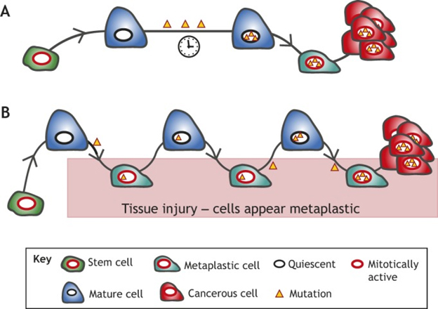
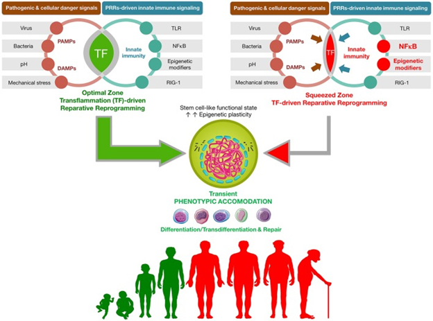
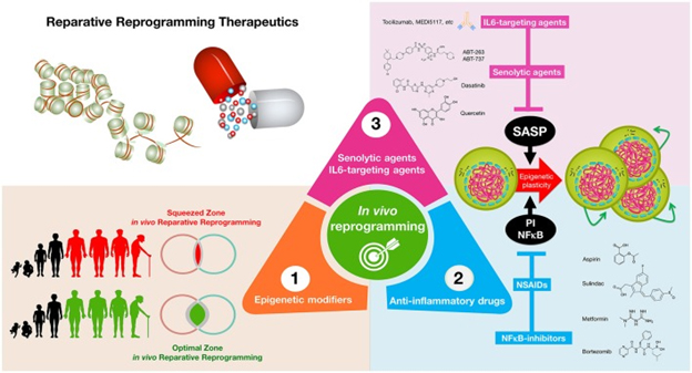
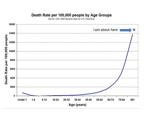
The house also demands that I exercise which is good: the deck, a large front walk, the stairs up to my study, and a 12-car driveway all require snow shoveling.  And if I need to go to the bathroom or want a snack while working in my study, I have to go out, use the stairs and traverse the deck.  My commute to work is up and down outside wood stairs, meaning most days I climb 8 or more flights of stairs.   About an acre of lawn needs mowing and fertilizing, and in the fall the trees shed vast carpets of leaves that demand raking.  It usually takes me a couple of days each Spring to pick up and haul away big branches that fall over the winter.  We have no central air conditioning, but gigantic trees provide shade and there are wall units in my study and bedroom.  All of this is conducive to a serene meditative state and mandatory exercise for me.

Our autos by our house in a NE winter storm


I like to think I am known for all-around generosity and acceptance of others

An unsung but most important skill I have is creating a context of family workability.  And central to this I believe is generosity, personal as well as financial.  So what about the 4 wives I had, mothers of my children?  We are all good friends, always have been, celebrated family holidays together, visit each other when we came, and for many decades shared time together in a camp on an island home on lake Winnipesaukee.  Back in 1983 I faced a dilemma: I had a small son with my current wife and another small son with my previous wife, ages 3 and 5.  We lived in separate houses but often shared meals together.  But I wanted the two boys to grow up in the same house with their mother and father and to know each other intimately as brothers.  The solution was to find and all move into a large house in 1984, our current one, on the lower floor a unit with my previous wife and my son by her, on the upper floor a unit with my current wife and our son.  Each unit had its own kitchen, dining and living room areas but the units were not closed off by doors so the kids and all of us could flow everywhere in the house, and usually we shared meals together.  Fast forwarding to today, it happened as intended.  Both boys grew up with their moms and dad, and they are close brother.  Both sons are happily married now and live in their own houses 26 minutes and 15 minutes from our house.  They are close friends and the source of active grandchildren: one has 3 delightful girls. 3 months, 30 months and 16 years, and the other had two  energetic  boys 4 and 6.   We visit with each other frequently. And my wife and previous wife and I still live in the old house and support each other personally in mutltiple ways.  This is a very happy story of family that goes on and on.  Someday I might seek to tell it in detail.

I take on challenges and projects that can keep my mind and body nimble even almost impossible ones.

Mine includes ever-deeper understanding human biology and aging, and how to create significantly longer healthy lifespans.  And it includes grasping at what the ultimate nature of reality is.  A major new challenge right now is starting a dietary supplement business where we will be promoting, marketing, selling and improving our 4-Herb Synergy supplement.

I wear devices that enable me to monitor a number of instantaneous health parameters – minute-to-minute, hourly, daily, weekly, month-to-month and year to year

I started this about 3 years ago wearing a Basis Peak watch, An Intel device that was unfortunately discontinued.  I now wear a second generation Oura Ring and a Fitbit Charge 3.  They both monitor a number of health parameters, some directly with sensors, some by computed combinations of sensor measurements, history and computed data-based inferences.  These include hour-by-hour and in some cases instantaneous measurements of steps taken, distance traveled, flights of stairs climbed, resting and instantaneous heart rate, HRV, body temperature, sleep stages (light, deep, REM), calories burned, frequency of movement, real time pacing and distance during exercising, routes traveled and much more   Both devices offer certain advantages and relative disadvantages, and I find it useful to gather and compare data from both.  Data is displayed on web interfaces with cell phone apps to access it and is saved to the cloud.  Oura data includes a few measurements which I have found very useful, such as numerical daily estimates of readiness  to handle stresses. So I can compare what is going on now when I have a bad cold with what went on three years ago under similar circumstances.  A FitBit communicating Aria scale gathers weight BMI and body fat percentage, which is also retrospectively available.

I am becoming more and more wired by microprocessor wearable devices

I regularly wear six of these right now.  Besides the Oura Ring and FitBit Charge 3 mentioned above, I wear:

  • Two quality hearing aids which communicate with each other and via Bluetooth to the next device and the cell phone cloud
  • A Resound Phone Clip device that allow me to make and receive hands-free phone calls via my hearing aids, listen to streaming music, and largely control my smartphone, all just with voice.
  • And of course my Pixel 2 smartphone itself with all its wonderful apps.

At this point these are all external devices; I have no implants, yet.  And I expect soon to add or substitute external device that measure more health parameters such as H2 oxygen level and blood glucose.

I pursue regular sleep and rest and monitor its quality. 

I love my regular sleep environment, the room and bed. There is an 8” memory foam mattress topper, a trusty electric blanket and 4 layers of comforters that can be added or peeled off.  Nighttime temperature around 61F, up to 66F in July-Aug with the aid of a quiet AC.  I seek 30% or more humidity with the aid of a vaporizer, more when I have a cold. I usually sleep 8-9 hours a night, more if I am sick.  I monitor the length of my sleep and the amount of time spent in sleep stages (deep, light, REM, and wake), and how these are staged using the sleep app from thr Oura ring.  The Oura Ring app offers a wealth of data about what goes on in my sleep like resting heart rate, HRV and body temperature.  This plus how I feel provides me with a good integrated picture every morning of what happened while I was in bed and my readiness to take on stresses in the coming hours.   If I feel sick, multiple indicators may be askew so as to confirm that such as disturbed sleep stages, elevated resting heart rate, unusually high body temperature and depressed heart rate variability.

I usually pursue a sensible diet, a Mediterranean-like Diet 

With lots of fish, fruits and vegetables and avoiding ultra-processed foods and red meats.  But I am not really religious about diet.  I will eat a 5-Guys cheeseburger every week or two and tend to eat what is put before me at meals at home.  Usually this is very wholesome stuff with lots of vegetables and fish.  Tonight it was old-fashioned tuna fish casserole, actually made with a 99-cent package of Tuna Helper. And sometimes I bring back large bags of potato chips when I grocery shop, I do consume a lot of olive oil, blueberries, nuts, fresh salad greens, olives, avocadoes and other “good for me” things.

I seek to keep moving

With regard to movement, my body seems to be a testament to the “use it or lose it” rule.  My Fitbit watch beeps every hour to remind me to get up from my computer and move around.  I like to do exercise equivalent of at least 6,000 steps daily, and many days this will happen as a result of my excursions out of the house or mowing the lawn or shoveling snow or chasing little grandkids around the mall. The Fitbit 3 watch and my Oura Ring give me good indicators of steps taken, floors climbed and calories burned, so evenings I will fill in to data quotes on the treadmill and with physical therapy exercises  Sometimes if I am ill I will do nothing,  The PT and stretching and weight lifting exercises can take up to 45 minutes and are very important for me too, for maintain good balance and physical control and avert lower back pain.  Every year for the last 8 years I Have done a 6-8 week round of physical therapy for lower back pain.  I tell a little story about this below. My daily home regimen of these exercises uses therabands and dumbbell weights, and varies day-to-day.

In addition to exercise Sensible exercise, I have included PQQ in my daily supplement list, for its ability to mimic exercise for the production of PGC 1alpha.

I monitor and seek to tightly control chronic (sterile) inflammation

This I believe is absolutely central for keeping me healthy and mobile and riding comfortably on the Gomparetz Curve.  I have made a specialty of studying the central role of chronic inflammation in all the diseases of aging, and what can be done to mitigate such inflammation.  And you can look at the blog entries in the Inflammation series to see what I have to say about the related science – ref, ref, ref, ref, ref.

There are several things I do to limit chronic inflammation, and these are supported by both much research and personal experience

  • First and foremost, for some 5 years now I have been consuming a home-made dietary supplement which is a liposomal formulation of standardized extracts of four traditional anti-inflammatory herbs – Curcumin, Boswellia, Ginger and Aswaghanda. There is extensive recent published research on the different anti-inflammatory actions of the chemicals in these herbs, and the liposomal nature of the concoction greatly enhances body absorbability.  I invented this concoction to deal with symptoms of arthritis and now several other family members are not also regularly taking it.  We have been working with a liposomal preparation manufacturing company for over three years to create a commercial marketable version of it.  This will be called 4-Herb Synergy and should no be on the market in 2-4 weeks now after a long period of development with many twists and turns and improvements along the way.  There is very much to say about this, so expect blog entries about it very soon.  They will be titled The Science Behind 4-Herb Synergy, and The Making of a Dietary Supplement – how I originally invented this supplement to help myself and what I think it has done for me.  I drafted these documents some time ago and plan to publish them two weeks before commercial availability of the product.
  • Avoiding inflammatory flares is another part of how I handle inflammation, and here is where many of the things I do work partially be controlling chronic inflammation. For example, daily physical exercise and physical therapy exercises can help avert disabling muscle and joint chronic inflammation that would work to shut down my ability to move freely and maintain balance.
  • Finally I need mention the critical roles of factors in fish oils for activating the all important resolution phase of natural inflammatory events. See my blog entry Inflammation Part 3: resolving inflammation – resolvins, protectins, maresins and lipoxins.

I seek to maintain healthy metabolism and body repair mechanisms

To do this, I supplement with resveratrol, pterostilbene, CO-Q10, and nicotinamide riboside.  If I don’t take the supplementation, I will generally feel sluggish during the day. Our bodies have multiple mechanisms for repair, renewal, and supporting high functioning in the presence of damage, ranging from deep and REM sleep to mechanisms for DNA repair to means for silencing dangerous sequences in our DNA including ancient retroviruses.  When a resource is required to deal with a crises or immediate survival situation and that same resource performs repair or renewal functions, inevitably priority is given to the immediate need.   I have written about how this is true for sirtuins which are employed for mitochondrial support, DNA repair, and generation of enzymes required for mitochondrial functioning.  NAD+ is critical both for support of both normal metabolism and endogenous production sirtuins.  So if there is not enough of it to go around, maintainance and repair at the cellular level is short-shifted.  Negative consequences are many including dysfunctional mitochondria, and autophagy, various illnesses and accelerated aging.  I have described this situation in various blog entries in the NAD World series.  Want a horrors list of what can happen if you don’t keep your NAD+ and sirtuins up?   Here is a horrors list from my TALES OF NAD+ POWERPOINT PRESENTATION.

The NAD- CHAMBER OF HORRORS tales are about some of the main things that can go horribly wrong if you don’t have enough NAD+ in your body or your NAD+/NADH ratio goes screwy. These include but are not limited to:

  • Inadequate production of sirtuins: SIRT1, SIRT6 and SIRT7
  • PARP starvation and compromised DNA repair; genomic instability
  • Inadequate production of key mitochrondial proteins, mitochondrial dysfunction and death
  • Extensive mitochondria-originated ROS flooding
  • Metabolic reprogramming to Warburg metabolism
  • Misfolded proteins don’t get cleaned up
    Compromised stress resistance
  • Histones don’t get adequately deacetylated
  • Reduced antioxidant defenses and oxidative damage to proteins
  • Deacetylated and inactivated tumor suppressor proteins
  • Microtubule railways hijacked, inflammasomes activated, and chronic destructive inflammation
  • Cell senescence
  • Impaired autophagy
  • Endoplasmic reticulum stress
  • As a very recent addition, age- related decimation of intestinal stem cell populations

Also, these horror tales include ones about the about downstream consequences of the above factors such as:

  • Genesis and persistence of most diseases of aging including cancers, atherosclerosis, diabetes and dementias
  • Low energy, tiredness, difficulty focusing
  • Hypertension, increased susceptibility to sunburn and skin cancer
  • Makes you fat and stupid
  • Poor sleep
  • Accelerated aging
  • Many many other unwanted consequences and forms of suffering

I use hormetic stresses to keep healthy

Utilization of sub-toxic dosses of stresses to numerous positive health effects is a fundamental area of longevity science I focused on 6-7 years ago, based on using a  property manifest in all living organisms known as hormesis.  See these blog entries,   I have developed several personal practices in this area.  Some of these practices are detailed in a fun PowerPoint presentation which can be accessed here.  Here are two slides from that presentation showing tiny hormetic interventions I pursued in a single day in 2013, identifying the biological pathways concerned.  I continue such practices today:


I also do several additional things to support cell and DNA repair and renewal functions, such as

  • Supplement with Quercetin and Fisetin to support body removal of senescent cells
  • Supplement with Acetyl-l-Carnosine, Acetyl-l-Carnitine, Alpha-Lipoic Acid, and melatonin for impact on mitochondria among other reasons
  • Manage stress, Use of stresses as hormetic interventions – heat shock and cold shock.  See the  PPT presentation mentioned above.

I seek to maintain full cognitive functioning via several approaches

  • One of these is regular physical exercising
  • Another approach is direct intellectual exercising, mainly focused on my two current areas of passion – longevity science and the nature of physical reality

With regard to longevity science I engage in multiple tasks.  One of these is watching emergent areas of science that might (or might not) lead to breakthrough innovations in human longevity.  I have written about any of these in this blog.  Examples of areas that haves recently grabbed my attention are:

Promote autophagy and senescent cell clearance

For example, I consume:

  • Curcumin, resveratrol for autophagy
  • Fisetin, pterostilbene, quercetin,a resveratrol for senolytic efects

Research on rejuvinating factors in young blood plasma

There is a 15-year history of this research that started with sewing two mice together so they shared a common blood stream, and old mouse and a young mouse.  You got it, the old mouse got a lot younger by multiple indicators, and the young mouse got older.  For a long time nobody knew why this process, called heterochronic parabiosis worked – obviously something in the bloodstream, but what?  Several candidates have been suggested for what is involved, GDF11 and TIMP2 being among them.  Recently research has suggested that the rejuvenating factors may be communicated in the plasma, not via cells – ruling out stem cells per-se as the communicating factors.  My personal favorite theory is that the good effects are communicated via transcription factors packaged in exosomes in the plasma.  Moreover it is thought by some and animal experiments suggest that the rejuvenating factors may be present in plasma derived from placentas or umbilical cords, bringing this closer to being a human therapy, not only because these are normally discarded at childbirth, but also because plasma is independent of blood types and being cell-free does not pose a threat for host-graft rejection.  In fact, the FDA has recently cracked down on fringe commercial clinics offering young blood anti-aging therapies for substantial fees This is an area of research I am watching.

Other initiatives to support continuation of my cognitive capabilities include:

  • Maintain good CNS vasculture, seeking ongoing reactivation of adult neural stem cells, and remodeling of their synaptic activity/plasticity
  • Supplements include DHA, pregnenalone, acetyl-l carnite, l-carnosine, alpha-lipoic acid, melatonin

I like to view pain as me good friend, not as my enemy

Pain is an evolutionarily-conserved signaling system.  Pain can be read it as “We have got some kind of possible problem here.  You should consider what this pain is likely due to and what you can do about it.”  I give two examples from my history.

  • Some 25 years ago I got some serious signs of rheumatoid arthritis: inflamed painful joints in my hands, elbows, wrists and knees. My rheumatologist put me on a massive initial dose of prednisone and told me I would have to be on stronger and stronger anti-arthritic medication for the rest of my life, Ike methotrexate.  Instead, I researched traditional herbal ant-arthritic substances, reading many books at the time because this was before Internet.  I identified four such substances (Curcumin, Boswellia, Ginger, and Ashwaghanda.  Whole books were devoted to each of these as an arthritis treatment.  All of these herbs were sold in capsule form in vitamin shops.  So I started taking substantial doses of each.  My arthritic symptoms soon went away.  When they started to come back some 4 years ago, based on more research I started to make and take a liposomal concoction of the same 4 herbs, for enhanced bio-availability.  Painful swelling and symptom went away again.  I am still brewing the concoction in my kitchen and taking it and the pesky inflammatory symptoms have not returned,  (Lab tests for CRP, IL6 and other inflammatory cytokines repeatedly confirm this, showing negligible values that might be typical of a 23-year old.) My radiologist indicated recently that the site of my quadrecept surgery appears now to be free of signs of osteo-arthritis.  That was not the case some 18 years ago after I had the surgery.  Finally, my wife, son Michael and I have been working intensively with a supplement manufacturer for over 3 years now and will soon be bringing to market 4-Herb Synergy, an advanced version of the product we have been making in our kitchen now for 4 years.  Pain led to remedy and now to product.
  • Some 9 years ago I started experiencing intense lower back pain, sometimes radiating to my right upper leg. Finally a MRI confirmed an awful-sounding diagnosis.  Spinal Stenosis.  You could actually see the bone spurs penetrating into the spinal column.  The pain and lack of sleep was awful.  Several orthopedic doctors indicated that my choices were few – tricky, dangerous and possibly unsuccessful spinal surgery or live with the pain best I can.  Ibuprofen anybody?  Not for me because a prescribed round of it had wrecked my stomach and given me ulcers years earlier.   Or even worse for many nowadays, opioid addiction from excessive use of opioid pain killers.  Then, based on the recommendation of a friend, I saw Dr. Rainville, a Physiatrist in the Spine Unit at New England Baptist Hospital. He was unimpressed by the MRI and said many people had such bone spurs.  He recommended a round of physical therapy, using a particular PT location and PT specialist who administered a portfolio of PT exercises Rainville had developed.  I was very skeptical; anybody could see the spinal stenosis on my MRI.  But I did the round of PT exercises, and like a miracle, in 6 weeks all back pain was gone – vanished.  Gone for 10 or 11 months that is and then the pains started to come back.   By then I had let up on the home counterparts of the PT exercises which depended on a number of machines like a rotary torso unit, a 4-way knee exercise machine, and a machine which made you heave large loads put on your back.  So I got my primary physician to write me a script for another round of exercises at the same place and therapist, and again, the pain went completely away for almost another year.  I successfully repeated this yearly process going back for a round of PT for 8 years in a row keeping me largely pain free, and then I learned that my specialized PT trainer had left the PT parlor I had been using.  So, 6 weeks ago I went back to see Rainville again, and this time he sent me to trainers he had worked with at NE Baptist Hospital.  And they put me on a regimen of 20-minute a day machine-free exercises that had been developed for the US Army and, I am told clinically validated for banishing lower back pain  I am now midway through  this regimen but now feel quite confident it will work, not only to be back-pain free but to restore my balance which I recently felt might be getting a bit iffy.  Again, pain led to action which now is leading me to increased mobility.

An example of how not to fall off the Gomperetz curve

A fairly common story about how elderly people fall off the Gomperetz curve does not apply to me but exemplifies what I am afraid of.  The story Illustrates the complex ways of interaction among multiple interventions.  A person in his mid 80s gradually stops working out and taking long walks because of severe arthritic and lower back pains.  His balance becomes poorer and in the middle of a New England blizzard he falls due to an icy patch and stretches and damages the quadracept muscles in one knee.  The strong doses of ibuprofen he was taking to control the arthritic pain and inflammation had made his balance all the worse.  Unable to walk he is hospitalized, subject to a minor quadrecept repair operation, finally sent to physical therapy where he learns to shuffle slowly with a walker, and is eventually sent home.  Still subject to arthritic pains, that sequence seems to mark the end of long walks or systematic physical exercise.  It also marked the end of his driving and relative independence, and the end of most of his social life.  Minus this exercise and sustained commitment to motion, his social and family life become more and more constrained and over a matter of months he begins to loose his cognitive capabilities.   About a year after the fall he is diagnosed with Alzheimer’s disease and about six months later is moved to a nursing facility for dementia patients.  Two and a half years later he is dead.  The event that visibly knocked him off the Gomperetz curve was the fall, everything else after this being more or less in expected downhill sequence.  Actually he was tottering dangerously on the curve before the fall with his severe arthritic pains, lessened movement and loss of balance – factors that made him vulnerable to slip and fall on the ice.  What he could have done when he started to feel the pain, instead of letting up on exercise, could include 1.  Going of a strong regimen of anti-inflammatory herbs (for me 4-Herb Synergy is what I do), and powerful fish oil supplements which contribute to the control of inflammation, and 2. getting into serious rounds of lower-back physical therapy designed to control the pain and enhance his balance, so he would not have to stop exercising (for me, thanks Dr Rainville at New England Baptist Hospital Spine Center) and 3.  Use daily feedback from a tracking device or two to make sure his exercise and movement regimen was  kept up (for me, my Oura Ring and Fitbit Charge 3), and 4.  Engaged the support of his wife to make sure he kept moving on a daily and hourly basis (for me, thank you Melody, who has just invited me  as I write this out to chop up sheets of ice formed on our driveway).  In other words, take proactive steps so the fall off the curve doesn’t happen.

PERSONAL DIETARY SUPPLEMENT REGIMEN  as of April 7, 2019

SUBSTANCE DOSE (1 pill) WHEN* NOTES (per pill)
4-Herb Synergy 1 tsp 1 AM,  1 Eve Proprietary liposomal preparation of Curcumin, Boswellia, Ginger and Aswaghanda**
Acetyl-l-Carnitine, Alpha-lipoic acid 1 AM, 1 Eve 500 mg Acetyl L-Carnitine HCl,  300 mg Alpha-lipoic acid
Alpha-Lipoic Acid 300 mg 1 AM, 1 Eve
Astragalus root 1,500 mg 1 AM, 1 Eve
Aswaghanda extract 300 mg 1 BB, 1 Eve KSM 66, Stand. to 5% Withanolides
Bacopa extract 300 mg 1 BB, 1 Eve Stand. to 45% Bacosides, with baCognizine
Benfotiamine 150 mg 1 AM, 1 Eve Benfotiamine (S-benzoylthiamine-O-monophosphate)
Bitter Melon Fruit 450 mg 1 AM, 1 Eve
Boswellia Serrata extract 75 mg 1 BB, 1 Eve 5_LOXIN, Stand to 50% AKBA
Calcium-Magnesium-Zink 500 mg 2 AM, 2 Eve 333 mg Calcium (as Cal Carbonate, Cal Asparate and Cal Citrate). 133 mg Magnesium (as Mag Oxide, Mag Asparate and Mag Gluconate, 5 mg Zink (as Zink Oxide, Zink Asparate, and Zink Gluconate)
Cat’s Claw 500 mg 1 AM, 1 Eve
Copper 3 mg 1 AM As Copper Gluconate
DHEA 50 mg 1 BB, 1 Eve Dehydroepiandrosterone
(Mega) EFAs 1,050 mg 2 BB, 2 AM, 1 Eve Molecularly distilled fish oils, 400 mg EPA, 200 mg DHA
Eleuthero Root 500 mg 1 AM, 1 Eve
Fisetin Flavinoid 100 mg 1 AM, 1 Eve From Wax Tree Extract, take for 5 consecutive days each month
Garcinia Cambogia extract 600 mg 1 AM, 1 Eve Stand to 50% Hydroxycitric Acid
Garlic Ultra 600 mg 1 Eve Stand to 2.5% Allicin
Ginger root 550 mg 1 AM, 1 Eve Ginger (rhizome)
Ginseng Extract 250 mg 1 AM, 1 Eve Americn Ginseng Extract (root)
Glucosamine, Chondroitin & MSM 1,000 mg 2 AM, 2 Eve 25 mg Sodium, 55 mg Potasium, 375 mg Glucosamine Sulfate, 300 mg Chrondroitin Sulfate, 250 mg Methylsulfonylmethane (MSM)
Grape Seed & Resveratrol 400 mg 1 BB, 1 AM,1 Eve 225 mg Grape seed extract Stand for 85% polyphenols, 75 mg grape skin extract Stand to 45% proanthocyanidins, 25 mg grape juice extract (whole fruit), 75 mg resveratrol
Green Coffee Bean Extract 200 mg 1 BB, 1 AM Stand to 50% chlorogenic acids
Green Tea Fat Burner 360mg 1 BB, 2 AM 200 mg GT extract Stand. to 50% EGCG (leaf),  160 mg Caffeine, 160mg other ingredients
Horny Goat Weed Extract 500 mg 1 AM, 1 Eve Stand to 10% Icarin
L-Carnosine 500 mg 1 AM,  1 Eve
Probiotic 15-35 100 mg 1 BB, 1 AM,1 Eve 15 Strains, 17.5 billion microorganisms per cap
Magnesium Citrate 200 mg 1 AM, 1 Eve
Melatonin 3 mg 1 Eve Before bed
Milk Thistle extract 300 mg 1 AM,  1 Eve Stand to 80% Silymarin
Carotene Complex 1 AM 3,000 mcg Vitamin A (as 100% beta-carotene and mixed carotenoids), 6 mg Food Carotenoid Blend: annatto (seed), carrot extract (root), West Indian lemongrass (aerial), spirulina
Nicotinamide Riboside 125 mg 2 BB Tru Niagen
Olive Leaf extract 500 mg 1 AM, 1 Eve Stand to 15% Eleuropain
Pterostilbene 50 mg 1 BB pTeroPure,
PQQ 20mg 1 AM, 1 Eve BioPQQ™ 
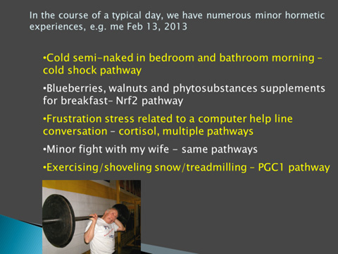
(Pyrroloquinoline quinone disodium salt)
Quercetin & Bromelain 406 mg 1 AM, 1 Eve 250 mg Quercetin dehydrate, 156 mg Bromelain
Saw Palmetto 540 mg 1 AM, 1 Eve Berry
Pregnenalone 50mg 1 AM
Selenium 200 mcg 1 AM As 1-selenomethionine
Stinging Nettle Leaf 480 mg 1 AM
Trans Resveratrol 500mg 1 AM, 1 Eve Knotweed Stand to 50% Trans Resveratrol
Turmeric extract 580 mg 1 BB, 1 Eve Curcumin C3 cmplx, Stand to 95% Curcuminoids, with Piperine
Ubiquinol 100 mg 1 BB, 1 Eve Quinol
Vitamin B-6 100 mg 1 AM, 1 Eve As Pyridoxine Hydrochloride
Vitamin B-12 500 mg 1 AM As Methylcobalamin
Vitamin B-100 complex 1,000 mg 1 AM 100 mg Thiamin, 100 mg Riboflavin, 100 mg Niacinamide, 100 mg B6, 400 mcg Folic Acid, 100 mcg B12, 1oo mcg Biotin, 100 mg Pantothenic Acid, 100 mg Choline Bitartrate 100 mg Inositol, 100 mg PABA
Vitamin C 1,000 mg 1 AM,  1 Eve As Ascorbic Acid
Vitamin D-3 5,000 IU 1 BB, 1 Eve 5,000 IU as  Cholecalciferol
Vitamin E 180 mg 400 IU as Alpha Tocopheryl Acetate
Ultra Vit K 2,400 mg 100 mg as Menaquinone-7, 1,300 mg as Menaquinone-4, 1,000 mg as Phytonadione


*  BB – before breakfast,  Morn – usually in AM, Eve – after supper before bed

** Own proprietary blend.  See blog entries On the Making of a Dietary Supplement and The Science Behind the 4-Herb Synergy Supplement and the disclosure and disclaimer notices.

DISCLOURE AND DISCLAIMER NOTICES

The writer of this blog has a proprietary interest in the 4-Herb Synergy supplement.

The above statements have not been reviewed by the FDA

From time to time, this blog discusses disease processes, and in the case of the present entry, the writer’s personal experience.  The intention of those discussions is to convey current research findings and opinions and personal experience, not to give medical advice.  the information in posts in this blog is not a substitute for a licensed physician’s medical advice. if any advice, opinions, or instructions herein conflict with that of a treating licensed physician, defer to the opinion of the physician. this information is intended for people in good health.  it is the reader’s responsibility to know his or her medical history and ensure that actions or supplements he or she takes do not create an adverse reaction.

The post UPDATE ON LONGEVITY INTERVENTIONS – MAINLY PERSONAL appeared first on AGINGSCIENCES™ - Anti-Aging Firewalls™.

INFLAMMATION PART 6: THE SCIENCE BEHIND THE 4 HERB SYNERGY DIETARY SUPPLEMENT

$
0
0

Ancient plant wisdom meets nanotechnology

By Vince Giuliano                                                                       May 28, 2019

 This blog entry is concerned with a new dietary supplement that my colleagues I have sourced, called 4 Herb Synergy. It is concerned with the scientific considerations we have had in mind in the process of creating the supplement, the molecular mechanisms we believe are responsible for how it works, and the vehicle preparation for enhanced bioavailability. After 4 years of development, the first commercial batch of the product has been produced and we should be selling it online in less than a week now.

This blog entry is focused on the science related to the 4 Herb Synergy supplement.  It is a companion to the simultaneously published blog entry On the Making of a Dietary Supplement, which is basically a highly personal story of the history of creation and fine tuning of the supplement.

Based on the preponderance of research described here, we can conservatively represent that 4 Herb Synergy supports a healthy response to chronic inflammation.  Please see the medical claims disclaimer at the end of this blog entry. These statements have not been evaluated by the Food and Drug Administration. Any new product described in this blog is product is not intended to diagnose, treat, cure, or prevent any disease.

 This is Part 6 of what will likely be a nine-part series of blog entries related to chronic  inflammation.  Being a central aspect of every degenerative disease of old age, chronic inflammation can be thought of as the Great Executioner, the most central machinery of most people’s ultimate illnesses and deaths. Parts 1 through 5 of this series are already published.  Part 1 of the series is the same as Part 5 of the NAD world.  That blog entry is concerned with The pro-inflammatory effects of eNAMPT (extracellular NAMPT, nicotinamide phosphoribosyltransferase).  Part 2 of the series relates a) the “master” pathway network of inflammation (NF-kB) to two other pathway networks clearly implicated in aging and disease processes, b) Genomic Instability (p53), and c) Oxidative stress (Nrf2).  Part 3  is concerned with the all-important resolution phase of inflammation, how acute inflammation goes away under ideal conditions instead of hunkering down to lingering and dangerous chronic inflammation. Part 4  of the inflammation series, is concerned with  PCSK9 inhibition – Also that blog entry is Part 1 of a series on interventions that reduce all cause mortality (ACM). Part 5 of the series is concerned with the basic science of inflammasomes and how they relate to a number of disease processes.

THE HERBAL INGREDIENTS IN 4-HERB SYNERGY

The supplement is a liposomal preparation of four herbal ingredients, extracts of traditional and well-researched herbs which we believe act synergistically: curcumin, boswellia, ginger and ashwaghanda.  The extracts are each standardized to multiply concentrations of the bioactive chemical component thought to be most beneficial.   For more rapid and complete body uptake, ingredients are encapsulated in liposome capsules made out of a phosphatidylcholine complex, a form of non-GMO sunflower lecithin, topped off with a bit of Vitamin C and natural cinnamon flavoring.

 CURCUMIN


Curcumin is the principal active ingredient in turmeric, a plant with a history of medicinal use going back over 4,000 years in the Ayurvedic tradition.  it has served as a principle spice in Southeast Asia and played an important role in religious ceremonies.  It showed up in China around 700 AD, in East Africa around 800 AD, in West Africa around 1200 AD, and in Jamaica around 1750.  In Chinese Medicine it is known at Jiang Huang

Contemporary medical researchers begin investigating curcumin over 25 years ago and the PubMed database of the National Library of Medicine currently contains 13,255 research publications related to curcumin (Up from 11,865 only seven months ago.)   In response to infection or cellular stress, inflammasomes are assembled, activated, and involved in Of these, 1599 articles are concerned with curcumin and inflammation.  (Note:  numbers of publications are as of latest date I reviewed this material before publishing this blog, and are constantly increasing, indicating how hot the field of these nutraceuticals is.) Many of these articles are related to curcumin’s capability to control inflammation and cancers via inhibiting the NF-kB pathway, the “master pathway” of inflammation.  Curcumin differentially regulates 67 mRNAs, inhibits unwanted apoptosis, induces autophagy and kills cancer cells.

The chemical composition of turmeric is very complex , including relatively large proportions of flavonoids, phenols and terpenoids, and moderate amount of tannins, alkaloids, saponins and steroids.  Its most positively-acting bioactive components appear to be curcuminoids and terpenoids (references).

The US government database clinicaltrials.gov lists 198 clinical trial studies for applications of curcumin in various disease conditions. (This number has increased from 152 in just the year since the first draft of this material.)

Curcumin is already a very popular dietary supplement.  A 2018 market research study indicates “The global curcumin market size was estimated at USD 44,246.3 thousand in 2016 and is projected to register a CAGR of 13.3% over the forecast period (to 2025).”

However, bioavailability of curcumin is very limited.  “Clinical trials in humans indicate that the systemic bioavailability of orally administered curcumin is relatively low (3-5) and that mostly metabolites of curcumin, instead of curcumin itself, are detected in plasma or serum following oral consumption (6, 7)(ref).”  That is why liposomal preparations of curcumin such as in 4 Herb Synergy are believed to have a significantly greater capability of reaching the body organs where the herbal chemicals can make a difference.

The turmeric extract in 4-Herb Synergy is standardized to 95% curcuminoids.

BOSWELLIA


In Ayurvedic medicine a gummy resin from a branching tree, Boswellia Serrata, is one of the most ancient and valued herbal remedies.   Extracts of Boswellia serrata have been traditionally used in folk medicine for centuries to treat various chronic inflammatory diseases.   As described in ancient Sanskrit texts 600 to 700 B.C., it is an effective remedy for skin and blood diseases, diarrhea, dysentery, ringworm, boils, fevers (antipyretic), cardiovascular diseases, asthma, bronchitis, liver maladies, chronic mouth and throat soreness, hemorrhoids, and a number of other conditions.

We know the gum from biblical times as Frankincense.  In Chinese medicine, it is known as Ru Xiang.

The National Library of Medicine research  database PubMed lists 577 research articles on Boswellia and 94 of these are related to Boswellia and control of inflammation.  Like the other herbal extracts used in 4 Herb Synergy, the chemical composition of the resinous part if Boswellia Serrata is very complex.  It contains four major pentacyclic triterpenic acids which are capable of inhibiting pro-inflammatory enzymes.  These are β-boswellic acid, acetyl-β-boswellic acid, 11-keto-β-boswellic acid and acetyl-11-keto-β-boswellic acid.  Out of these four boswellic acids, acetyl-11-keto-β-boswellic acid, known as AKBA, is the most potent inhibitor of 5-lipoxygenase, an enzyme responsible for inflammation.   The extract used in making 4 Herb Synergy is particularly enriched for the presence of AKBA.  Research shows that the enzyme 5-lipoxygenase inhibited by AKBA plays a major role in the formation of leukotrienes, which stimulate and perpetuate inflammation.  This inhibition results in significantly reduced levels of inflammatory mediators (IL-1β, IL-6, TNF-α, IFN-γ and PGE2), and increased level of IL-10.

The US government database clinicaltrials.gov lists 11 clinical trial studies for applications of boswellia in various disease conditions.

Bioavailability of boswellia is quite limited, as is the case for curcumin.  That is why we think a liposomal preparation of boswellia such as in 4 Herb Synergy has a significantly greater capability reaching the body organs where the herbal chemicals can make a difference.

The Boswellia extract in 4-Herb /synergy is standardized to 30% AKBA.

GINGER


Ginger, the “root” or the rhizome, of the plant Zingiber officinale, has been a popular spice and herbal medicine in the Ayurvedic tradition for thousands of years. It plays a central role in many other traditions of folk medicine as well, in China, in ancient Greece, throughout the Middle East and Asia. Ancient texts suggest that its use has been strongly associated with wealth, power, and commerce.  Explorers like Marco Polo and Vasco da Gama were concerned about where it was grown and how to get it back to European capitals.  In Europe you had to be very rich to get the herb back when they were alive.

In folk medicine the medicinal applications of ginger are widespread and vary to some extent by society and culture.  In several ancient societies, ginger was viewed to have spiritual, aphrodisiac, general healing, and life-extending powers.   Ginger is mentioned in ancient religious texts like the Quran; “Allah says ginger will be one of the drinks that will refresh the believers in paradise.”  In ancient ayurvedic texts ginger is recommended for painful and inflamed joints, and improving digestion and elimination.   In China, ginger has been used to help digestion and treat stomach upset, diarrhea, and nausea for more than 2,000 years.

The PubMed database of the National Library of Medicine currently contains 3394 research publications related to ginger.  Of these, 223 articles are concerned with ginger and the control of inflammation.

The primary active ingredients in ginger are known as gingerols.  These suppress the production of pro-inflammatory molecules (TNF-alpha and interleukin subtypes including IL-1β and IL-6).   They inhibit PI3K/Akt and NF-κB signaling, decreases inflammatory biomarkers NO and hs-CRP, and reduce levels of oxidative stress in several cell types.

The US government database clinicaltrials.gov lists149 clinical trial studies for applications of ginger in various disease conditions.

The Ginger extract in 4-Herb /synergy is standardized for its content of gingerols..

ASHWAGHANDA


Ashwaghanda (Withania somnifera) is another highly esteemed Ayurvedic herb with history of use going back more than 4,000 years.  The plant’s root has been used in traditional remedies for inflammation, ulcers, fatigue, cough, rheumatism, gynecological disorders, emaciation, sore eyes, and mental disorders.  In the Ayurvedic tradition, Ashwagandha is classified as a rasayan, a rejuvenating or life-extending agent. 

The PubMed database of the National Library of Medicine currently contains 1099 research publications related to ashwaghanda.  Of these, 67 are concerned with ashwaghanda and control of inflammation.  The active ingredients in ashwaghanda are known as withanolides.

Withanolides down-regulate iNOS and suppress the production of pro-inflammatory molecules (TNF-alpha and interleukin subtypes).  The anti-inflammatory properties of withanolides have been researched extensively, including as they affect the NF-κB, JAK/STAT, AP-1, PPARγ, Hsp90 Nrf2, and HIF-1 pathways. Aswaghanda appears to upgrade presence of the longevity factors FOXO3A and SIRT3 in serum.  There has been continuing investigation of the clinical application of withanolides in inflammation-mediated chronic diseases, including arthritis, other autoimmune diseases, cancers, and neurodegenerative diseases.

The US government database clinicaltrials.gov list  three clinical trial studies for Aswaghanda.

The proprietary ashwaghanda extract in 4-Herb /synergy is standardized to 10% withanolides.

The inflammation-related science behind the 4 Herb Synergy concept

4 Herb Synergy has not yet been subject to clinical trials that would be necessary to justify any medical claims made for the substance.  So I have to declare again that This product is not intended to diagnose, treat, cure, or prevent any disease. And, these statements have not been evaluated by the Food and Drug Administration.

Inflammation is a generic term for a large number of bodily reactions that differ in detail by cause, by organ and in impact.  Inflammation is a bodily reaction to stresses that is ancient, present in all animals, natural, key to wound healing and essential for us to keep living in health.  It is a phase of stress responses that is transitory and in healthy circumstances self-resolving. The last thing we would want is to get rid of all inflammation, for that would soon kill us.  Chronic inflammation on the other hand is a different situation, triggered by some diseases and serious stresses, increasingly common with advanced aging, persistent and unresolving.  And it is itself the cause of many dangerous and deteriorative processes.  So the objective of any intervention to control inflammation, including any dietary supplement, is to limit and control chronic but not acute inflammation.  A series of blog entries already referenced expands on these points and explores the extreme complexities of inflammation.  4 Herb Synergy is targeted to inhibit many common forms of chronic inflammation, but far from all forms of inflammation.  And I see this as good.

Synergy in the control of inflammation

Because the herbal ingredients in 4_Herb Synergy act through different molecular mechanisms as indicated above, we believe they are likely to act synergistically in the control of chronic inflammation.  Yet, both acute and chronic inflammation are very complicated.  There are a large number of risk factors and potential sources of chronic inflammation, including disease processes, environmental causes and advanced aging itself. And there are a large number of circumstances in which it can become manifest, gum infections, trauma injuries, joint pains and some dementias being only a few items on the list.  And similarly there are various approaches to controlling inflammation which vary in their effectiveness.  Because inflammation is a critical body mechanism for dealing with threats and diseases we would definitely not want to wipe it out completely if we could.  Acute attacks of inflammation can justify the use of steroids like prednisone, and the medical; establishment should be entrusted in such situations.  And even for the resolution of chronic inflammation, it may be wise to pursue additional dietary and other approaches.  Again, our point here is not to provide medical advice.  People experiencing serious inflammatory conditions should consult with a medical professional.


Image source

There is a large number of causes for initiation of inflammation as a response of the innate immune system.  These include numerous disease conditions, pathogens and toxins, and insect bites, all associated with numerous PAMPs (pathogen associated molecular patterns) and DAMPs (danger-associated molecular patterns).  Likewise there are numerous manifestations of inflammation, many of which are organ-specific or disease-specific with their own etiologies and manifestations.   Inflammation of the brain as in Alzheimer’s disease shows up and acts very differently than does inflammation of the joints in osteo-arthritis.   What all forms of inflammation have in common, however, is activation in cells of a protein NF-kB, and its translocation to the nucleus of cells where it triggers the production of pro-inflammatory cytokines.  Research has established that each of the herbal ingredients in 4 Herb Synergy in its own ways inhibits the expression of NF-KB.  This is the simplest explanation of how it works.

While there is no published scientific literature yet on 4 Herb Synergy itself, there are significant bodies of research on each of its ingredients, as linked to above, and on bioavailability and liposomal formulations.  While I could generate a lengthy and detailed blog entry on these facts, I confine myself here to a few summary and highlight statements.  In particular, Jim Watson and I have written extensively about two key pathways that are central to health and vitality, NRF2 and NF-kappaB.  In a couple of nutshells:

NRF2

Each of the herbal components in 4 Herb Synergy promote the expression of NRF2, a transcription factor that plays a key role in activating some 240 genes that are parts of the body’s own antioxidant and stress-protective systems. These genes up-regulate production of endogenous anti-oxidant proteins, stress response mechanisms, and drug metabolism and detoxification enzymes.   The science related to NRF2 is well-established and supported by hundreds of research studies and publications.  Check out these blog articles we have written on NRF2:   –

–  The pivotal role of Nrf2. Part 1 – a new view on the control of oxidative damage and generation of hormetic effects  

–   NRF Part 2 – Foods, phyto-substances and other substances that turn on Nrf2, and

  NRF Part 3 – Is promotion of Nrf2 expression a viable strategy for human human healthspan and lifespan extension?

NF-kB

As already mentioned, each of the herbal components of 4 Herb Synergy also inhibits the activation of NF-kB (Nuclear factor kappa-B), another pathway involving activation or suppression of multiple genes and often called “the master pathway of inflammation.”  Control of chronic inflammation is critical in maintaining health, particularly in the face of stresses and aging.  Chronic inflammation can involve a very complex set of processes and management of it has been of concern for years to us creators of 4 Herb Synergy


Image source    Conditions associated with NF-kB expression and chronic inflammation

Among the articles recently written by or contributed to by me on inflammation are:

–  Inflammation series Part 1 (also Part 15 of the NAD world Series, The pro-inflammatory effects of eNAMPT(extracellular NAMPT, nicotinamide phosphoribosyltransferase)

–  Inflammation Part 2. Relating the “master” pathway network of inflammation (NF-kB) to two other pathway networks clearly implicated in aging and disease processes, 2) Genomic Instability (p53), and 3) Oxidative stress (Nrf2). 

–  Inflammation Part 3: resolving inflammation – resolvins, protectins, maresins and lipoxins

Inflammation Part 4 – PCSK9 inhibition

Inflammation Part 5: Inflammasomes – science of and disease implications

AGING, CELL AND TISSUE REPAIR, RENEWAL AND REGENERATION, INFLAMMATION AND THE SASP

Aging, Inflammation, Disease Processes, and Longevity

A great many researchers appear to have reached a consensus that constitutional inflammation increases with advanced aging and that this inflammation sources most if not all of the diseases characterizing the final stages of human life: cancers, diabetes, cardiovascular issues, dementias, etc. The process has been characterized by the term “inflammaging.”  A recent contribution to the extensive literature on this topic is the 2018 publication Inflammation, inflammaging and cancer, from which L have extracted the following quote:

“Numerous evidences show how apparently different age-related pathologies, including cancer, cardiovascular diseases and type 2 diabetes reveal a common inflammatory background [78]. Epidemiological studies demonstrate the relationship between increased levels of inflammatory mediators like Interleukin(IL)-6 or C-reactive protein (CRP) to multiple age-related diseases [9]. In fact, inflammaging is characterized by the establishment of a systemic proinflammatory state with increased level of circulating interleukins such as IL-6, IL-1 and Tumor Necrosi Factor(TNF)-α and inflammatory markers, such as CRP [6]. This results from the activation of signalling networks critical to inflammation, such as those regulated by the Nuclear Factor (NF)-kB transcription factor, along with a variety of different sources of the inflammatory stimuli triggering and sustaining inflammaging, such as senescent cells, the meta-inflammation, the gut microbiota and nutrition [101112]. — In the nineteenth century Rudolph Virchow was the first to hypothesize a connection between inflammation and cancer, but only in the last two decades researchers have produced striking evidences on the role played by the inflammatory process in promoting cancer [1314]. Indeed, not only cancer can arise on sites of chronic inflammation but also a pro-inflammatory microenvironment, supported by inflammatory cells and mediators, is an essential component of cancer and one of its hallmarks [151617]. — Chronic inflammation is, thus, associated with all stages of cancer development increasing its risk, supporting cancer initiation, promoting cancer progression, and supporting metastatic diffusion [10]. Recently, it has been demonstrated that preventive treatment with anti-inflammatory drugs like aspirin reduce the incidence and mortality for colorectal cancer [18]. This leads the way to the potential preventive and therapeutic role of the modulation of cancer-associated inflammatory microenvironment [19].  — The aim of this review is to explore the role of the main actors contributing in the development of inflammaging and cancer.”

Bioavailability and Liposomal formulation of 4 Herb Synergy

Powerful as the herbal extracts that I’ve discussed above are, there are serious limits to their cellular absorption when they are consumed orally.  This seriously limits their biological effectiveness. The central innovation of 4 Herb Synergy is combining a group of synergistically operating anti-inflammatory herbal extractds in a liposomal formation to greatly enhance bioavailability/cellular absorption.

Limit of bioavailability of herbs

For many herbs as well as for certain drugs, limited cellular absorption may very seriously limit the degree of control of chronic inflammation or pain that can be realistically achieved with an ingested substance.   This has to do with a consumed substance getting to where it is needed in the body.  An orally consumed substance targeted to a cell organelle such as mitochondria in the brain has to get:

  • Past the gut bacteria
  • Past the stomach acid
  • Through the intestinal wall and into the bloodstream via the portal vein
  • Past the liver whose job is detoxification and removal of unrecognized substances
  • Avoid the kidneys so it is not peed out
  • Through the blood-brain barrier
  • Through cell membranes
  • Through nuclear and mitochondrial membranes within cells

For some herbal substances like resveratrol and curcumin, only a tiny fraction of what is ingested can get past these barriers.  Studies in rats for example have shown that less than1% of orally consumed curcumin make it into the plasma, less for humans.  See the 2014 research publication Recent Developments in Delivery, Bioavailability, Absorption and Metabolism of Curcumin: the Golden Pigment from Golden SpiceEven lesser amounts of orally consumed curcumin can get through into the brain, and much less into brain cells and cell organelles.  Drastically upping the ingested dose is not an option because that leads to liver toxicity.  So for example, curcumin which is highly effective in killing certain deadly cancer cells in a test tube like gliablastoma, cannot be given orally in doses high enough to impact on such cancers in the brain.  Such bioavailability limitations exist to varying extent for other herbs administered to control inflammation and associated pain.  One very common approach to enhancing bioavailability and supplement pills has been to add substances, such as adding black pepper extract to curcumin, but the resulting increase in bioavailability tends to be quite limited.

Nano and liposomal drug and supplement delivery

The limits on cellular absorption has led the pharmaceutical industry, and more recently some researchers in the dietary supplement industry (including us), to investigating and pursuing means for increasing cellular absorption.  In fact, enhancing cellular absorption is probably the main frontier insofar as enhancing the capabilities and effectiveness of herbal substances is concerned. A number of approaches have been shown to be effective for enhancing cellular absorption of the herbal substances. See for example Bioavailability enhancers of herbal origin: An overview. (2013). “Nowadays with the advancement in the technology, novel drug delivery systems open the door towards the development of enhancing bioavailability of herbal drug delivery systems. For last one decade many novel carriers such as liposomes, microspheres, nanoparticles, transferosomes, ethosomes, lipid based systems etc. have been reported for successful modified delivery of various herbal drugs.“ We believe the greatest promise lies in using nanotechnology approaches where the effective bio availability might be increased by a significant factor. In these approaches the active substances are encapsulated in extremely tiny nano-sized delivery packages which can evade or pass directly through most of the body barriers mentioned above.

The 2014 publication Nanotechnology-based drug delivery systems and herbal medicines: a review describes the situation: “Herbal medicines have been widely used around the world since ancient times. The advancement of phytochemical and phytopharmacological sciences has enabled elucidation of the composition and biological activities of several medicinal plant products. The effectiveness of many species of medicinal plants depends on the supply of active compounds. Most of the biologically active constituents of extracts, such as flavonoids, tannins, and terpenoids, are highly soluble in water, but have low absorption, because they are unable to cross the lipid membranes of the cells, have excessively high molecular size, or are poorly absorbed, resulting in loss of bioavailability and efficacy. Some extracts are not used clinically because of these obstacles. It has been widely proposed to combine herbal medicine with nanotechnology, because nanostructured systems might be able to potentiate the action of plant extracts, reducing the required dose and side effects, and improving activity. Nanosystems can deliver the active constituent at a sufficient concentration during the entire treatment period, directing it to the desired site of action. Conventional treatments do not meet these requirements. The purpose of this study is to review nanotechnology-based drug delivery systems and herbal medicines.”

The nanotechnology approach used in 4 Herb Synergy is to encapsulate herbal ingredients into liposomes, extremely tiny pouch-containing particles of a phospholipid material, these particles are typically between 80 and 250 nm in size, too small to be seen through an optical microscope. The advantages of this approach are:

  • Liposomes are “natural.” They are used in several biological systems and are found, for example, in human mother’s breast milk.
  • The pouch- containing vehicles are made of a completely natural substance commonly found in cell walls, phosphatidylcholine (aka lecithin)
  • Lecithin is also known to be health conveying, and is it commonly sold as a dietary supplement.
  • Compared to other approaches liposomes are relatively easy to make and the technology for making liposomes is relatively familiar.

The use of liposomal delivery to increase bioavailability has been researched extensively in the pharmaceutical industry, and has been of growing importance. About 20 years ago the first liposomal drug was approved for clinical use, the anti-cancer drug doxorubicin. It is believed that liposomes are longer retained in blood circulation and accumulate at pathological sites (tumours or inflamed tissues), leading to higher efficacy and lower systemic toxicity compared to the free drugs or herbal substances they encapsulate. “The application of liposomes to assist drug delivery has already had a major impact on many biomedical areas. They have been shown to be beneficial for stabilizing therapeutic compounds, overcoming obstacles to cellular and tissue uptake, and improving biodistribution of compounds to target sites in vivo. This enables effective delivery of encapsulated compounds to target sites while minimizing systemic toxicity(ref).” Liposomal dietary supplements delivering herbal ingredients have been much slower to appear in the marketplace, only in the last few years. To our knowledge 4 Herb Synergy is unique in the number and combination of ingredients it combines.

Testing 4 Herb Synergy for liposome content and size distribution

A key issue in evaluating any commercial supplement that is represented to be liposomal, Is Is it really that? Answering this is not easy. The naked eye cannot tell a liposomal product from a simple emulsion, and a powerful optical microscope can’t help either.  Fortunately there is a sophisticated optical light scattering technology that can be brought to bear to tell the actual size distribution of the liposomes if they indeed exist in the product.  We have had our product tested for liposomal content and size distribution on two occasions.  Here is the size distribution according to a test of our original homemade product:

Following is a graph of the liposome size of a product batch (not of our own product) produced by our contract manufacturer using the same liposome-generating technology used in making our product.

Bottom scale in both graphs is nanometers, logarithmic for our homemade sample, linear for the manufactured one. A personal objective for the original creation of 4-Herb Synergy was the control of my personal chronic inflammation underlying a painful arthritic condition.

Note that for both tests, the distributions peak at about the 200-300 nanometer size range, thought to be very good for bioavailability.  The blip about 6,000 nm in our homemade product is seen by microscopic examination to be for herbal fragments due to impurities in the ginger  extract we used consisting of tiny fragments of the original ginger.  (Such contaminants are harmless and do not show up in the graph for the manufactured product.)

Evidence for efficacy

As of this time, evidence of efficacy of 4 Herb Synergy for controlling constitutional inflammation is limited and anecdotal.  I list a few pieces of personal data for  me for whatever they may be worth, well-established indicators of inflammation as measured after three years of consuming teaspoons full of 4 Herb Synergy twice a day:  a: CRP of .2, b. Neutrophil  to Lymphocyte ratio of 1.1, c. very low presence of the pro-inflammatory cytokines IL-1beta, IL-6 and IL20 as indicated in an inflammatory panel conducted in December 2017   and d. continuing absence of inflammatory stiffness or associated joint pains.  Each of these is remarkable for someone of my age of 89 with documented earlier history of rheumatoid and osteo-arthritis.

Normal CRP, the most common test for inflammation, is 0.0 to 9.9 mg/L, and .2 is at the extreme low end.  Neutrophil to Lymphocyte ratio is also a very sensitive biomarker of inflammatory state and has high predictive value for outcomes in inflammatory diseases.  There are 495 published papers on this biomarker, most appearing recently.  It is regarded to be a better predictive measure than CRP for mortality for several disease conditions including cancers, coronary artery disease and infectious diseases. My score of 1.1 is on the very low end with 82% of death risk for coronary heart disease associated with a value of this ratio >3.  A second person who has been consuming 4 Herb Synergy shows similar scores, to the extent he/she has been tested.

MEDICAL DISCLAIMER

THE PURPOSE OF THIS BLOG ENTRY IS TO PRESENT SCIENTIFIC FACTS RELATED TO A DIETARY SUPPLEMENT RECENTLY DEVELOPED BY THE AUTHOR AND HIS COLLEAGUES.  OVER THE 10-YEAR HISTORY OF THIS BLOG, WE HAVE REFRAINED FROM ACCEPTING ANY ADVERTISING OR ADVOCATING ANY COMMERCIAL PRODUCT.  WE HEREIN WISH TO CONTINUE THE LONG-ESTABLISHED POLICY OF THIS BLOG , WHICH IS TO PRESENT ESTABLISHED SCIENTIFIC FACTS RELATED TO WELLNESS AND HEALTH, NOT TO  PROVIDE MEDICAL ADVICE OR ADVOCATE ANY TREATMENT FOR CURE, PREVENTION OR DIAGNOSIS OF ANY DISEASE.   

THE STATEMENTS IN THIS BLOG ENTRY HAVE NOT BEEN REVIEWED OR APPROVED BY THE FDA.

FROM TIME TO TIME, THIS BLOG MAY DISCUSS RESEARCH DEVELOPMENTS RELATING TO DISEASE PROCESSES.  THE INTENTION OF THOSE DISCUSSIONS IS TO CONVEY CURRENT RESEARCH FINDINGS AND OPINIONS, NOT TO GIVE MEDICAL ADVICE.  THE INFORMATION IN POSTS IN THIS BLOG IS NOT A SUBSTITUTE FOR A LICENSED PHYSICIAN’S MEDICAL ADVICE. IF ANY ADVICE, OPINIONS, OR INSTRUCTIONS HEREIN CONFLICT WITH THAT OF A TREATING LICENSED PHYSICIAN, DEFER TO THE OPINION OF THE PHYSICIAN. THIS INFORMATION IS INTENDED FOR PEOPLE IN GOOD HEALTH.  IT IS THE READER’S RESPONSIBILITY TO KNOW HIS OR HER MEDICAL HISTORY AND ENSURE THAT ACTIONS OR SUPPLEMENTS HE OR SHE TAKES DO NOT CREATE AN ADVERSE REACTION.

The post INFLAMMATION PART 6: THE SCIENCE BEHIND THE 4 HERB SYNERGY DIETARY SUPPLEMENT appeared first on AGINGSCIENCES™ - Anti-Aging Firewalls™.


The making of a dietary supplement – the long and short histories of it

$
0
0


  By Vince Giuliano  June 3 2019

This blog entry tells the stories of how our new dietary supplement 4 Herb Synergy came into being.  As such, this blog entry tells two short histories: a universal history over more than a billion years, and a highly personal history during my adult lifetime.

Regular readers of this blog have hopefully come to view me as a truth-seeking scientist who has been willing to share his views on a large number of scientific topics related to the nature of human life and longevity.  I have been doing this for over 11 years now, pursuing a pro-bono career of researching topics key to longevity, writing about them in this blog and from time to time speaking about them as an invited speaker at a professional conference, podcast or webcast.   Commercial activities did not enter my mind and I have consistently rejected suggestions that I accept advertising here or feature particular drugs, supplements, or commercial services,  Now, as I approach age 90, I am starting up a commercial venture.  Probably starting in a week from now, my LLC will be selling my 4 Herb Synergy product.  This blog entry seeks to explain how that shift in a commercial direction happened and my motivations for pursuing it.

The first history below is about important events for life itself, ones that almost all happened before I existed, events that were pre-requisite for the second personal history, or for any of us to exist for that matter.  This second history in this blog entry is my personal story related to creating 4 Herb Synergy.  Then, I discuss my motivations for doing so and address a few related questions.

I think that the science basis for the 4-Herb Synergy supplement is very strong.  I cover that in a companion blog entry to this one, Inflammation Part 6: The Science Behind the 4 Herb Synergy supplement.

First, The Billion Year Story

  • More than One billion years ago, plants began evolving sophisticated chemical responses for them to be able to maintain their health given a wide variety of stresses. The evolution of plants made them living biochemical research laboratories.


So, if you ask when the R&D leading to 4 Herb Synergy was first started, it is no exaggeration to answer it was over 1 billion years ago.

  • 600 million years ago or so, primitive animals began to separate from plants and evolve, each new species always adopting whenever it could the same genes and strategies used for healthy survival by earlier more-primitive species – including incorporating many of the original plant-based defenses.

Images source: Wikepedia, Britannaca Kids

EON ONSET MAJOR EVENTS
Hadean 4.5 Ga formation of earth and continents, chemical evolution
Archean 3.8 Ga origin of life, prokaryotes flourish
Proterozoic 2.5 Ga eukaryotes evolve, development of oxygenated atmosphere, some animal phyla appear
Phanerozoic 540 Ma most animal phyla present, diverse algae; explosive evolution of multicellular life forms

    Ga = billion years ago, Ma = million years ago  Table source

  • 150,000 years ago or so, these genes and plant survival-based strategies were finally passed on to us humans, via our countless intermediary biological ancestors.


Image source

  • 4000 years ago, strong traditions of folk medicine were evolving in India and throughout the world, based mainly on the use of plant-based substances. These provided multiple forms of biochemical support for our health and well-being – often working through the very same biological pathways that were active hundreds of millions of years ago in plants.


Image source

  • 4000 years ago until today, certain key herbs have been used for their powerful anti-inflammatory properties, among these Turmeric, Boswellia, Ashwagandha and Ginger. Writings about the medicinal properties of these herbs can be found in the ancient Chinese and Aryuvedic literature and they were used in many other areas of the world as well.


For the last 40+ years these herbs and herbal extracts of them have been sold in pill form as dietary supplements, representing a current aggregate market of hundreds of millions of dollars a year.  The safety of these supplements is unquestioned.

  • In the last 30 years the same herbs have been extensively studied for the biological pathways that they activate. The mechanisms they employ to control inflammation have been discovered and explained in tens of thousands of research publications in the biomedical literature. The four herbs we mentioned utilize somewhat different biological mechanisms and pathways and we believe they operate synergistically.


Image source                                                               Image source
Around 20 years ago it was becoming clear that the biggest limit to these dietary supplements for creating health and limiting inflammation was insufficient  bioavailability – ability of the injested substance to transverse numerous bodily barriers and challenges to get to the important interior areas of cells where they could make a difference.


Image source

  • Particularly in the last 15 years it is becoming increasingly clear that all of the major diseases that kill people with aging – cancers, dementias, cardiovascular issues, diabetes, etc. – are characterized by chronic unresolvable inflammation. And that control of such inflammation could have major implications for healthy aging and extending lifespans.


Image source                                                                    Image source

  • Particularly in the last 5 years there have been major developments in the practical application of nanotechnologies for enhancing the bioavailability of drugs and herbs such as those mentioned. This involves using high technology approaches to encapsulate drugs or herbs in tiny containers, ones too small to see by an optical microscope, containers that can easily pass through bodily barriers that limit bioavailability. An important category of nano-delivery techniques is the use of liposomes, tiny vesicles that are used in nature. For example mother’s milk contains liposomes, probably to enhance the bio availability of its ingredients for tiny infants.


Image source                                                        Liposome

This has been the billion-year impersonal history, background to the personal history story which follows.

My personal 47-year story

The prehistory and history of 4 Herb Synergy

I go on here with some highly personal material, namely a history of what led to 4 Herb Synergy and my personal motivations now for wanting to see it on the marketplace.   This involves telling a quite personal story. A N=1 story.  To be clear here I am making no medical claim for 4 Herb Synergy or any actual product I may be associated with. The statements in this blog have not been evaluated by the Food and Drug Administration. 4 Herb Synergy or any similar dietary substance being sold is not intended to diagnose, treat, cure, or prevent any disease. I intend that at some point it will be possible to have a clinical trial for 4 Herb Synergy with results that allow medical claims to be made for it.

About 1972

Some 47 years ago, when I was in my early 40s, my professional focus was related to the unfolding computer/information revolution.  This was a long time before I got seriously into the health sciences.   I was getting morning bouts of joint stiffness and pain, particularly in my joints, arms, legs, knees and hands.  At first these symptoms were mild but gradually they became more serious and debilitating.  I was an early follower of Adelle Davis back then, an early exponent of nutritional approaches to health, and looked up what was she said in her books about arthritic pains.  My look was superficial but I soon learned that the problem could be related to my super-pressured life and work style and could be due to overexpression of the stress hormone cortisol.  I was typically making 2-3 business trips a week, often to the West Coast or overseas.   I could not change my lifestyle but found a suggestion that I cut back on my coffee load.  I cut back from 5-8 cups a day to 2 cups a day.  The arthritic symptoms became very mild, worsening if I went back onto too much coffee.  Problem was solved and my active life went on.

Gradually, however, as the years rolled by, the pain and swelling symptoms came back.


About 1987

So, about 32 years ago I hit the alternative medicine books again, and this time settled on an approach of taking 1gm of vitamin C a day. Vitamin C was regarded to be a nostrum for almost anything back then in the heyday of Linus Paulings advocacy of it.   Again, my inflammatory joint problems improved a lot.  I figured that between the Vit C and the coffee limitation I had solved the problem again.  And I had plenty of other matters to be occupied about.


This seemed to work fine for another 10 years or so.

About 1997

Yet again, the joint pains very slowly worsened over the years until about 20 years ago, when I had an inflammatory flare of joint pain and stiffness so severe that I rushed to an emergency appointment with a rheumatologist.  He immediately put me on 75 mg prednisone and Plaquenil, ordered a variety of blood tests, and soon definitively diagnosed me as having rheumatoid arthritis (RA).  He told me I would have to be on increasing doses of anti-arthritic drugs the rest of my life.  He suggested I start with methotrexate, which I did not do.  I hated that idea.  Emboldened by my earlier successes, I hit the herbal science books again.  This was before serious research on Internet was possible. I read several books on traditional herbs and other supplements for the control of RA, and came up with a dietary supplement regimen which I have essentially followed since.  Central to this regimen, in addition to the usual suspects like vitamin C and D, were four herbs of Ayurvedic origin that were also each touted by the book authors to be powerful anti-inflammatories that could control RA – Curcumin, Ginger, Ashwagandha and Boswellia.  These were readily available in pill form as dietary supplements, so I decided to take them all. After taking this regimen for about 3 months my arthritic symptoms had all but vanished again and I was off of prednisone and on a reduced dose of Plaquinil.  My rheumatologist was amazed but pleased and my inflammatory/RA indicators like CRP and sed rate where nearly down to normal.  But he still thought I should start on methotraxate, which I still did not do.

I give a lot of credit to this supplement regimen for keeping me agile for my age and largely free  of inflammatory  aches and pains for some 16 years after then, until about six years ago when I was about 83).


About 2007

Backtracking a bit here to about 12 years ago (2007, age 77), I decided about then to enter a whole new career trajectory that would involve deep study of the sciences associated with longevity. The personal health experiences I had had up to this point including the ones with inflammation were strong motivating factors.  After publishing the initial version of an online treatise in 2008 featuring my “anti-aging firewall” dietary supplement regimen, I started this blog in 2009.  Now my research was no longer based on books but mainly on the Internet – the Internet I had helped create in an earlier reincarnation of my career.  Through the years as I studied the various topics covered in this blog, I retained a strong interest in herbal substances and on the topic of chronic inflammation.  Among other matters I delved into the current research in the same four Aryuvedic herbs and into the different biologic inflammatory pathways they impacted.  I characterize some of this research in the companion blog entry Inflammation Part 6: The Science Behind the 4 Herb Synergy supplement.

.About 2014

Some five years ago (age 84), I noticed yet-another initial re-appearance of the old inflammatory joint condition.  It was threatening to re-arise yet again!  I was beginning to experience a bit of “trigger finger” in the smallest finger on left hand.  That is, pain and discomfort as it is popped from open to closed position.  A symptom I had noticed and then had vanished 40 years before.  The typical pro-inflammatory constitutional effects of aging where starting to present themselves in me!  This was despite controlling the coffee, despite taking the vitamin C, despite my taking the four key anti-inflammatory herbs, and despite my exercising regularly and observing other anti-aging protocols.  I thought I had to do something further to stay ahead of inflammation and rheumatoid as well as osteo-arthritis.  Fortunately this time around I was better prepared to take up the challenge.  In the course of my research:

  1. I had learned that the books I had read years before about the four Aryuvedic herbs and arthritic inflammation were correct according to quite current research studies. There are large bodies of contemporary research on the anti-inflammatory and anti-arthritic effects of each of these 4 herbs.  There are thousands of research publications in the government database pubmed.org relating to the properties of medicinal effects of these herbs  Some of the herbal components of 4 Herb Synergy have been in clinical trials for disease treatments, some 150 clinical trials for curcumin alone
  2. I also learned how the healthful biological impacts of the herbal substances were restricted when you ate them in pill form by their limited bioavailability – limited ability to survive the journey to get to the insides of the cells of the body where they are needed.
  3. And I had learned that there is a high-technology nanoscience approach to multiplying the bio-effectiveness of the four herbal ingredient, through encapsulating herbal extracts in nano-sized liposomes, a system used in nature.
  4. Best of all, my colleague and frequent co author Dr. James Watson pointed out to me that there is an approach to making liposomal preparations in your own kitchen. In fact there were amateur YouTube videos on how to make liposomal Vitamin C.  All you needed was a very powerful mixer and a tool-cleaner ultrasound unit.

So, over five years ago (early 2014, age just turned 84) I started making and taking a liposomal preparation of the very same four herbs.  This was at start only for my personal use. In the family we called it “Lipomix,” and we often still call it that,

You guessed it – almost all of the inflammatory symptoms yet-again soon vanished.  The trigger finger went away again and it or other arthritic symptoms have not so far reappeared again.  After about a year (2015) my wife also developed some inflammatory symptoms and she too started helping me to make Lipomix and to take it.

2016

By 2016, we were providing our home-made product to a selected number of other people as well who had heard about it via word-of mouth. Unfortunately we were unable to manufacture in our kitchen on a scale that could meet the demand. So it dawned on us that it would be good if there was a commercial version of the product that people could simply order.  We sought to find a manufacturer who could develop a commercial version of the product: one with a small form factor, good taste, and long shelf life.  Our first two tries at this with supplement manufacturers were not successful.  Otherwise reputable supplement manufacturers simply lacked the depth of experience with making liposomal products to be able to reliably make our relatively complex 4-ingredient product.  In 2016 we finally established a relationship with an established manufacturer of liposomal supplements, a small but very reputable company with proprietary liposome-making technology.  This manufacturer already had developed several liposomal products; some were being sold under its own name, others produced on a contract bases and sold under different names

2017

By 2017 my colleagues and I (my wife Melody Winnig and my son Michael Giuliano were actively engaged in steps to be in business, with intention to make our Lipomix available as a commercial product.  A long period of R&D ensued with the manufacturer tweaking samples, and sending to us for review.  We formed a formed a LLC, now called Synergy Bioherbals LLC, got trademark protection for our product, finally renamed the product as 4 Herb Synergy, took out a provisional patent on the product and have applied for a regular patent on the product which is now being reviewed by the Patent Office.  But most of our active energy went into the process of R&D. We participated in multiple rounds of sample product evaluation, for example,and comparisons with existing commercial products.


Product samples during development period.  On left: early samples showing ingredient separation.  Center:  samples showing unwanted variation in smoothness and taste.  Right: bottles for some of the samples and other products we examined.

June 2019

In the course of the last four years until now (June 2019, age 89), we have shared Lipomix with perhaps 30 people collecting their anecdotes, had 4 Herb Synergy tested using very sophisticated light scattering technology on two occasions to verify that it is indeed liposomal and not just an emulsion.  We have learned about problems with it, and taken steps to rectify the problems.  We have made many small improvements to it.  And we have seen what it does for our own health and vitality.

As part of my research reported in this blog, I have dug much deeper into learning about inflammation and its associated pathways.  I have come to appreciate how extensive the topic of inflammation is.  I have learned how universal and important the inflammatory response is, about the roles of chronic inflammation in all of the age-related degenerative diseases and conditions that kill older people, the key biological involved in inflammation.  I learned about NRF2, NF-kappaB, resolvins, protectins, histone acetylation and de-acetylation.  I learned how NF-kB, the “master pathway of inflammation” is inhibited by the four Aryuvedic substances as well as by a number of other plant-based substances.  To read about these check my recent blog entries in the inflammation series and earlier ones on closely related topics.

Only now do we believe the R&D on 4-Herb Synergy is sufficiently complete that we can go live in selling the product, which we expect to happen within a week or so.  Compared to our home-made Lipomix, the 4 Herb Synergy product:

  • Uses the most concentrated standardized extracts available for the most bio-active ingredients in Curcumin, Boswellia and Ashwagandha.
  • Is in a concentrated liposomal emulsion form factor, with a 30-day supply being in a slim 180 mg plastic bottle instead if filling a quart jar.
  • Has a pleasant citrus flavor instead of tasting like strong herbs.
  • Has an expected shelf life of 30-45 days, once opened if refrigerated.
  • Is made in a GMP-certified manufacturing facility that has had its products tested in a number of ways for safety and authenticity of its liposomal content.

We have been making and taking our homemade Lipomix every day until a few days ago when we received some bottles of the first batch of the 4 Herb Synergy manufactured product, and weswitched to that.

We call the manufactured product 4 Herb Synergy because of multiple improvements and enhancements to it, some made by us, some by the manufacturer.  4 Herb Synergy was developed through collaboration with a company that has long focused on making liposomal products and is produced under contract for our LLC by that company.  We own trade secrets and the patent pending on the product.  The manufacturing company own trade secrets and patents relative to manufacturing it.


About 2 years ago (2015, age 85) we decided to make Lipomix into a commercial product, a dietary supplement.  Others could do as we do and make it at home, but the process is rather fussy and messy. And the original kitchen process leaves tiny herbal fragments in the liposomal product, albeit that these are harmless. It takes us about two hours to make a couple of quarts of it, including utensil cleanup time.  The process is probably beyond what most older people could or would be willing to do.  We established a close relationship with a manufacturer experienced in making liposomal supplement products, and started developing a commercial version   I call it 4 Herb Synergy here because of multiple improvements and enhancements to it, some made by us, some by the manufacturer in close collaboration.  The commercial 4 Herb Synergy is more compact than the original version, better flavored, free from herbal fragments, easier to take and has longer shelf life.   We are only new switching our personal consumption from the home-made lipomix version to 4 Herb Synergy.

On my motivations

Before wrapping up the personal part of this blog entry I want to say a few words about the motivation for going in a commercial direction after ten years of purely volunteer effort in my longevity science career.  And also, state the motivation for making it a dietary supplement instead of a pharmaceutical product.

Why do I want to get into a commercial business this late in my career?

To make money?  Frankly yes, hopefully, ultimately, in time.  Though that’s not the main reason I tell myself.  An 89 year-old intellectual researcher-blog writer guy starting a risky new venture business and putting my limited retirement resources at risk to do so?  Give me a break!  Why?   In heavens name why?  The reason is that I think the marketplace is a potentially very powerful way to reach a lot of people, and my family and I might make a significant difference that way, a different kind of difference than made by the blog.  I love you loyal blog readers, but let’s face it.  Most people out there can’t follow the technospeak of the biological and medical sciences that we have to serve up, and will never read and understand many of the entries in this blog. So the social impact of this blog is likely to be very limited.  But most everybody is willing to buy things that they think may make a difference in their lives even if they have no inkling how those things actually work.  And they do that all the time, like with their cars, refrigerators and cell phones.  So that’s why this 89 year-old is doing the business thing.  Because I think just possibly (and even likely), some of you who get to taking 4 Herb Synergy may be healthier and lead longer productive lives.  And I am wise enough or crazy enough to think that might turn out to be a whole lot of people in time.

What is the nature of the business?

Our involvement with 4 Herb Synergy is a family business, involving a limited Partnership of my son Mike aged 39 who has a business background and managerial capabilities, my wife and fellow longevity researcher Melody Winnig, and I.  You may know from my early bio that I have a good deal of business and business consulting experience.  So does Melody who is currently acting as CEO and who has additional background in marketing.  We are the sole owners of the LLC, Synergy Bioherbals, and holders of the patent pending on the product.  And Mike was in business for himself for a long time too.  As to the money part, my work in research for this blog and writing it has been strictly pro-bono for 10 years now.  No advertising or commercial touting on this blog, no university appointments or research grants, no sugar daddies in or venture biotech guys in the background.  I had a fairly good retirement bundle from my earlier careers and have been living on that and supporting my longevity activities for some 16 years now.  However, one of the consequences of living a long long time and being generous to family is that over time the retirement bundle has been getting smaller.  Since I expect to keep up my longevity science career through my 90s and perhaps even beyond 100, it would be very useful to establish an additional income source. So, we can make very good use of any additional income from the business to keep up my family’s present quality of lifestyle.

Why a dietary supplement?  This is an easy question.  Because to play in the “legitimate pharmaceutical substance” game would require tens of millions or even billions of dollars, would require at least 10 years for clinical trials, and, worst of all, would require turning over control of the business and 4 Herb Synergy’s future to money-hawk venture investors or to a big pharma company.   Gone would any ability to influence scientific integrity.  A supplement company on the other hand can be started on a shoestring with lots of sweat energy, and that is exactly what we have been doing. And, for now at least, we can stay in complete charge.

Again, for a review of the science behind 4 Herb Synergy, you can check out the companion blog to this one The Science Behind the 4 Herb Synergy supplement.

MEDICAL DISCLAIMER

THE PURPOSE OF THIS BLOG ENTRY IS TO PRESENT GENERIC AND PERSONAL INFORMATION RELATED TO A DIETARY SUPPLEMENT RECENTLY DEVELOPED BY THE AUTHOR AND HIS COLLEAGUES.  OVER THE 10-YEAR HISTORY OF THIS BLOG, WE HAVE REFRAINED FROM ACCEPTING ANY ADVERTISING OR ADVOCATING ANY COMMERCIAL PRODUCT.  WE HEREIN WISH TO CONTINUE THE LONG-ESTABLISHED POLICY OF THIS BLOG , WHICH IS TO PRESENT ESTABLISHED SCIENTIFIC FACTS AND PERSONAL COMMENTARIES RELATED TO WELLNESS AND HEALTH, NOT TO  PROVIDE MEDICAL ADVICE OR ADVOCATE ANY TREATMENT FOR CURE, PREVENTION OR DIAGNOSIS OF ANY DISEASE.  

THE STATEMENTS IN THIS BLOG ENTRY HAVE NOT BEEN REVIEWED OR APPROVED BY THE FDA.

FROM TIME TO TIME, THIS BLOG MAY DISCUSS RESEARCH DEVELOPMENTS RELATING TO DISEASE PROCESSES.  THE INTENTION OF THOSE DISCUSSIONS IS TO CONVEY CURRENT RESEARCH FINDINGS AND OPINIONS, NOT TO GIVE MEDICAL ADVICE.  THE INFORMATION IN POSTS IN THIS BLOG IS NOT A SUBSTITUTE FOR A LICENSED PHYSICIAN’S MEDICAL ADVICE. IF ANY ADVICE, OPINIONS, OR INSTRUCTIONS HEREIN CONFLICT WITH THAT OF A TREATING LICENSED PHYSICIAN, DEFER TO THE OPINION OF THE PHYSICIAN. THIS INFORMATION IS INTENDED FOR PEOPLE IN GOOD HEALTH.  IT IS THE READER’S RESPONSIBILITY TO KNOW HIS OR HER MEDICAL HISTORY AND ENSURE THAT ACTIONS OR SUPPLEMENTS HE OR SHE TAKES DO NOT CREATE AN ADVERSE REACTION.

The post The making of a dietary supplement – the long and short histories of it appeared first on AGINGSCIENCES™ - Anti-Aging Firewalls™.

FUNNY THINGS ARE HAPPENING TO ME ON THE WAY TO 100

$
0
0

By Vince Giuliano

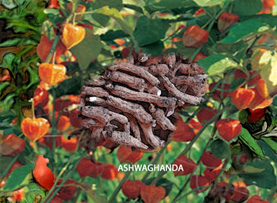
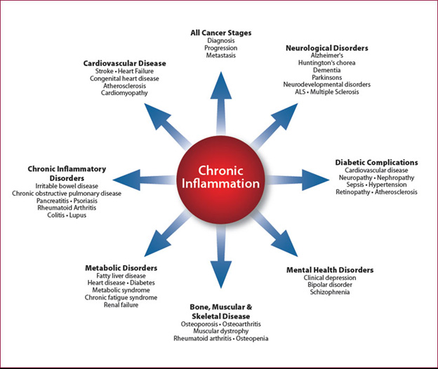
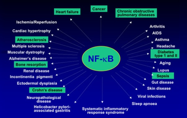
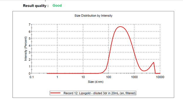
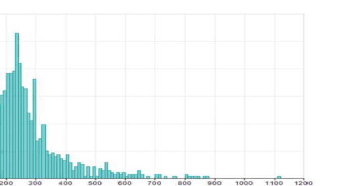
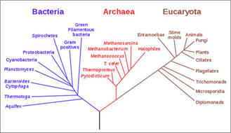
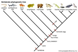
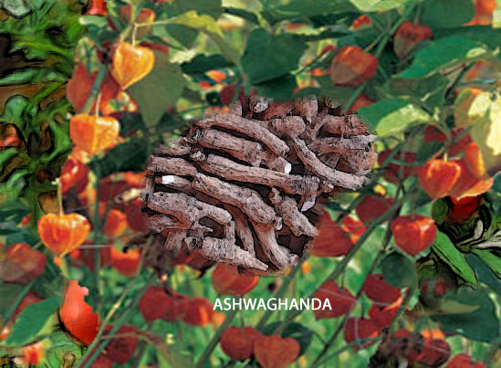
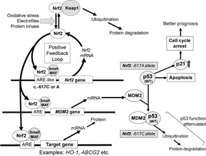
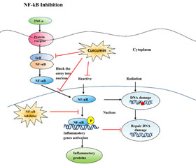
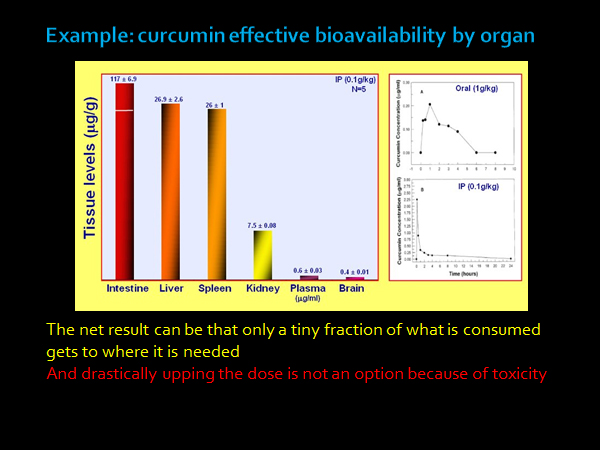
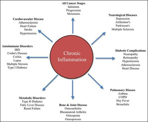
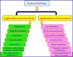
This is a highly personal blog entry, generally light in its approach but containing some relevant scientific content.  It raconteurs a variety of topics, and is intended to provide context and information about me for blog readers.  It is especially for those who follow what I have been saying about keeping healthy and active and who would like to track how I am actually doing.  The adage “Pay more attention to what he is doing than to what he is saying” is good advice too-often applicable to politics and also to any published advice on health and longevity.  I write this from the perspective of a longevity researcher who is about to turn 90 and is projecting out how he thinks things will go between now and 100 and perhaps even until he reaches 110.  In the process I disclose a few matters about myself that might be new and surprising even to my longstanding readers.  It describes my recent journey from being just an intellectual-teller to also being a craftsman-entrepreneur.

First of all, I continue to be healthy, active, productive and professionally involved and expect to continue doing so until at least 100.

  • I can still wrestle and twist my 100-pound lawnmower over the rugged, rocky and rooted 1.5-acre terrain that is my house front and back yard. And dig up heavy rocks and chainsaw big fallen tree branches. Just did some of that.
  • I am getting deeper and deeper involved in managing my new dietary supplement business, 4 Herb Synergy, believing that it offers the best single intervention I have now for contributing to my own health and longevity.
    • I can still manage to pursue my 4-year-old grandson around and outside the house with hope of catching him, and pick up and hold my wiggly 3-year old granddaughter while paying attention to my wife asking me to take out the trash NOW.
    • I am still to my knowledge free of any of the degenerative diseases of old age that kill people – diabetes, dementias, cancers, inflammatory disorders of various kinds. And I have the temerity to believe I can keep myself that way.
    • I am still intending to pursue my longevity science career toll I am 100 or more, always out to acquire new insights, writing what is on my mind, talking at professional meetings when invited, and doing podcasts.
  • I am still waking up in the middle of the night with possibly crazy ideas on biological science topics and staggering off to a computer to capture them. Sometimes my wife wakes up and catches me doing this and asks “Are you all right?”  I can feel a bit sheepish when I explain what I am doing to her.
  • Just about everything I said in the previous personal blog update   ON LONGEVITY INTERVENTIONS – MAINLY PERSONAL published in April of this year is still valid.  This present blog is also highly personal, but mainly relates to additional topics.

I seem to be taking constantly longer and broader perspectives.  This applies to my own life as well as my views on society.

Here are some old-man reminiscences.  I state them to convey some messages:  In the past I was fairly good at identifying new trends and movements that I thought would have a big impact on society.  And then, joining them. This seems to be an ongoing lifelong pattern for me right up to this moment.  I am still the same me.

I have living memories of life in the Great Depression, World War II, The Cold War, Sputnik, the Civil Rights marches and many other epochs and events known to others only through history. I can watch historical films with a perspective that I was alive then and went through it before.  I heard about the Pearl Harbor bombing on the radio an hour after it happened and, the next day, Franklin D. Roosevelt’s famous “Day in Infamy” radio speech.  I saw newsreel movies of the sunken battleships in Pearl Harbor, Hitler rallies with 10,000 people giving the “Sig Heil” salute, saw Mussolini addressing adoring crowds from a balcony.  I heard Kennedy’s promise to land a man on the moon and then saw it happen on a fuzzy black-and-white TV.  So, I see many of today’s happenings from a personal historic viewpoint, perhaps a tad less anxiously than do many younger contemporaries.  This period of time seems to be characterized by denegation of science, political and commercial greed, dismantling of social and health safety nets, corruption and rising danger of nuclear war.  These too should pass.

We are in the middle of a century which is for biology like the 20th century was for physics and electronics – one of amazing discoveries and practical applications.  I think we now know less than 20% of what there is of importance to know about biology.  And our patterns related to life and longevity are still mainly driven by tradition, personal greed, commercial greed, and longstanding social habits.  We need to recognize and deal with these factors to free ourselves to move forward. That is a long process that I am seeking to contribute to.  But I cannot expect too much to happen over a year, or even over 10 years.  Patience is essential.  I have to be content with contributing only as I can, even if I think my vision enables me to see a wonderful potential future for our human species.  It must be all about the species, not me.

I have a lifelong pattern of looking to my future more than going over my past, but now I am allowing my past to contribute more to my future outlook.  As the years continue to roll by, I have more and more direct experience to help guide my future actions.

  • Many dreams or projections when I was much younger were highly prophetic.
    • My best friend from when I was 7 was Jerry Colet. In 1944 his family acquired one of the very first TVs sold after WW2.  I think it had a 3-inch screen like the Pilot TV set shown in the picture.  We lived in the same apartment building and I would often watch TV with Jerry, crowding close to the tiny screen to see anything more than a moving blur.  The black-and-white pictures were smeary, jerky and so poor you could hardly make out faces.   I had a very vivid dream then when I was about 14.  Of a gigantic color TV with a picture so clear and vivid it was completely real.  A window on the world, showing beautiful scenes of natural wonders.   I knew someday that dream would reflect reality, as it does now.  The HD TVs in our house now are much bigger than the one in the dream and the picture quality just as detailed and vivid.  And with streaming 4K video I can choose to see on them just about everything there is to see in the world.
  • In 1944 at age 14, I read an article in Popular Science about a completely new thing – a “Giant Brain,” created by Howard Aiken, the man who built the first general-purpose computer at Harvard during WW2, the Harvard Mark 1 shown in the picture. The computer was 51 feet long, 8 feet high and weighed 5 tons.  All the logic depended on electromagnetic relays since the computer was built before transistor or even vacuum tube digital circuits existed.  Later in 1950 I started learning about computers from another computer pioneer Arthur Burks at the University of Michigan, and in 1953 was creating and running programs on the first Burroughs computer, the UDEC at Wayne State University, working for another computer pioneer Arvid Jacobson.  Howard Aiken enrolled me as a Ph.D. student in his lab at Harvard in 1953.  The Harvard Mark 1 was still running in the same big room shown.   I did my research for a PhD late at night running programs on a Univac 1 installed there in the empty space shown.  While working there, from time to time somebody turned off all the lights plunging us and the enormous computer room and into darkness.  I finally discovered that was this was to debug the Mark 1 by allowing technicians to look for tiny sparks from faulty relay units.  A Google search on Vincent E. Giuliano will reveal many of the accomplishments and publications in my computer-related career following getting that PhD in 1959.

At age 72 I decided to transition to a new career in longevity science, and that is still my main career.  That is because I saw similarities between this 21st century and the last 20th century.  I perceived that a pattern of research, discoveries, inventions, commercial and social developments existed in the last century related to electronics.  And that a similar pattern of research, discoveries, inventions, commercial and social developments exist now related to life sciences.  Just as the 20th century was one of electronics, the 21st century will be one of the life sciences.  So I wanted to tie my future to the life sciences.  And I picked longevity as a sub-focus because it would be inconvenient for me to die soon in the process.  Growing ever older, I needed to know for myself what I had to do to keep going. And going.  And going further.

  • Now, a little more than a month ago, I started up a new entrepreneurial career with the Synergy Bioherbals family business. My first business began when was I dropped out of high school at age 14 and opened up a storefront radio repair business on 12th street in Detroit.  That was in 1944 when World War II was ending. I spent a fair amount of time then waiting for the next customer to come in the door of the shop.  Now I am constantly going online to see if any new customers for 4HerbSynergy have come in to my online shop.  Fortunately, this time around I have plenty of valuable things to do while waiting.

Some of my personal characteristics formed early in my youth persist to today

For example, I am frugal in funny ways., I will keep and wear torn and stained shirts and pants on days when I don’t go out, and will always strive to eat everything on my food plates.  I may keep jars of creamed herring or olives in the fridge long after their expiration dates.  If I see a decent-looking rubber band or paper clip on a sidewalk, I will pick it up and put it in my pocket to take home.  And I am notorious for never throwing anything out that I think may ever be used again.  Including chainsaws and dehumidifiers and toaster ovens that need repair.  And I keep racks of ancient clothing in my attic to feed the moths.  I still keep my college textbooks and class notes, and I have my first and only tuxedo from 1950.  I have a “depression-era thrift mentality” this way, learned in part from my mother Pearl and her father James who lived nearby while I was a child.  James would often shop for us since my mother, a single mom, worked for something like $25 a week as a secretary in the Wayne County Welfare Department.  From when I was 8, James would knock on the wire-glass fire-safety door of our apartment and when my mother opened it, he would say something like “Pearl, here is the rutabagas you wanted me to buy.  You owe me three cents.”  My mother might say “I only have two pennies; I have to give you a nickel.”  He would take it and fifteen minutes later he would knock on the door again “Pearl, here are the two pennies I owe you.” In his childhood in Piedmont Italy, James’ parents could not afford to raise him. So they apprenticed him when he was 8 to a tinkerer, a man who had a little push-cart and went up and down the cobbled streets shouting his presence and sharpening knives and repairing pans with solder.  James saved what little money he could, and about by age 18 had enough to buy a steerage-class steamship ticket to the USA.  So James came over on a boat from Italy about 1905 without money or education and was recruited at the dock to go to work in the Calumet copper mines.  Later he worked at other mines and at the original Highland park factory of Ford motors   In the 1930s during the Great Depression, he saved his pennies and nickels and invested in the stock market.  He died a millionaire in the 1940s.  He was a very gruff man.  He did not like me and was sometimes cruel to me, but I learned very important lessons from him.

Kids often bullied me as a child in grade school.  They questioned my patriotism   I had an Italian name and, after all, Italians were our enemies during WW2.  And at the time there was still much prejudice against Italian immigrants just like there is today against Hispanic immigrants.  I did not matter to kids that my father was born in Detroit and my mother was born in Salt Lake City.  Suffice it to say, I had a lot of emotional wound-healing and growing to do to get to where I am today.

Emergence of key Motivators

Through my life but especially in recent years, my actions are governed by a small set of key motivators.

These include Mission and Purpose, Identity Commitment, Health, Goals, Priorities and Plans, Learning, Compassion, Urgency, and Congruity.  I will devote only a few words to each here though a book could be written on them.

Mission and Purpose: My Mission and Purpose in life is to be of service to other living entities and life itself. 

Identity Commitment:  I identify myself basically as a member of the human species and my strongest commitment is to the survival, continuity, wellbeing, and long life of that species.  I am committed secondarily but still strongly to other usual entities including family, nation, and to my continuing being alive.  My commitment extends to factors necessary for the wellbeing of our species, including the health of the planet itself, the wellbeing of countless supporting species, peaceful and sustainable economic development and humane social conditions.

Health:  Health is a personal necessity for me to be successful in any of my endeavors, and my commitment to health extends outwards from me to family members closest to me, to my communities, to our species, to members of other species, and to the planet in general.

Goals and Plans; I depend on these for behavior beyond that which is endowed in me via genetics, epigenetics and social force fields.   I consciously set my own goals, priorities and plans.  Or at least, I think I do.  And I seek to behave in ways that reflects these priorities. I have been doing this since I was a teenager.  Am I always successful in behaving in such a deliberate manner?  No, but I am slowly getting better at it due to the urgency that comes with old age.

Learning:  I don’t know most of what there is to know.  But ignorance for me is rarely a good reason for inaction.  I believe I must be constantly learning to keep functioning.  I have long thought that anything worth doing is still worth doing even if this means doing it badly at first.  All great achievements are based on improvements after first attempts.  The important thing is to keep trying things and to learn from feedback from earlier actions.  Wheels on cars require ball bearings for them to function reliably, but ball bearings in wheels came only thousands of years after the first wheels were used.

Compassion:  My fellow human beings are doing as well as they can give their backgrounds, capabilities and priorities.  I need to be compassionate to them even if they are very different from me or if I strongly disagree with them about important things.

Urgency: As I grow ever-older and the needs of our species loom bigger and my probable remaining lifespan becomes shorter, urgency of each of these motivators increases.

Congruity; Given the urgency my actions need to be congruent with these motivators and respond to them with integrity.  Such could likely not be the case, for example, if I had a strong drug addiction or if I spent most of my time playing video games.

My latest thinking on longevity interventions

Currently, I am very excited about certain streams of both theoretical and practical actionable developments; the first is controlling chronic inflammation for allowing us to live up to and possibly beyond 100 years of age with vitality, good health, and full functionality.  The second is on regenerative medicine, on regenerating problematic or diseased cells, organs and bodily systems like salamanders can grow new limbs or tails.  These are visions medicine recently told us were impossible.  I also continue to be very interested in circadian regulation.  This poorly-understood area is of great importance because the efficacy or lack of efficacy of just about any health intervention we can think of depends crucially on when that intervention takes place. I say a few words on each of these streams of theory and practice here, and provide a few links to where the topics are explored more thoroughly.

  • Controlling chronic inflammation

Regular readers of this blog know I have focused on chronic inflammation and its control over the last 3-4 years, writing a number of blog entries on or related to this topic.  I understand chronic inflammation as a key concomitant to all the degenerative diseases if old age (cardiovascular problems, cancers, diabetes, dementias, sclerotic diseases, etc.) and as a Great Killer of older people – if not actually Their Greatest Killer.   The exciting message is that we now know some practical ways to control and limit chronic inflammation.

(DISCLOSURE: I INVENTED THIS PRODUCT AND WITH OTHER FAMILY MEMBERS AM AN OWNER OF THE COMPANY SELLING IT.)

  • Regenerative Medicine: cell, organ and body system renewal

I can recall one summer when I was perhaps 6 I found a brightly-colored and slimy salamander under a wet rock.  I grabbed it by the tail.  And then I found myself just holding the wiggling tail with the rest of the salamander scampering off.  I felt very sorry for the harm I had done to the little beast.  But a few days later I looked again under the same rock and saw what looked like the same salamander, complete with a new tail.  An uncle explained to me that salamanders can grow new tails and legs, but we can’t do that ourselves.  I have wondered about the WHY of that ever since and in recent years have been excited by breakthroughs in the science of regenerative medicine. “Regenerative medicine may be defined as the process of replacing or “regenerating” human cells, tissues or organs to restore or establish normal function. This field holds the promise of regenerating damaged tissues and organs in the body by replacing damaged tissue or by stimulating the body’s own repair mechanisms to heal tissues or organs(ref).”  I think it will be increasingly relevant as we seek longer and longer health spans and lifespans.

  • When the first breakthroughs related to epigenetically reversing the age of body cells into IPSCs occurred about 10 years by applying the OSKM “Yamanaka Factors,” these excited me and I generated a series of posts on the topic(ref)(ref)(ref),

Regenerative medicine has already had a profound impact on my personal life.  Let me tell you about corneal ulcers and NGF (nerve growth factor).  For over a year my wife was treated for recurrent corneal ulcers – infections in the cornea of one eye which were treated by antibiotics but kept coming back.  These were very painful and negatively impacted her vision and quality of life, despite that she was being treated by nationally famous practitioners -researchers in corneal health at Mass Eye and Ear Hospital.  Finally, with their recommendation, my wife was insurance-approved for a very expensive treatment that would regenerate her corneal nerves, actually grow new nerves, which would enable migration onto the cornea of stem cells which would in turn prevent the infections and ulcers.  The nerve-growth factor medication is called Oxervate, This drug was approved by the FDA only in 2018 Dr. Reza Dana, my wife’s senior physician at Mass Eye and Ear hospital, helped get it approved for use in the US.  The first administration to a US patient was reported in 2019 and my wife’s  treatment started soon thereafter.  “Cenegermin is the active ingredient in Oxervate. It is the recombinant version of human nerve growth factor (NGF), discovered by biochemist and neurologist Dr. Rita Levi-Montalcini of Italy. Her groundbreaking work on cenegermin earned her the Nobel Prize(ref).“  For more details, see Topical recombinant human nerve growth factor an innovation in ocular surface disease treatment

The Oxervate arrived weekly for 8 weeks at our garage door in 70 pound boxes which were actually small insulated refrigerators. Most of the weight was dry ice, solid frozen carbon dioxide there to make sure the Oxervate stays cold. Oxervate payload in each box weighed less than an ounce, less than the weight of a pickle slice.    My wife finished her Oxervate treatment over a week ago, and she and her physicians and I so far think that the regenerative treatment is working, though it’s far too early to be sure.  This is a real example of high technology regenerative medicine that is available right now.   So regenerative medicine is already beyond being just something on-the-come which I think will impact our future.  I will do my best to stay on top of this area in this blog and fully expect to experience other personal impacts of it soon.

Circadian regulation

I have written about this topic in the past: Shedding new light on circadian rhythms in 2014, and in January 2012, my colleague-writer Victor posted a blog entry Circadian Regulation,NMN, Preventing Diabetes, and Longevity

I now see circadian regulation to be of increasing importance and I started reviewing the newer literature of it.  Living organisms have mostly evolved where there are night-and-day cycles driven by earth’s rotation around its axis.  Accordingly, most biological systems are tuned to respond to times in those cycles when survival has dictated they need be most and least responsive.  As a result, mammals have evolved both central and peripheral clocks that respond to time of day, season and other factors such as recent food consumption.

“The central clock in mammals stems from a network of neurons in the suprachiasmatic nucleus (SCN); these neurons, in addition to maintaining their own cell-intrinsic clock, receive photic cues from the retina to synchronize peripheral day and night cycles throughout the body using a variety of mechanisms, including nervous system signaling, body temperature regulation, hormonal signaling, and regulation of metabolism [2,3]. Peripheral tissue circadian rhythms are synchronized by the central clock, but they also contain their own cell-intrinsic circadian rhythms [4].(ref)”  The central clock has as key actors two genes called CLOCK and BMAL1, which interact in transcriptional feedback loops.

My new interest in circadian regulation is stimulated by recent research in natural regenerative processes, which indicate that these too are under tight circadian regulation.  I hope to report more on this in this blog soon.  Each body system involved in regeneration contains a complex network of cell types with different circadian mechanisms contributing to that regeneration. I plan to lay that out for you in further detail.  And a practical matter I hope to be able to report on is, for various classes of dietary supplements, what is generally the best time of day to take them?

Why start selling supplements at my age?

Invention of the wheel My motivation for doing this is complex and includes urgency.   I tell you a little imagined story to give you an idea:  Imagine a time way back in pre-history when we humans were just settling down from nomadic existence to primitive agriculture, around 3,500BC.  There were a few tribal wise men or tellers, and some even foretold a future where things could be moved around more easily – in contraptions they described which we would now call primitive wheelbarrows and carts.  The tellers would talk about these to anybody who would listen. Important to these imagined contraptions are things now known as wheels.  But that telling by itself made no difference in peoples’ lives.  They still hauled dead animals or pots of water on their backs or animal backs or by dragging them on the ground. This is sort of how I regard many of the writings in this blog – interesting but making little real difference in peoples’ lives.   As a blog writer I have been a teller.

Then, here and there, there were a few farmers who were also amateur craftsmen, or perhaps had sons or daughters who were taken to woodworking.  I am talking about the individuals who started making the earliest most primitive wheelbarrows and carts with wheels.    People started using these first wheelbarrows and carts to haul soil, rocks, dead animals, vegetables, and animal feeds.  Probably the first such vehicles were made by such farmer-craftsmen for their own use.  Perhaps some of these people were tellers who grew frustrated that others did not listen to them.  Perhaps they had suffered injuries that prevented them carrying enough to get by.  Other saw these contraptions and some wanted their own vehicles. Cart craftsmanship became a specialty, and craftsmen built better and better carts. In time, wheeled vehicles enabled vastly better commerce and were demanded by people engaged in commerce.  As the iron age came along, iron-mongers made metal rims for wheels, improving their ability to maintain roundness and endure road wear.  The key step here was the craftsmen building actual vehicles, not just the tellers talking about them.

My journey from teller to craftsman.  So, going back to my own story, four years ago I knew I could go on and on telling my blog readers about the incredible complexity of inflammatory processes, NF-kB the master inflammatory pathway, NRF2, the multiple pathways herbal substances use to inhibit inflammation, etc, etc.  I would be like the village teller.  Except that I knew that for all but a few highly trained specialists, my blogs were too technical to be followed. Most people could not read and understand passages like this one from a blog of about average technical complexity: “An inflammasome is a cytosolic high molecular weight protein complex that can detect and be activated by any of a number of signal sources that could initiate a possible problem for the cell, including microbial toxins, live bacteria, xeno-compounds, viruses, fungi, mycobacteria, or Protozoa exposure.  These activators are also known as Pathogen-Associated Molecular Patterns (PAMPs) and Danger-Associated Molecular Patterns (DAMPs).  A number of different inflammasomes have been identified including a number named because of the NLR protein they contain, including NLRP1, and the most-studied one NLRP3.  Others include the IPAF (NLRC4) inflammasome.   Not all inflammasomes contain a member of the NLR family, such as the recently-discovered AIM2 (absent in melanoma) inflammasome).  The primary purpose of the Inflammasome is to trigger the activation of the mature form of Caspace-1, IL-1 and IL-18, potent inflammatory cytokines that are secreted by the cell after activation by the Inflammasome.  Another purpose can be to accelerate cell death via pyroptosis.  The Inflammasome components are mostly proteins encoded by NF-kB transcription factor regulated genes. However the JAK-STAT pathway also plays a role.”  Can you really follow that or are you willing to take the time and effort to do so to understand it?  This, knowing that many other paragraphs of equal or greater complexity will follow? If the answer is “no” than you probably understand why I decided to go on from being a teller to also being a craftsman builder.  I also knew four years ago that I had developed a dietary supplement that was working well for me.

What I have been preaching to others in this blog is understanding, but not necessarily easily actionable understanding, not necessarily understanding that would make much of a difference.  So, I decided to add the craftsman role to my existing teller role, and continued crafting a science-based dietary supplement that I thought could provide many of the benefits I was writing about.  At first and for a long time this supplement was only for my own use.  Then, as we improved the home-made product, family members and selected friends started using it.  After we became convinced that it works and after years of improvement, we decided to get it out there as a commercial product.  It is now on the market and called 4HerbSynergy.  A much more complete history of 4 Herb Synergy is in a recent blog entry The making of a dietary supplement – the long and short histories of it.

The idea of packing knowledge into a product that people can use to improve their lives without understanding the knowledge itself is very well established.  Do you understand how your cell phone really works?  Your auto engine?  Your refrigerator?  Your GPS?  Your medicines?  —- Probably not but that is OK.  You can take advantage of the knowledge by using the product without actually knowing the knowledge itself.

I tend to be both a theoretician and an experimenter, with a lifelong pattern of puttering to create useful things

My interest on making things is also nearly lifelong.  When I was perhaps 7, a friend of my mother who was in the radio repair business gave me two large cardboard boxes of what for him was largely junk, radios that did not work anymore, tubes, circuit boards of the times, condensers and resistors, a working soldering iron, wires – all things that could be recycled to build all kinds of interesting devices.  This pile of junk occupied a large table in my bedroom which I called “my lab,” and soon I was reading about how to make things, and then making them.  The first was a RF oscillator, a tiny AM broadcasting station that was an ugly mess of dangling wires; but it actually worked.  I kept adding to my lab – used telephones and surplus WW2 military electronic devices, more and more radio junk.  Later as a teen I moved my lab to a much larger space in the 3rd floor of my grandmother’s immense house.  There, I acquired some heavier equipment like an obsolete dental x-ray machine and a ponderous war-surplus 2-way military tank radio.  I joined the Institute of Electronic Engineering when I was in the 9th grade.  I was ever building Tesla Coils, and more and more sophisticated contraptions. In my later University years, I went back to that lab during weekends and vacations several times to build things.  Some were radio jammers used to silence the loud radios in the college dormitory rooms next to mine that kept me up at night and made it very hard to study.  One was demonstration digital circuits on a large display board that demonstrated how Boolean logical operators could be realized in hardware.  They were built from surplus relays used in shoe x-ray machines.  This was in a graduate seminar taught by Arthur Burks, a computer pioneer and that in 1950 marked my entrance to the world of computers.

There are other examples of my lab puttering, including ones that led to radical career-shifts. I crafted an impressive PC personal-psychiatrist computer program called SENSAI over a period of more than a year in the late 1980s, working late nights at home after my regular consulting job.  That led me to a career shift to where I was founding COO and Chief Scientist of Mirror Systems, a software company.

Fast forwarding to the latest wave of creative physical endeavor, it was repurposing my kitchen into a lab almost 5 years ago where I first I started making our 1st-generation herbal anti-inflammatory goop.  I continued to make and improve this precursor, later working with a supplement manufacturer to end up with the 4HerbSynergy supplement we are selling today.    The lab equipment included a very powerful Vitamix blender, a 2-quart ultrasound tool cleaner, and a digital micro-scale for measuring ingredients.    I have been the primary lab rat.   How do I know that it works to control inflammation?  All along from the start I have been taking the supplement myself.  Two or three times during the 4+ years I went off of it for a period of up to a few weeks –  perhaps because I thought I was too busy to make it.  Each time I did this, sure enough, I would feel arthritic inflammatory symptoms creeping back, ones like trigger-finger and knee pains.   When I got back on the supplement again, these symptoms would go away again.  Hmnn! My wife is our 2nd main lab rat.  She has been taking the supplement for over 3 years now and also has her own stories about being on and off it.  Perhaps 30-40 people have tried our home-made supplement over the years.  Many kept asking for more and a secondary motivation for the business is that we had some trouble keeping up with such requests.

For years, we called the home-made stuff “Lipomix.” We shifted over to taking the commercial version, 4HerbSynergy, about 6 weeks ago, but the ultrasound unit still waits on standby in a cardboard box in our living room.  That long period of puttering and personal experimentation was critical to developing our 4HerbSynergy product.  I would never consider the costs, aggravation and risks of going into a new business without feeling a strong familiarity with a supplement product and knowing that it really works for me and others.  I got that through our kitchen lab work.

Life for me continues to be an adventure requiring constant learning

As do all other human beings, I face many challenges and questions born of uncertainty like:

  • Can I maintain my health and full vitality for another 10, 20, 30 years or even longer? All history suggests the answer is certainly NO for the longer periods.  My science background suggests “Perhaps, it is worth a try.  Keep after it and we will see.”  The part of me that practices reality creation says “Yes, sure, that is already settled.”  Again, for insight into this part of me you can read my treatise ON BEING AND CREATION, and have a look at the associated  ON BEING AND CREATION blog.
  • Can I help build up sales for our new 4hebsynergy business to the point where the business is viable?  Clearly, this challenge is of our own making, but it is necessitating a lot of new behavior taking me outside my comfort zone.  It is requiring me to interact differently with many new people and learn a lot more about the dietary supplement industry – and how electronic media marketing can be utilized for our business.  In other words, I just signed up for an Entrepreneurial University-level education
  • Can I simultaneously build a new business and all that it entails, further myself as role of sage and blog-writer in the area of longevity science, continue to deepen my insights into the nature of reality, and do the other things required for me to lead a full and happy family and social life?   I really don’t know but based on my faith in reality creation, I say YES, certainly.

—————————————————————————————————-

  • THE STATEMENTS IN THIS BLOG ENTRY HAVE NOT BEEN REVIEWED OR APPROVED BY THE FDA.  THE DIETRY SUPPLEMENT MENTIONED HERE IS NOT INTENDED FOR THE DIAGNOSIS, PREVENTION OR TREATMENT OF ANY DISEASE.FROM TIME TO TIME, THIS BLOG MAY DISCUSS RESEARCH DEVELOPMENTS RELATING TO DISEASE PROCESSES.  THE INTENTION OF THOSE DISCUSSIONS IS TO CONVEY CURRENT RESEARCH FINDINGS AND OPINIONS, NOT TO GIVE MEDICAL ADVICE.  THE INFORMATION IN POSTS IN THIS BLOG IS NOT A SUBSTITUTE FOR A LICENSED PHYSICIAN’S MEDICAL ADVICE. IF ANY ADVICE, OPINIONS, OR INSTRUCTIONS HEREIN CONFLICT WITH THAT OF A TREATING LICENSED PHYSICIAN, DEFER TO THE OPINION OF THE PHYSICIAN. THIS INFORMATION IS INTENDED FOR PEOPLE IN GOOD HEALTH.  IT IS THE READER’S RESPONSIBILITY TO KNOW HIS OR HER MEDICAL HISTORY AND ENSURE THAT ACTIONS OR SUPPLEMENTS HE OR SHE TAKES DO NOT CREATE AN ADVERSE REACTION.

The post FUNNY THINGS ARE HAPPENING TO ME ON THE WAY TO 100 appeared first on AGINGSCIENCES™ - Anti-Aging Firewalls™.

SELECTED NOTICES AND BOOK REVIEW

$
0
0

From time to time I will be listing a few notices that I think might be of particular interest to my readers, starting here.  I also review a new book I think to be of special importance.

1.  PHONE CALL-IN SESSIONS – TALK LONGEVITY WITH VINCE

I will be experimenting with weekly call-in group conversations, Tuesday evenings 7:00 – 8:00 pm EDT.  First regular Tuesday session will be, October 8.  However since people may be celebrating Yom Kippur then, I am also scheduling a call in, same time, on Monday Oct 7.  The agenda of the conversations will be up to you. Any topic of health or longevity is fair game.   And all who will have joined the phone conversation will be free to join in.   Be aware though that I plan to record the conversations and might want to use parts of them on this or other blogs.  I plan to keep the conference line open for conversations during the 7-8 pm time frames no matter how many call in, even if it is just to chat with a single caller.  I have so far scheduled these through Nov 11th but will keep them going after that if there is sufficient interest.

Vince Giuliano is inviting you to a scheduled Zoom meeting.

Join Zoom Meeting
https://us04web.zoom.us/j/500754984?pwd=Rm5lNmxYVkZnTSttVlRXd0I2MVBmQT09

Tuesday Meeting ID: 500 754 984   Tues mtgs Password: 004871

Mon Oct 7 Meeting ID 409-297-457  Mon Oct 7 mtg password:  725408

One tap mobile
+16468769923,,500754984# US (New York)
+14086380968,,500754984# US (San Jose)

Dial by your location
+1 646 876 9923 US (New York)
+1 408 638 0968 US (San Jose)
+1 669 900 6833 US (San Jose)
Tuesdays Meeting ID: 500 754 984  Mon Oct s Meeting ID:  409-297-457

Find your local number: https://us04web.zoom.us/u/fisIuyStX

(If you have a problem calling from the country you are in, please let me know and I will see if I can add that country to the list for incoming calls — Vince)

2.  BOOK REVIEW

I am still listening to the final chapter of the audiobook version of David Sinclair’s new book Lifespan: Why We Age – and Why We Don’t Have To.  The excitement engendered by this reading combined with that of my current research continues to resonate in me, taking over my thoughts not only by day but in bed trying to go to sleep, and sometimes in dreams.  There have been a few nights when I wake up at 3 AM to rush in the cold to my nearest computer to make notes of new insights – or at least what I hope will continue to be seen the next day as new insights.   I am 98.5% aligned with David with respect to the sciences of longevity and his views and implications.   I  think this book might – just might – turn out to be a landmark writing of Western History, a full Manifesto right up in importance with Charles Darwin’s On the Origin of Species, Karl Marx’s Das Kapital, and Thomas Jefferson’s Declaration of Independence.

The book is a Manifesto because it comes out and clearly argues for certain central tenants that we in the aging community may believe privately, but are so radical that we dare share them only with a few colleagues in our aging sciences community, or with close friends, or with family members.  And when we do share them, we are prepared for a reaction of “Please pass the potatoes.”  Or “Maybe he is a little nuts, but I will ignore this because he is a good guy with his heart in the right place.”   Here are some of David’s Manifesto declarations, in my phrasings of them:

  • Aging is not inevitable, nor is it noble nor is it something we should be complacent about. It is a scourge that ultimately causes most diseases that lead to untold pain, suffering, and deaths
  • We are putting our medical research dollars in the wrong places, such as in trying to cure diseases like cancers which are basically downsteam consequences of aging. It would be much cheaper and we would be better off putting the dollars into aging research where we could quickly learn how to stop or reverse aging and avert the diseases happening in the first place. (A bit self-serving for a person who makes his living doing aging research, but I think is absolutely correct.  NIH allocates only a tiny sliver of its medical research budget to aging,)
  • It is time we start treating aging as what it is, a terrible disease that leads to death for everyone instead of an inevitable condition that just is and goes along with being human.
  • It is probably easier to cure aging than, say, cancers.

These are views of a Professor in the Harvard Medical School and head of a key program of aging research there, and a prize-winning pioneer who led research on several key molecular biology aspects of aging, such as the key roles of sirtuins, a person who merits careful listening to.  That is why the book could end up being an important Manifesto related to the human condition.

Beyond these manifesto points, there are many other points I profoundly agree with like

  • We are quickly moving to having more and better interventions to forestall important aspects of aging or reverse them.
  • Perhaps the central new anti-aging technology will be cell and organ renewal involving some of the Yamanaka factors (The topic of one of the podcasts mentioned below).

I don’t want to over-emphasize the relatively minor quibbles I have with a few of David’s points:

  • I disagree with David’s statement that there is no scientific reason for aging. I think that reason is the Second Law of Thermodynamics, that entropy (the degree of disorganization) in any (closed) system can in time only increase.  David differentiates between digitally-encoded information in our genes, and analog information encoded in our epigenome.  The digital genetic information is passed on reliably mostly error-free from generation to generation via sexual reproduction in humans and is identical in all cells of an individual. While much (but not all) of the analog epigenetic information is acquired in the developmental, and experience processes of individual cells and varies by cell and organ type.  A way I put it is that the species-related information is encoded digitally to preserve the species, but the information related to individual members of a species and to their organs is encoded in analog form, where it is subject to entropy degradation.  This is another example of the old observation that evolution cares about species, not so much about members of a species.  So the fight against entropy (the principle that in a system everything tends with time to become more disorganized) is mainly on an individual level, not on a species level.  Entropy is a very fundamental law of nature.  Increasing entropy is why you will never see smoke gathering itself together in the sky and going down a smokestack, or the pieces of a fallen shattered teacup putting themselves together again, the little puddles of tea on the floor gathering themselves together and going back into the teacup, and the intact and full teacup jumping back on the table.  Increasing entropy is why time runs in only one direction and why we age.
  • I think it is unclear that we can indefinitely fight entropy in our bodies to extend our healthy years of being alive. Of course, we might go on for a long time since our bodies are not closed systems and we can mobilize energy for the purpose, – so our bodies need not, in fact, be absolutely subject to the Second Law of Thermodynamics.  I am betting on anti-aging – up to some still-unknown point.
  • David compares aging to dirt on a DVD, which can be wiped away to restore the original digital data. I have a large collection of DVDs, and some of them have acquired not only dirt, but deep scratches causing irretrievable loss of digital data.  Favorite parts of some of my favorite movies on DVDs are now gone because of these scratches.  I am concerned that aging may also seriously damage our digital non-germline information, putting absolute age reversal in question.
  • David’s definition of aging is, Aging is loss of “analog” information such as is information encoded in the epigenome. I see the situation in slightly different terms.  The definition I suggest is “Aging is effective loss of access to information due to age-related increases in systems entropy (e.g. worsening signal-to-noise ratio).  The issue with David may be simply due to his over-simplification in a publication for a general audience since most people don’t know what entropy is.

There are a lot of supporting discussions in this book, and the narrated version carries me along nicely.  The audio version is supported by PDF data files which you can purchase separately.  Read this book.

3.  PODCASTS OF MINE

Besides reading my blogs, you can listen to me.  I have done a number of podcasts.  Among the most recent ones are:

a.  Podcast interview November 2018 with Tara Smith, on New Paradigm for Cellular and Tissue Regeneration

“Vince Giuliano from Aging-Sciences.com explains the new paradigm of how cells can revert to their previous cell state through epigenetics and inflammation for tissue regeneration. We also discuss the potential downside of senolytics and how senescent cells can indirectly help promote cellular regeneration, as well as his opinion on human connection and strong relationships.”  I highly recommend this one as it heralds what I believe will be a central thrust in aging research in the coming 20 years.

b.  A podcast interview recorded June 18, 2018, on Anti-aging and Reversing Degenerative Diseases, on Youtube sponsored by Self-Hacked. Posted on the Self-Hacked blog.  The interviewer is Joe Cohen.  the interview was over a year ago, but I just came across it.

The discussion runs over a variety of topics, ranging between the personal and scientific and sharing what I consider to be wisdom as well as information.  I consider myself to be a self-hacker, this part of me being integrated with the health and longevity scientist part.  And this is reflected in the conversation where I relate relevant personal anecdotes as well as make a few science-based assertions. I discuss selected epigenetic effects. I describe some of my main personal longevity interventions, talk about sleep, exercise, management of stress, inflammation, and my personal freedom from age-related diseases.  I respond to questions about a few “anti-oxidants” and other selected dietary substances.  I do not discuss the leading-edge topics that interest me today.  I describe the early history of arthritis that led me to the self-hacking that led to the early development of the 4 Herb Synergy supplement.  I touch on the benefits of fish and fish oils.

c.  Podcast Interview on Healthy Longevity with Susan Downs February 2018 on

Deals with a multiplicity of topics, including changing lifespans and why this has been happening, the nature of aging, aging programs, multidimensional nature of health and health preservation, epigenetics and aging clocks, caloric restriction, certain key dietary supplements, and multiple suggestions for maintaining health and extending our personal lifespans.

4.  AGING MATTERS BLOG BY JOSH MITTLEDORF

If you follow this blog and don’t know about Josh’s Blog Aging Matters, you might want to check it out.  It contains many facts and a number of interesting views, though I do not necessarily agree with all of them.  The most-recent postings in this blog are:


Inflammation Part 7: Neurohormesis, neuroinflammatory diseases, and their treatment by mushroom substances (Section 1)

$
0
0

Can a mushroom play an important role in the health and anti-aging picture?  Recent research says Yes, and this blog entry tells the story

Current medical treatments for age-related neurological brain diseases like Alzheimer’s and Parkinson’s remain largely ineffective, despite all the medical interventions that have been thrown against them.  It may sound crazy, but mushroom-based substances could possibly change that.   This blog entry, in two Sections,  is about recent research linking up seemingly disparate topics that have been discussed over the history of this blog.  :  1.  Neurohormesis, the hormetic stress response, as particularly focused on neuronal/microglial processes, 2.  How neurohormesis is likely to be broadly applicable as a strategy for preventing or treating hitherto intractable neuroinflammatory diseases like Alzheimer’s, and 3 how certain mushroom-derived substances, such as from the traditional Lion’s Mane mushroom might be used to elicit hormetic responses for neuroprotection and to prevent or cure neuroinflammatory diseases.  4.  Possible implications for cell, organ renewal, and consequent longevity. And finally, 5.  Implications for self-biohackers.

In the course of the discussion I will touch on many topics discussed in previous blog entries, including xenohormesis, inflammasomes, microglia-neuron interactions, NRF2, Alzheimer’s and Parkinson’s diseases, the role of stress responses in the evolution of plants, cell-regeneration, lipoxins, nerve growth factor, BDNF, and yet another role for sirtuins.. I am particularly inspired here by the diligent research work on two of my colleagues, Dr. Edward Calabrese of the University of Massachusetts, the long-standing world-leader of hormesis research, and Dr, David Sinclair at the Harvard Medical School who wrote the original publication introducing me to xenohormesis.

I tell the first part of this story in this blog entry.  But there is much more of interest for those who want to fight the entropy of Aging.  I will round out and complete the story (at least, for now) in the following soon-to-be-published blog entry Inflammation Part 8: Neurohormesis, neuroinflammatory diseases, and their treatment by mushroom substances (Section 2).

This is Part 7 of what is likely to become a 10 or more-part blog series related to chronic inflammation. Being a central aspect of every degenerative disease of old age, chronic inflammation can be thought of as the Great Executioner, the most central machinery of most people’s ultimate illnesses and deaths. Parts 1 through 6 of this series are already published.  Part 1 of the series is the same as Part 5 of the NAD world.  That blog entry is concerned with The pro-inflammatory effects of eNAMPT (extracellular NAMPT, nicotinamide phosphoribosyltransferase).  Part 2 of the series relates a) the “master” pathway network of inflammation (NF-kB) to two other pathway networks clearly implicated in aging and disease processes, b) Genomic Instability (p53), and c) Oxidative stress (Nrf2).  Part 3  is concerned with the all-important resolution phase of inflammation, how acute inflammation goes away under ideal conditions instead of hunkering down to lingering and dangerous chronic inflammation. Part 4  of the inflammation series, is concerned with  PCSK9 inhibition – Also that blog entry is Part 1 of a series on interventions that reduce all cause mortality (ACM)Part 5 of the series is concerned with the basic science of inflammasomes and how they relate to a number of disease processes.  Part 6 of this series is concerned with the scientific considerations behind 4 Herb Synergy, a dietary substance I have invented to control chronic inflammation.

PREAMBLE

In researching the literature related to this topic, like looking for wild mushrooms in the woods,  I found potential trails of molecular biology branching off in every-which direction.  And my attempts to go off on some of them just led to many more interesting such trail choices, which led to yet-more paths.  So, it has been difficult in this exploration to distinguish between what is of central importance and what is just very interesting.  I have sought to confine my coverage here to subtopics that are both, but I am hampered by not knowing what the overall landscape is.  I will seek to tell the story as if I were telling a mystery murder novel – element by element, with the hope that by the end the elements will have come together enough for you to get the storyline.

ELEMENT A: A MAGIC MYSTERY MUSHROOM


Lion’s mane (Hericium erinaceus)  Image source

The active ingredients of Lion’s Mane mushrooms (Hericium Erinaceus) appear to be able to activate pathways of regenerative medicine, according to recent research findings.  Impacts of consuming the mushroom’s extract include ones related to the brain, CNS and vision, including enhancing neurogenesis(ref), enhancing neuronal plasticity(ref), regenerating neuronal axons(ref), and reducing neuroinflammation(ref).  The observed reduction in beta-amyloid plaque accumulations resulting from consuming the mushroom extract(ref), possibly offers an approach to treating Alzheimer’s disease(ref)(ref).

Lion’s Mane mushrooms have been used in China for thousands of years as both a food and as a medicine.   This mushroom is commonly found on dead and decaying hardwood logs, most often in the fall throughout North America.  By our former summer home in New Hampshire we would often see it on dead and rotting birch tree branches. For an introduction,  See Hericium erinaceus: an edible mushroom with medicinal values. (2013). “Mushrooms are considered as nutritionally functional foods and source of physiologically beneficial medicines. Hericium erinaceus, also known as Lion’s Mane Mushroom or Hedgehog Mushroom, is an edible fungus, which has a long history of usage in traditional Chinese medicine. This mushroom is rich in some physiologically important components, especially β-glucan polysaccharides, which are responsible for anti-cancer, immuno-modulating, hypolipidemic, antioxidant and neuro-protective activities of this mushroom. H. erinaceus has also been reported to have anti-microbial, anti-hypertensive, anti-diabetic, wound healing properties among other therapeutic potentials.”

Hericium Erinaceus active ingredients can penetrate the blood-brain barrier and offer excellent bioavailability(ref).  Much of the research so far has been on rats, but because of the similarity of pathways, there is a significant probability that findings will carry over to humans.  There is lots more to say about Lion’s Mane mushrooms and its components and the possible role they could play in healthy aging.  For example, see the 2019 publication Hericium erinaceus Improves Recognition Memory and Induces Hippocampal and Cerebellar Neurogenesis in Frail Mice during Aging

In fact, in researching this blog, I became so impressed with the substance that 10 days ago I added Lion’s Mane extract to the list of dietary supplements I am taking daily.   I am now free of brain fog all day long, and I think it likely helps improve my mental clarity and capability to think-through very complex matters like wrestled with in this blog. 

Lion’s Mane extract contains many interesting compounds which could themselves be the subjects of essays. I mention only a couple as examples  The 2012 publication Amycenone, a nootropic found in Hericium erinaceum 2012 reports: “The current paper describes the physiological and nootropic actions of Amycenone, which is an activator of brain function that is obtained from extracts of the Yamabushitake (Hericium erinaceum).  — Kawagishi and his group have studied compounds that are derived from medicinal mushrooms and their use in the treatment of dementia since 1991. They have found that H. erinaceum exerts important bioactivities, including the induction of nerve growth factor (NGF) synthesis, the inhibition of the cytotoxicity of beta-amyloid peptide, and the protection against neuronal cell death caused by oxidative or endoplasmic reticulum stress. — Since NGF was first discovered in the 1940s, it has garnered attention as a substance in the brain that curbs the degeneration and loss of neurons and that promotes the repair and regeneration of nerve function. However, NGF cannot pass through the blood–brain barrier. — Amysenone (Amyloban®3399, which contains a standardized extract of H. erinaceum) has been found to pass through the blood–brain barrier, and its safety as a health food is currently being ascertained. — On the basis of the author’s first-hand experiences, Amyloban®3399 was found to clearly increase alertness. The actions of Amyloban®3399 in treating sleep-related breathing disorders were examined. Amyloban®3399 was effective in improving sleep apnea and hypopnea syndrome.  The use of Amyloban®3399 has been noted to result in the obvious restoration of cognitive function in mild cognitive disorder.”

Another of  Lion’s Mane key bioactive ingredients, Erinacine S, has been a particular focus of recent research. Erinacine S is so-far known to be produced only in Hericium Erinaceus mycelia.

And, to top it off Lion’s Mane component substances are also anti-hyperglycemic.  The story is told in the 2017 review article Anti-hyperglycemic property of Hericium erinaceus – A mini review.  But Lion’s Mane is only Element A of the story I am telling, so I will go on but will return to it repeatably.

Lion’s Mane is not the only interesting medicinal mushroom, though it is the one I focus on here.  Back ten years ago, one of my first-published blog entries was about another important medicinal mushroom Cordyceps militaris and cancer,  Other mushrooms in addition to Lion’s Mane are known to be capable of exercising  therapeutic actions against nervous systems disorders. 


Cordyceps Militaris Image source             Coriolus versicolor  Image source

One of these is is Coriolus versicolor, a mushrooom, well known in China as Yun Zhi.   See “Neuroinflammation and Mitochondrial Dysfunction in the Pathogenesis of Alzheimer’s Disease: Modulation by Coriolus Versicolor (Yun-Zhi) Nutritional Mushroom In fact, there are a number more of promising mushroom candidates,  See An Overview of Culinary and Medicinal Mushrooms in Neurodegeneration and Neurotrauma Research 2017, and Edible and Medicinal Mushrooms: Emerging Brain Food for the Mitigation of Neurodegenerative Disease, also 2017.

ELEMENT B NEUROTROPHINS

Neurotrophins appear to be central mediators of nervous system cell and organ renewal processes.  Researchers need to understand what neurotrophins are about if they want a shot at finding a way for us old folks to renew the brainpower we once had as youths. The first neurotrophin was identified in the mid-1950s, and they have been intensively studied since the early 1990s.  Even back then they were known to regulate development, function and maintenance of vertebrate nervous systems. “Neurotrophins activate two different classes of receptors, the Trk family of receptor tyrosine kinases and p75NTR, a member of the TNF receptor superfamily. Through these, neurotrophins activate many signaling pathways, including those mediated by ras and members of the cdc-42/ras/rho G protein families, and the MAP kinase, PI-3 kinase, and Jun kinase cascades. During development, limiting amounts of neurotrophins function as survival factors to ensure a match between the number of surviving neurons and the requirement for appropriate target innervation. They also regulate cell fate decisions, axon growth, dendrite pruning, the patterning of innervation and the expression of proteins crucial for normal neuronal function, such as neurotransmitters and ion channels. These proteins also regulate many aspects of neural function. In the mature nervous system, they control synaptic function and synaptic plasticity, while continuing to modulate neuronal survival.(ref).”

Pubmed.org currently lists 139,458 research articles on neurotrophins. Neurorophins of particular interest to me have included BDNF, NGF and GDNF.  A little about each of these:

BDNF (Bain-Derived Neurotrophic Factor). 

Way back in 2010 I created a blog entry which is worth reviewing BDNF gene – personality, mental balance, dementia, aging and epigenomic imprinting“BDNF stands for brain-derived neurotrophic factor, the protein generated by the BDNF gene, a substance that has been drawing a lot of attention recently in neuropsychiatric research circles.  I review some basic facts about BDNF here, recent research on how BDNF relates to personality, mental balance, and aging and, finally, current research on how BDNF expression is conditioned by epigenomic imprints. — BDNF acts on certain neurons of the central nervous system and the peripheral nervous system, helping to support the survival of existing neurons, and encourage the growth and differentiation of new neurons and synapses.[4][5] In the brain, it is active in the hippocampuscortex, and basal forebrain—areas vital to learning, memory, and higher thinking.[6] BDNF itself is important for long-term memory(ref).[7] ”  BDNF is an neurotrophic, meaning that it plays an important role in neurogenesis, the important process in parts of the brain of neural stem cells differentiating into neurons. “Neurotrophins are a family of proteins that induce the survival,[1] development and function[2] of neurons. — Brain-derived neurotrophic factor (BDNF) is a neurotrophic factor found originally in the brain, but also found in the periphery. More specifically, it is a protein which has activity on certain neurons of the central nervous system and the peripheral nervous system; it helps to support the survival of existing neurons, and encourage the growth and differentiation of new neurons and synapses through axonal and dendritic sprouting. In the brain, it is active in the hippocampuscortexcerebellum, and basal forebrain—areas vital to learning, memory, and higher thinking. BDNF was the second neurotrophic factor to be characterized, after NGF and before neurotrophin-3. — Despite its name, BDNF is actually found in a range of tissue and cell types, not just the brain. Expression can be seen in the retina, the CNS, motor neurons, the kidneys, and the prostate(ref).” — According to the December 30 2009 paper BDNF Val66Met is Associated with Introversion and Interacts with 5-HTTLPR to Influence Neuroticism. “Brain-derived neurotrophic factor (BDNF) regulates synaptic plasticity and neurotransmission, and has been linked to neuroticism, a major risk factor for psychiatric disorders.”

There are many interesting things to say just about BDNF, such as about how it promotes neural plasticity and neurogenesis, and is itself promoted by exercise(ref).  Interesting for the present discussion is the link with Element A.  Extracts of our mushroom friend Hericium Erinaceus upregulate BDNF signaling.  See these articles for examples and discussions.  Is unprading BDNF a mechanism through which Lion’s Mane extract does its good stuff?  Yes, and I describe additional mechanisms below well.  Lion’s Mane extract also activates other neurotrophins – and it can jump across the blood-brain barrier to do so.

NGF (nerve growth factor)

“Nerve growth factor (NGF) is a neurotrophic factor and neuropeptide primarily involved in the regulation of growth, maintenance, proliferation, and survival of certain target neurons. It is perhaps the prototypical growth factor, in that it was one of the first to be described. Since it was first isolated by Nobel Laureates Rita Levi-Montalcini and Stanley Cohen in 1956, numerous biological processes involving NGF have been identified, two of them being the survival of pancreatic beta cells and the regulation of the immune system(Wikipedia).”  By now you have probably guessed that our Mushroom friend Lion’s Mane is also a powerful stimulator of NGF, as attested to by these recent professional publications.

I tell a personal story of how a new recent treatment with NGF appears to have helped my wife in the recent blog entry Funny Things Are Happening To Me On The Way To 100.   The treatment, using a recombinant NGF, regenerated neurons in the cornea of one of her eyes and averted constant infections, much pain, and probable blindness in that eye. The neurons in that eye were depleted, meaning stem cells could not freely migrate to the cornea, renewing it.  Thinking traditionally, these lost neurons were supposed to have been gone forever.  Her vision in that eye is quite good now, and she is awaiting previously impossible cataract surgery on it to bring it up to full potential.  This was a powerful up-close example for me of regenerative medicine.  Not yet-another “coming to you in the future” story.  My life-partner actually restored her seeing, being able to drive, to read comfortably and the quality of her life, I surmise for the rest of her life.   Already done-did as far as we can tell.  Remember the Zoltar machine and its statement that “Your wish is granted” in the movie Big?  That is what NGF said in response to a wish of mine.

GDNF (Glial cell line–derived neurotrophic factor)

This neurotrophic factor relates to glial cells and appears to be particularly relevant in the case of Parkinson’s disease. “Glial cell line‐derived neurotrophic factor (GDNF) was first discovered as a potent survival factor for midbrain dopaminergic neurons and was then shown to rescue these neurons in animal models of Parkinson’s disease. GDNF is a more potent survival factor for dopaminergic neurons and the noradrenergic neurons of the locus coeruleus than other neurotrophic factors, and an almost 100 times more efficient survival factor for spinal motor neurons than the (other) neurotrophins(ref).”  I have published blog entries in the past on the roles of glia and microglia.  See Key roles of glia and microglia in age-related neurodegenerative diseases, and New views of Alzheimer’s disease and new approaches to treating it

And yes, Hericium Erinaceus extracts also promote GDNF.  See the publications in this list. Neurotrophins regulate development, maintenance, and function of vertebrate nervous systems.

Image source “This image has drawings of three different types of glial cell — astrocytes, microglia and oligodendrocytes. Each has its own important function. — An adult man has about 85 billion glia and about 86 billion Neurons — cells in the brain that conduct electrical signals.” — Astrocytes provide other brain cells with food and oxygen. Oligodendrocytes wrap around long parts of neurons to help their electrical signals move faster. And microglia help protect the brain from dangerous infections or damage.”

So, the major bottom line of Element B is that neurotrophins are essential for maintenance of healthy essential nerve functioning and that consuming our magic mushrooms activate the expression of multiple neurotrophins facilitating nerve injury repair and avoidance of pathologies associated with nerve loss or dysfunctioning.

ELEMENT C: NEURAL INFLAMMATION AND LIPOXIN AX4

“It has been demonstrated that inflammation cascade is linked to neurodegenerative diseases, particularly, Alzheimer’s disease (AD) [34].” From the publication Neuroinflammation and neurohormesis in the pathogenesis of Alzheimer’s disease and Alzheimer-linked pathologies: modulation by nutritional mushrooms 2018.   Neural inflammation is a central aspect of the pathologies associated with all major age-related progressive nervous system diseases, including Alzheimer’s disease, Parkinson’s disease and Lou Gehrig’s disease (Amyotrophic lateral sclerosis  (ALS).  These are major debilitators and killers of older people.   Of course, how neural inflammation plays out in each of these diseases is somewhat different and the scientific literature related to them is far too vast to be characterized here.  In short, a summary of salient points:

Neuroinflammation in Alzheimer’s disease 

An important theory states “Increasing evidence suggests that Alzheimer’s disease pathogenesis is not restricted to the neuronal compartment, but includes strong interactions with immunological mechanisms in the brain. Misfolded and aggregated proteins bind to pattern recognition receptors on microglia and astroglia, and trigger an innate immune response characterized by release of inflammatory mediators, which contribute to disease progression and severity. Genome-wide analysis suggests that several genes that increase the risk for sporadic Alzheimer’s disease encode factors that regulate glial clearance of misfolded proteins and the inflammatory reaction. External factors, including systemic inflammation and obesity, are likely to interfere with immunological processes of the brain and further promote disease progression. Modulation of risk factors and targeting of these immune mechanisms could lead to future therapeutic or preventive strategies for Alzheimer’s disease.”  (From the 2015 review article Neuroinflammation in Alzheimer’s disease.)

Following the suggestion of this hypothysis, we could surmise that approaches that dampen down chronic inflammation might lessen the probability of onset of AD might include:

  1. To combat systemic inflammation, anti-inflammatory herbal dietary supplements that can penetrate the blood barrier. Blog readers will recognize that I have invented a liposomal preparations of curcumin, ginger, boswellia, ashwagandha explicitly for this purpose(ref)(ref).
  2. Dietary substances that upregulate the expression of lipoxins that can accelerate the resolution phase of inflammation, so that it goes away by itself. The blog entry Inflammation Part 3: resolving inflammation – resolvins, protectins, maresins and lipoxins discusses substances found in Fish Oils which are lipid mediators of inflammation, leading to resolution of chronic inflammatory situations, that is to natural disappearance of an inflammatory situation such as what we would expect in recovery from a minor infection or insect bite.

Here is a diagram of how Lipoxin A4 is thought to control inflammation.  The activating mushroom, in this case, is shown to be Coriolus Versicolor.  It could just as well be Lion’s Mane.


Image source: Neuroinflammation and Mitochondrial Dysfunction in the Pathogenesis of Alzheimer’s Disease: Modulation by Coriolus Versicolor (Yun-Zhi) Nutritional Mushroom .

What is new here is that Lion’s Mane (or Coriolus Versicolor) can provide a second mechanism for treating neural disorders.  That is,

  • Lion’s Mane active ingredients upregulate the expression of lipoxin A4 (LXA4), accelerating the resolution phase of chronic neuroinflammation, and limiting neuropathic inflammation and the pain associated with such inflammation(ref).
  • Lion’s Mane extract can cross the blood-brain barrier(ref).

Each of these following links leads to a collection of research literature establishing the point mentioned, among many other points

Herici um Erinaceus lipoxin LXA4– Lion’s Mane extract is efficacious in upregulating this protectin, important for bringing an end to chronic inflammation.

LXA4 neuropathic inflammation – In a variety of disease and pathological situations, the lipoxin LXA4 reduces neuropathic inflammation, reduces microglial activation, and limits neuropathic pain.

From the 2015 publication Lipoxins: nature’s way to resolve inflammation: “An effective host defense mechanism involves inflammation to eliminate pathogens from the site of infection, followed by the resolution of inflammation and the restoration of tissue homeostasis. Lipoxins are endogenous anti-inflammatory, pro-resolving molecules that play a vital role in reducing excessive tissue injury and chronic inflammation. —  Lipoxins regulate components of both the innate and adaptive immune systems including neutrophils, macrophages, T-, and B-cells. Lipoxins also modulate levels of various transcription factors such as nuclear factor κB, activator protein-1, nerve growth factor-regulated factor 1A binding protein 1, and peroxisome proliferator activated receptor γ and control the expression of many inflammatory genes.”

ELEMENT D  CONTROL OF NEUROPATHIC INFLAMMATION-RELATED PAIN

One of the quality-of-life stakes in the situation we have been discussing is how to bring the persistent and debilitating chronic pain associated with neuroinflammatory conditions under effective control.

LXA4 is triggered by aspirin among other substances, and aspirin-triggered lipoxins play important roles in the resolution of inflammation as laid out in these publications, among others.  Also, these aspirin-triggered lipoxins can play important roles in the management of neuropathic inflammation-related pain.

Hericium Erinaceus neuropathic pain – References for Lion’s Mane extract is efficacious in controlling neuropathic pain.

Our old friends (or sometimes enemies), inflammasomes and neuropathic pain

The 2013 publication Involvement of the spinal NALP1 inflammasome in neuropathic pain and aspirin-triggered-15-epi-lipoxin A4 induced analgesia suggests a mechanism through which neuroinflammation leads to neuropathic pain.  “Neuroinflammation plays an important role in nerve-injury-induced neuropathic pain, but the explicit molecular mechanisms of neuroinflammation in neuropathic pain remain unclear. As one of the most critical inflammatory cytokines, interleukin-1β (IL-1β) has been regarded as broadly involved in the pathology of neuropathic pain. The inflammasome caspase-1 platform is one primary mechanism responsible for the maturation of IL-1β. Lipoxins, a type of endogenous anti-inflammatory lipid, have proved to be effective in relieving neuropathic pain behaviors. The present study was designed to examine whether the inflammasome caspase-1 IL-1β platform is involved in chronic constriction injury (CCI)-induced neuropathic pain and in lipoxin-induced analgesia. After rats were subjected to the CCI surgery, mature IL-1β was significantly increased in the ipsilateral spinal cord, and the inflammasome platform consisting of NALP1 (NAcht leucine-rich-repeat protein 1), caspase-1 and ASC (apoptosis-associated speck-like protein containing a caspase-activating recruitment domain) was also activated in spinal astrocytes and neurons, especially at the superficial laminae of the spinal dorsal horn; The aspirin-triggered-15-epi-lipoxin A4 (ATL), which shares the potent actions of the endogenous lipoxins, was administered to the CCI rats. Repeated intrathecal injection with ATL markedly attenuated the CCI-induced thermal hyperalgesia and significantly inhibited NALP1 inflammasome activation, caspase-1 cleavage, and IL-1β maturation. These results suggested that spinal NALP1 inflammasome was involved in the CCI-induced neuropathic pain and that the analgesic effect of ATL was associated with suppressing NALP1 inflammasome activation.”

This discussion related to neuroinflammation was started with particular reference to Alzheimer’s disease.  Very similar discussions could be framed starting with any of the other neuroinflammatory diseases.  I strongly suspect that the preventative and therapeutic implications for use of extracts of Lion’s Mane and possibly other mushrooms are applicable across the board for most of these maladies.

ELEMENT F NEUROHORMESIS

Members of every known biological species experiences stress threats to their wellbeing, and there are some stresses that threaten their members very existence.  And all species have developed their own biological responses to commonly occurring stresses.  The more complex and sophisticated the organism, the more numerous and more complex these stress responses tend to be, and the more levels they operate on, e.g. cellular, cell organelle, system of molecular biology, organ, major body system, etc. A quite universal property of these stress responses is that they exhibit hormesis; they are hormetic.  That is, for mild levels of the stress up to a certain point, the natural stress response is sufficient to make the organism better off, more robust and healthy, than if there were no stress in the first place.  So, low doses of many stresses can be used to induce health and in some cases, possibly longevity.  There are countless examples of hormesis such as surgical pre-conditioning to improve outcomes by restricting blood flow, heat stress in saunas and cold streses via ice baths, to psychological stress in basic military training. Calorie restriction, the best documented intervention for extending the lifespan of animals, is a hormetic interventions.  If the stress is above a certain level, of course, the organism may become damaged or die.  Hormesis is fundamental to biology and the need for it can be understood by thinking of biological process in terms of systems control theory.  Jim Watson and I have published a number of articles related to hormesis and hormesis-related health interventions in this blog, most of which can be found in this listing.

Neurohormesis involves generating sub-toxic stresses on nervous tissues, and can be used as a disease-preventative and health and longevity-inducing intervention.  Exercise itself is an example applicable to nervous and muscle tissues, as are cognitive and physical challenges.  Neurohormesis induced by certain herbal substances like curcumin or boswellia, another kind of example, can work through generating stresses that downregulate the NF-kB pathway and upregulate NRF2 pathway, not only inhibiting the expression of inflammatory cytokines in nervous tissues, but also upgrading the expression of hundreds of stress responsive ARE (antioxidant response element) proteins and limiting microglial and inflammasome activation(ref)(ref).  And the actions discussed and illustrated above of Lion’s Mane and other mushroom substances in controlling neuroinflammation via upgrading the LXA4 lipoxin are also hormetic effects.  Again, these are topics that have been discussed extensively in this blog(ref)(ref).

RECAP OF THE STORY SO FAR

  • Recent research confirms that extracts of a traditional medical mushroom, Lion’s Mane (Hericium erinaceus) appear to have amazing clinical powers to affect nervous tissues, including enhancing neurogenesis, enhancing neuronal plasticity, regenerating neuronal axons, reducing neuroinflammation, and reducing beta-amyloid plaque accumulations. Further, it appears that key compounds in the extract have the capability of crossing the blood-brain, so they can get to where they could be needed in the brain and CNS. Research publications suggest that the same may be said of certain other mushrooms, e.g. coriolus versicolor.
  • Recent research has also pointed to key mechanisms for the neural implications of Lion’s Mane being the upregulation of endogenous molecules known as Neurotrophins, including nerve growth factor (NGF), brain-derived neurotrophic factor (BDNF) and glial cell line–derived neurotrophic factor (GDNF). Neurotrophins are essential for maintenance of healthy essential nerve functioning.  Consuming Lion’s Mane extract upregulates the expression of multiple neurotrophins facilitating nerve injury repair and avoidance of pathologies associated with nerve loss or dysfunction.
  • A key aspect of neurodegenerative diseases like Alzheimer’s, and Parkinson’s is chronic neuroinflammation, which gets “locked in” via inflammasome activation as part of the disease process and then plays important roles in furthering progression of the disease. So, a second mechanism of Lion’s Mane extract for dealing with neuroinflammatory diseases is the upregulation of an inflammation-resolving lipoxin LXA4.

WHERE I WILL BE GOING IN THE NEXT Section 2 BLOG ENTRY

Yes, I have been pointing out why extracts of a couple of ancient and traditional mushrooms may possibly be useful right now by health biohackers to help control neuropathic inflammation and awful neuropthic back and other pains, and may lead us to simple, practical and effective means for averting and controlling neurodegenerative diseases  I will cover this topic listing additional elements in the following Section 2  blog entry which I will likely publish in about another week.  In that blog entry I will discuss neural regeneration and the roles of lipoxins in initiating that, address the critical roles of glia and microglia in nervous system health, say more on control of neuropahic lower back pain, relate implications for wound healing, more on Alzheimer’s and Parkinson’s diseases and the prospectus for averting or controlling them with mushroom substances.  And I will hazard projecting how long it will take for this information to seep into medical practice. And I will speculate on what self-health hackers can do to keep themselves going happily in the interim.

REMINDER:  PHONE CALL-IN SESSIONS – TALK LONGEVITY WITH VINCE

I am experimenting with weekly call-in group conversations, Tuesday evenings 7:00 – 8:00 pm EDT.  Second regular Tuesday session will be, October 15.  The agenda of the conversations will be up to you. Any topic of health or longevity is fair game.   And all who will have joined the phone conversation will be free to join in.   Be aware though that I plan to record the conversations and might want to use parts of them on this or other blogs.  I plan to keep the conference line open for conversations during the 7-8 pm time frames no matter how many call in, even if it is just to chat with a single caller.  I have so far scheduled these through Nov 11th but will keep them going after that if there is sufficient interest.

If these times don’t work for you and you would like me to schedule different times or additional phone meetings, please e-mail me to that effect.

Vince Giuliano is inviting you to a scheduled Zoom meeting.

Join Zoom Meeting
https://us04web.zoom.us/j/500754984?pwd=Rm5lNmxYVkZnTSttVlRXd0I2MVBmQT09

Tuesday Meeting ID: 500 754 984   Tues mtgs Password: 004871

One tap mobile
+16468769923,,500754984# US (New York)
+14086380968,,500754984# US (San Jose)

Dial by your location
+1 646 876 9923 US (New York)
+1 408 638 0968 US (San Jose)
+1 669 900 6833 US (San Jose)
Tuesdays Meeting ID: 500 754 984  Mon Oct s Meeting ID:  409-297-457

Find your local number: https://us04web.zoom.us/u/fisIuyStX

(If you have a problem calling from the country you are in, please let me know and I will see if I can add that country to the list for incoming calls — Vince)

MEDICAL DISCLAIMER

FROM TIME TO TIME, THIS BLOG DISCUSSES DISEASE PROCESSES.  THE INTENTION OF THOSE DISCUSSIONS IS TO CONVEY CURRENT RESEARCH FINDINGS AND OPINIONS, NOT TO GIVE MEDICAL ADVICE.  THE INFORMATION IN POSTS IN THIS BLOG IS NOT A SUBSTITUTE FOR A LICENSED PHYSICIAN’S MEDICAL ADVICE. IF ANY ADVICE, OPINIONS, OR INSTRUCTIONS HEREIN CONFLICT WITH THAT OF A TREATING LICENSED PHYSICIAN, DEFER TO THE OPINION OF THE PHYSICIAN. THIS INFORMATION IS INTENDED FOR PEOPLE IN GOOD HEALTH.  IT IS THE READER’S RESPONSIBILITY TO KNOW HIS OR HER MEDICAL HISTORY AND ENSURE THAT ACTIONS OR SUPPLEMENTS HE OR SHE TAKES DO NOT CREATE AN ADVERSE REACTION.

Inflammation Part 8: Neurohormesis, neuroinflammatory diseases, and their treatment by mushroom substances (Section 2 of this discussion) Can a mushroom play an important role in the health and anti-aging picture? Recent research says Yes, and this blog entry tells another part of the story.

$
0
0

By Vince Giuliano

This blog entry continues the discussion of the previous blog entry, Part 7 of the Inflammation series.  So you might want to read that Part 7 before this one if you have not already done so.  I continue element-by-element.

ELEMENT E: REVIEW OF SCIENTIFIC ASPECTS RELATED TO NEUROINFLAMMATION: REACTIVE OXYGEN SPECIES, MITOCHONDRIAL FACTORS, INFLAMMASOMES, PPARs, METBOLIC PATHWAYS, SIRTs, DNA DAMAGE AND ROLES OF NRF2 AND NF-KB, AND ANOTHER MAGIC MUSHROOM, YUN-ZHI


Yun Zhi (Turkey Tail) mushrooms Image source

About neuroinflammation

Like so many phenomena in biology, neuroinflammation has both good and bad sides.  The good sides are essential for human life, The bad sides come with certain disease processes and aging.   This duality is outlined in the 2016 publication Neuroinflammation: The Devil is in the Details.  “There is significant interest in understanding inflammatory responses within the brain and spinal cord. Inflammatory responses that are centralized within the brain and spinal cord are generally referred to as “neuroinflammatory”. Aspects of neuroinflammation vary within the context of disease, injury, infection or stress. The context, course, and duration of these inflammatory responses are all critical aspects in the understanding of these processes and their corresponding physiological, biochemical and behavioral consequences. Microglia, innate immune cells of the central nervous system (CNS), play key roles in mediating these neuroinflammatory responses. Because the connotation of neuroinflammation is inherently negative and maladaptive, the majority of research focus is on the pathological aspects of neuroinflammation. There are, however, several degrees of neuroinflammatory responses, some of which are positive. In many circumstances including CNS injury, there is a balance of inflammatory and intrinsic repair processes that influences functional recovery. In addition, there are several other examples where communication between the brain and immune system involves neuroinflammatory processes that are beneficial and adaptive. The purpose of this review is to distinguish different variations of neuroinflammation in a context-specific manner and detail both positive and negative aspects of neuroinflammatory processes.”

Image source: Neuroinflammation: The Devil is in the Details

Chronic neuroinflammation

Comprehending the negative aspects of chronic neuroinflammation and effectively dealing with them leads us to considering many aspect of the longevity sciences treated in the history of this blog.  The best article I have seen bringing these matters together with respect to neuroinflammation is the 2017 article Neuroinflammation and Mitochondrial Dysfunction in the Pathogenesis of Alzheimer’s Disease: Modulation by Coriolus Versicolor (Yun-Zhi) Nutritional Mushrooms.  I list some of the highlights from this article here, which is about much more than Alzheimer’s disease and Yun Zhi (Turkey Tail) mushrooms.  But if you really want to understand these matters fit together, I suggest you read this article; then keep re-reading it reading its references until you really understand it.

I believe many if not most remarks here related to neuroinflammation regarding Alzheimers Disease are in fact applicable to all neurodegenerative diseases.: Parkinson’s Disease, ALS, and others.  All are characterized by high levels of neuroinflammation.

The abstract of the article reads “Abnormal redox homeostasis and oxidative stress have been proposed to play a role in the etiology of several neuropsychiatric disorders and emerging interest has recently focused on markers of oxidative stress and neuroinflammation in neurodegenerative disorders as well as in different forms of chronic mental illness. Oxidative stress and altered antioxidant systems have been considered an important factor underlying the pathogenesis of Alzheimer’s disease (AD). Altered expression of genes related to oxidative stress, oxidative damage to DNA, protein and lipids, as well as alterations in the redox state in central and peripheral tissues could act synergistically, and contribute to the course of the disease. Specifically, we discuss the emerging role of lipoxinA4 and inflammasome in neurodegeneration. However, the notion that low levels of stress can induce responses that may be protective against the pathogenic processes is a frontier area of neurobiological research focal to understanding and developing therapeutic approaches to neurodegenerative disorders. Herein, we discuss the potential therapeutic role of Coriolus versicolor, a mushrooom, well known in China as Yun Zhi. We propose a potentially innovative treatment for AD and, possibly, other neurodegenerative conditions associated to neuroinflammation.”

Some of the important points are:

·       “Brain inflammation has been linked to many of these diseases, including amyotrophic lateral sclerosis (ALS), multiple sclerosis (MS), Parkinson’s disease (PD) and, particularly, Alzheimer’s disease (AD)5.”  Yes my strong intuitive hunches are a) mitigating brain inflammation may be the best approach to controlling or even curing these diseases, b) if we can do this with one of these disease, we can also probably do it with all of them. And c) we are already on our way now of being able to do this.

·       “To adapt to environmental changes and survive different types of injuries, brain cells have evolved networks of responses that detect and control diverse forms of stress6,7. Consistent with this notion, integrated survival responses exist in the brain, which are under control of redox-dependent genes, called vitagenes (Figure 1), including heat shock proteins (Hsps), sirtuins, thioredoxin and lipoxin A4.” To the vitagene list I would add heme oxygenase (HO-1) which is induced by NRF-2 expresssion.

Image source: Neuroinflammation and Mitochondrial Dysfunction in the Pathogenesis of Alzheimer’s Disease: Modulation by Coriolus Versicolor (Yun-Zhi) Nutritional Mushrooms.

You will recognize many topics illustrated here from earlier blogs.  As explained in Part 7 of the inflammation series, I think you could swap mushrooms, that is substitute Hericium Erinaceus for Coriolus Versicore in the above diagram and have the diagram be still valid.  That is Lipoxin A4 seems to be the key mediator of inflammation for both mushrooms.  Of course, the popular mushroom name for Hericium Erinaceus is Lion’s Mane.  And, the popular mushroom name for Coriolus Versicore is Turkey Tail.  “These proteins actively operate in detecting and controlling diverse forms of stress and neuronal injuries7-9. LXA4, a metabolic product of arachidonic acid, is considered an endogenous ‘‘stop signal’’ for inflammation, and shows potent anti-inflammatory properties in many inflammatory disorders, such as nephritis, periodontitis, arthritis, inflammatory bowel disease10.”  The  Lipoxin A4 may be very important.  But other lipoxins, maresins, protectins and resolvins are also important for seeing to the resolution phase of inflammation.  See the blog entry Inflammation Part 3: resolving inflammation – resolvins, protectins, maresins and lipoxins

·       “The mitochondrial free radical theory of aging postulates that the damage caused by accumulating ROS produced by mitochondria is the driving force behind aging26. This theory is corroborated to some extent by the inverse correlation between mitochondrial ROS production and lifespan in mammals27.  Current evidence points to mitochondrial dysfunction as an overarching mechanism of aging and age-related disease. It is implicated in an extensive list of aging pathologies such as cancer, intestinal barrier dysfunction, depression, chronic obstructive pulmonary disease (COPD), diabetes, and others28-30.”  Yes I agree what goes on in mitochondria provides a special view on inflammation, but is very important.  And there are several critical relationships between mitochondrial, cytosolic, and nuclear events that drive health or lack of it.  Just one simplified example is that if mitochondria are damaged and give off too much ROS into the cytosol and then into the nucleus, this will result in DNA damage, causing the DNA  repair machinery to kick into gear.  This machinery has a priority for consumption of SIRTs.  This means fewer of the sirtuins will be available for production of critical mitochondrial proteins.  Starved of such proteins the mitochondria will function less well and give off even more ROS.  A vicious negative cycle can thus ensue.

·


Image source

“ROS are not the only aspect of flawed mitochondria that contributes to degenerating health, the emerging picture is that mitochondrial dysfunction in human aging and aging-associated diseases are not limited to accumulation of mtDNA mutations, but extend to abnormalities in mitochondria biogenesis, turnover, dynamics, and proteostasis. Mitochondria undergo constant fusion and fission to maintain a balance between mitochondrial biogenesis and mitochondrial autophagy (mitophagy) or apoptosis31.”  Absolutely right.  More on mitochondrial biogenesis to follow here.  Another key factor is mitochondrial transportation within nerve cells using kinesin and dynesin motor proteins.   Nerve cells can be amazingly long and the nerve cells need mitochondria to be positioned where they are needed for energy production,  See Transporting mitochondria in neuronsNerve cell’s internal axonal railway systems breaking down can be part of the neurodegenerative picture(ref).

·      “The nuclear protein, peroxisome proliferator-activated receptor-γ co-activator-1α (PGC-1α), is a key mediator of mitochondrial biogenesis and an inducer of Mfn2. Perturbations of the IMM and OMM during ROS leakage are exacerbated by disrupted fusion and fission regulatory pathways. Thus, a deleterious feedback loop between ROS and dynamics leads to mitochondrial ROS dysfunction and subsequent apoptosis. Mitochondrial Sirtuins are modulators of energy metabolism, DNA repair and oxidative stress. Sirt-1 is both a nuclear and cytoplasmic protein and has been observed in mitochondria, while Sirt-3, 4, and 5 are mitochondrial proteins33.”  Yes, relevant to the example I cited above.  See the blog entry Mitochondria Part 2: Mitochondrial Responses to Stress: Mitochondrial Signaling: Survival and Death Pathways.

·      “Recent studies have demonstrated that the inflammasome modulates neuroinflammatory processes at the initial stage, with a secondary cascade of events inclusive of oxidative stress, having been shown to possess the ability to activate the inflammasome60. — AIM2 is a cytoplasmic sensor that recognizes dsDNA of microbial or host origin. Upon binding to DNA, AIM2 assembles inflammasomal multiprotein complex, which induces pyroptosis and proteolytic cleavage of the proinflammatory cytokines pro-IL-1β and pro-IL-18. Release of microbial DNA into the cytoplasm during infections activates the AIM2 inflammasome. For instance, inappropriate recognition of cytoplasmic self-DNA by AIM2 contributes to the development of a number of autoimmune and inflammatory diseases, as well as in neurodegenerative disorders63,64.”  Yes, inflammasomes are responsible for perpetuating inflammatory processes.  To learn about them see the blog entry Inflammation Part 5: Inflammasomes – science of and disease implications.

So in this article we have links between to neroinflammation and many of our old friends we have talked so much about in this blog like the mitochondria, the exercise protein PGC-1α, roles of ROS, the sirtuins, the NAD world and inflammasomes.

The 2018 article Neuroinflammation and neurohormesis in the pathogenesis of Alzheimer’s disease and Alzheimer-linked pathologies: modulation by nutritional mushrooms  picks up on the same themes. This article reinforces and expands on the contents of the previous one and contains much more information on the bioactive properties of Hericium erinaceus.  “ A growing number of studies have demonstrated that dietary interventions regulate mitochondrial ROS production, detoxification and oxidative damage repair. Many (but not all) of these nutritional interventions are related with extension of lifespan, or protection against diseases related with age, in mammals. Emerging nutraceuticals are today showing promise as modulators of mitochondrial redox metabolism capable of eliciting beneficial outcomes. Mushrooms, known for their strong antioxidant properties, have attracted interest due to their potential in neuroprotection, antioxidant, and anti-inflammatory effects, as well as in proteome and mitochondrial homeostasis restoration as a  basic mechanism to withstand mitochondrial dysfunction-associated neuroinflammatory disorders.”

ELEMENT F NEURAL REGENERATION

I believe an important facet of this overall story is endogenous mechanisms of neural and nervous system regeneration via the Yamanaka OSKM factors.  I am picking up here on the theme of my earlier blog article this year AGING, CELL AND TISSUE REPAIR, RENEWAL AND REGENERATION, INFLAMMATION AND THE SASP. ‘We are entering an era where organ repair, renewal and regeneration has been demonstrated in laboratory fish and small animals, where we are understanding the molecular mechanisms through which these can take place, and we know how to trigger them.”

In the early days after discovery of the Yamanaka factors and learning about how the OSKM factors could be used to regress just about any somatic cell into pluripotent stem-cell like status, investigators naively thought that regeneration of somatic tissue cells would be a relatively simple matter: a. we would collect some skin cells from an individual, b. then we would regress these cells back to pluripotent  status using the OSKM factors, and c. we would introduce those cells back into the tissues of that person where we wanted to induce regeneration, be this in any organ, including in the brain or elsewhere in the CNS.  D.  There, we thought, the pluripotent cells  would act like natural stem cells.  Because the cells where originated in the individual in the first place, there would be no host-graft allergic reaction.  This approach did not work and sometimes led to teratomas, cancer-like cysts of mixed body tissues growing around the re-injection site.

The approach was wrong in several central respects: 1.  Regression back to full pluripotent status carried the cells back epigenetically too far.  When you introduced pluripotent cells into a place of the body, they did have proper signaling to tell them what kind of tissues to become, so they often became several kinds mixed together.  Thus, the teratomas.  If we want to regenerate nerve cells, it is better to start out with somatic cells of the nerve cell lineage, and regress them only to some earlier epigenetic status in which they retain their lineage information.  And 2,  The OSKM regression and re-differentiation can best take place in the body right in the place where you want the regenerated nerve tissue,  that is where the proper niche signaling conditions can exist for the re-differentiation to best take place.  It is also where there is signaling indicating which kinds of cells of the lineage are needed (e.g. for the nerve cell lineage, neurons, oligodendrocytes, astrocytes and micro-glia) and how they can differentiate in a way to re-integrate with and renew existing tissues.

The big breakthrough for me about all of this was a little more than a year ago when I learned that for several kinds of tissue (including nervous tissues):  a.  such OSKM tissue renewal goes on in the body all the time, and b.  that that regeneration is prompted by and requires cell-senescence signaling for its triggering.

Here I want to get back to the specific topic of neural regeneration such as discussed the 2016 publication Direct reprogramming of somatic cells into neural stem cells or neurons for neurological disordersBy 2016 we were beginning to identify specific roles of the individual Yamanaka factors, SOX2 in particular, identify how neural cells of different types can trans-differentiate (e.g. epigenetically regressed glial cells re-differentiate into neurons), and begin understanding factors relating to in-vivo renewal of neural tissues. “The ultimate goal of cell reprogramming is to seek an innovative approach for cell therapy in human diseases. With understanding the property of induced cells in vitro and establishing transgenic reporter system, it is important to determine if the conversion is applicable in vivo. Recent studies transfected human fibroblasts and astrocytes with lentiviral tet-on system expressing regulatable neural reprogramming genes (BAM). After transplanted into the rat brain, these non-neuronal cells transdifferentiated into neurons when transgene expressions were turned on by doxycycline administration (Torper et al., 2013). Also, the same study reprogramed endogenous astrocytes into neurons using the same factors. This is the first study showing that direct neuronal conversion in vivo is achievable. — Glia in the central nervous system tissue can be directly converted to neurons. It was demonstrated that a single transcription factor, Sox2 alone or together with Ascl1, converted NG2+ glia into induced doublecortin (DCX)+ neurons in the adult mouse cerebral cortex following brain injury (Heinrich et al., 2014). However, this did not happen in unlesioned cortex, indicating the necessity of signal from injury. Similarly, over-expression of a single transcription factor, Sox2, in the injured adult spinal cord, directly transformed resident astrocytes into DCX+ neuroblasts (Su et al., 2014). Another single transcription factor, NeuroD1, is also able to convert reactive glial cells into functional neurons in the cortex of stab-injured or Alzheimer’s disease mouse models (Guo et al., 2014). In this study, astrocytes were transdifferentiated into glutaminergic neurons whereas NG2+ cells were reprogrammed into glutaminergic or GABAergic neurons. Besides residential glia in the central nervous system, cochlear non-sensory epithelial cells can be re-programmed into functional neurons using Ascl1 alone or Ascl1 and NeuroD1, which may be a good indicator for the regeneration of auditory neurons in the mammalian cochlea (Nishimura et al., 2014).”  Personally, I am impatiently awaiting for a practical approach for the regeneration of my auditory neurons.  So I can put my hearing aids in a drawer and gradually forget that I once needed them.  My guess is that this may come to be in the next 3-5 years.

In the last three years, progress has been accelerating. Specifically, as examples:

The road to understanding how effectively to induce in-situ neurogenesis when therapists think it is needed has been a rocky one.  The 2017 publication Methods of reactivation and reprogramming of neural stem cells for neural repair reports “Research on the biology of adult neural stem cells (NSCs) and induced NSCs (iNSCs), as well as NSC-based therapies for diseases in central nervous system (CNS) has started to generate the expectation that these cells may be used for treatments in CNS injuries or disorders. Recent technological progresses in both NSCs themselves and their derivatives have brought us closer to therapeutic applications. Adult neurogenesis presents in particular regions in mammal brain, known as neurogenic niches such as the dental gyrus (DG) in hippocampus and the subventricular zone (SVZ), within which adult NSCs usually stay for long periods out of the cell cycle, in G0. The reactivation of quiescent adult NSCs needs orchestrated interactions between the extrinsic stimulis from niches and the intrinsic factors involving transcription factors (TFs), signaling pathway, epigenetics, and metabolism to start an intracellular regulatory program, which promotes the quiescent NSCs exit G0 and reenter cell cycle. Extrinsic and intrinsic mechanisms that regulate adult NSCs are interconnected and feedback on one another. Since endogenous neurogenesis only happens in restricted regions and steadily fails with disease advances, interest has evolved to apply the iNSCs converted from somatic cells to treat CNS disorders, as is also promising and preferable. To overcome the limitation of viral-based reprogramming of iNSCs, bioactive small molecules (SM) have been explored to enhance the efficiency of iNSC reprogramming or even replace TFs, making the iNSCs more amenable to clinical application. Despite intense research efforts to translate the studies of adult and induced NSCs from the bench to bedside, vital troubles remain at several steps in these processes. In this review, we examine the present status, advancement, pitfalls, and potential of the two types of NSC technologies, focusing on each aspects of reactivation of quiescent adult NSC and reprogramming of iNSC from somatic cells, as well as on progresses in cell-based regenerative strategies for neural repair and criteria for successful therapeutic applications.”

The 2018 publication Cell Signaling in Neuronal Stem Cells deals with the complex mechanisms involved in neurogenesis.  It reports “— The process by which new neurons are generated is called neurogenesis; this involves multiple and complex pathways [3]. The NSCs give rise through asymmetric cell divisions, to the neural precursor cells which in turn by this same type of cell division, give rise to new functional neurons, both in the embryonic neural development and in the adult CNS. This creation of a new functional neuron includes the self-renewal of neural stem cells and neural precursor cells, the generation of neuroblasts that differentiate into young neurons that migrate, mature, and integrate into the pre-existing neuronal circuit, processes regulated by the dynamic interaction between the genome, epigenetic mechanisms, and extrinsic signals (Figure 1) [4].”  — The management involving stem cell (SC) therapy along with physiotherapy offers tremendous chance for patients after spinal cord injury (SCI), traumatic brain injury (TBI), stroke, etc. However, there are still only a limited number of reports assessing the impact of stem cells (SCs) on the rehabilitation process and/or the results of the simultaneous use of SC and rehabilitation. Additionally, since there is still not enough convincing evidence about the effect of SCT on humans, e.g., in stroke, there have been no studies conducted concerning rehabilitation program formation and expected outcomes. It has been shown that bone marrow-derived mesenchymal stem cell (BMSCs) transplantation in rats combined with hyperbaric oxygen therapy (HBO) can promote the functional recovery of hind limbs after SCI. An anti-inflammatory effect has been shown. One case study showed that, after the simultaneous use of SCT and rehabilitation, an SCI patient progressed from ASIA Grade A to ASIA Grade C. Such promising data in the case of complete tetraplegia could be a breakthrough in the treatment of neurologic disorders in humans. Although SCT appears as a promising method for the treatment of neurological conditions, e.g., complete tetraplegia, much work should be done towards the development of rehabilitation protocols.”

Doxycycline, a neurogenerative longevity drug?

 The drug Doxycycline keeps coming up in the scientific literature associated with longevity, but never in a way that had particularly grabbed my attention.  Most of the references seem to have been to uses of the Tet-on Tet-off system whereby the drug can be used as a switch to activate transcription to turn genes off and on.  A week ago I was prescribed a 21-day course of doxycycline for a sinus and minor prostate condition, so I decided to pay a little attention to what is known about the drug.  Being in the middle of the course of taking the drug I was amazed with what I found yesterday, as outlined here.

Doxycyline Image source

First of all, “Doxycycline works by inhibiting bacterial protein synthesis by binding to a ribosomal subunit, preventing amino acids from being linked together. Without proteins, bacteria are unable to function(ref).”  By this mechanism, doxycycline also inhibits certain forms of normal human protein synthesis, creating many actions in addition to slowing or stopping reproduction of unwanted bacteria. Doxycycline is a synthetic antibiotic of the tetracycline inhibitors group.  Its reported effects are on pain, inflammation and neuroprotection (Clark et al., 1997; Cho et al., 2009; Jantzie and Todd, 2010; Yoon et al., 2012)(ref).”

Let’s next consider the 2013 publication Doxycycline increases neurogenesis and reduces microglia in the adult hippocampus.  “Adult hippocampal neurogenesis results in the continuous formation of new neurons and is a process of brain plasticity involved in learning and memory. Although inducible-reversible transgenic mouse models are increasingly being used to investigate adult neurogenesis, transgene control requires the administration of an activator, doxycycline (Dox), with unknown effects on adult neurogenesis. Here, we tested the effect of Dox administration on adult neurogenesis in vivo. We found that 4 weeks of Dox treatment at doses commonly used for gene expression control, resulted in increased neurogenesis. Furthermore, the dendrites of new neurons displayed increased spine density. Concomitantly, Iba1-expressing microglia was reduced by Dox treatment. — The mechanisms regulating adult neurogenesis are highly relevant for our understanding of brain plasticity and for the potential use of these cells as therapeutic targets. Indeed, although their role remains unclear, increasing evidence suggests that new neurons are involved in mechanisms of learning and memory (Van Praag et al., 19992002; Saxe et al., 20062007; Dupret et al., 2007; Trouche et al., 2009; Massa et al., 2011; Gu et al., 2012; Shors et al., 2012; Tronel et al., 2012) as well as in depression and mood control (Santarelli et al., 2003; Samuels and Hen, 2011).”  OK,  The “adults” in the article title are adult mice.  But because our relevant pathways are so similar, there is a good chance doxycycline upgrades neurogenesis in us as well.

Neurogenesis is also key for averting or treating neural diseases(ref)(ref).

A 2019 publication directly relevant to the subject of this blog entry is The Neuroprotective Effect of Doxycycline on Neurodegenerative Diseases.  It suggests that doxycycline is not only interesting for working with animal disease models, but it might be of direct relevance for treatment or prevention of human neuroinflammatory diseases.  “Neurodegenerative diseases (NDs) are a group of chronic, progressive disorders characterized by the gradual loss of neurons that affect specific regions of the brain, which leads to deficits in specific functions (e.g., memory, movement, cognition). The mechanism that drives chronic progression of NDs remains elusive. Among the proposed underlying pathophysiological mechanisms, aggregation and accumulation of misfolded proteins and neuroinflammation have been credited to contribute to extensive neural loss. Therapeutic agents that confer neuroprotection by downregulating these shared characteristics could therefore have beneficial effects on a wide range of NDs. In this regard, a commonly used antibiotic, doxycycline (Doxy), has been shown to reduce the progression and severity of disease in different experimental models of neurodegeneration by counteracting these common features. This review will focus on the effects reported for Doxy regarding its neuroprotective properties, the “off-target” effects, thereby supporting its evaluation as a new therapeutic approach for diseases associated with a neurodegeneration.”

Another highly relevant 2019 article is Doxycycline for Alzheimer’s Disease: Fighting β-Amyloid Oligomers and Neuroinflammation.  “Alzheimer’s disease (AD) is the most widespread form of dementia, affecting about 45 million people worldwide. Although the β-amyloid peptide (Aβ) remains the most acknowledged culprit of AD, the multiple failures of Aβ-centric therapies call for alternative therapeutic approaches. Conceivably, the complexity of the AD neuropathological scenario cannot be solved with single-target therapies, so multiple-target approaches are needed. Core targets of AD to date are soluble oligomeric Aβ species and neuroinflammation, in an intimate detrimental dialogue. Aβ oligomers, the most neurotoxic species, appear to induce synaptic and cognitive dysfunction through the activation of glial cells. Anti-inflammatory drugs can prevent the action of Aβ oligomers. Neuroinflammation is a chronic event whose perpetuation leads to the continuous release of pro-inflammatory cytokines, promoting neuronal cell death and gross brain atrophy. Among the possible multi-target therapeutic alternatives, this review highlights the antibiotic tetracyclines, which besides antimicrobial activity also have pleiotropic action against amyloidosis, neuroinflammation, and oxidative stress. A particular focus will be on doxycycline (Doxy), a second-generation tetracycline that crosses the blood-brain barrier more easily and has a safer clinical profile. Doxy emerged as a promising preventive strategy in prion diseases and gave compelling pre-clinical results in mouse models of AD against Aβ oligomers and neuroinflammation. This strongly supports its therapeutic potential and calls for deciphering its exact mechanisms of action so as to maximize its effects in the clinic.”

Coupling what is said in the above three articles. It appears that Doxycycline can contribute to neurogenesis, reduction of neuroinflammation, and even possibly to reduction of misfolded proteins.

So is doxycycline an important longevity drug, or simply another entry in a long list of substances that appear to have some longevity benefit?  I think it is too soon to say yet.  I will be looking for unusual results of my current doxycline treatment in coming weeks and may get back to this topic in subsequent blog entries.  When I started writing this blog, I had no idea that doxycycline would come up!

 So where does this leave us?

There is a great deal more that can be said about the subjects of this and the previous blog, and I will likely return to some of these topics soon.

As a reader of advancing age you might well ask “What are the practical things this and the previous blog entry tell me I might do to prevent or resolve chronic neuroinflammation?  I plan to address important aspects of that question in a soon-to-come blog entry on what I actually have been and am doing for myself.  Specifically I plan to re-list the dietary supplements I am taking and, in each case, state what benefits I am expecting from that supplement and how it relates to my current health.  And yes, you will now find certain mushroom extracts on that list.

MEDICAL DISCLAIMER

FROM TIME TO TIME, THIS BLOG DISCUSSES DISEASE PROCESSES.  THE INTENTION OF THOSE DISCUSSIONS IS TO CONVEY CURRENT RESEARCH FINDINGS AND OPINIONS, NOT TO GIVE MEDICAL ADVICE.  THE INFORMATION IN POSTS IN THIS BLOG IS NOT A SUBSTITUTE FOR A LICENSED PHYSICIAN’S MEDICAL ADVICE. IF ANY ADVICE, OPINIONS, OR INSTRUCTIONS HEREIN CONFLICT WITH THAT OF A TREATING LICENSED PHYSICIAN, DEFER TO THE OPINION OF THE PHYSICIAN. THIS INFORMATION IS INTENDED FOR PEOPLE IN GOOD HEALTH.  IT IS THE READER’S RESPONSIBILITY TO KNOW HIS OR HER MEDICAL HISTORY AND ENSURE THAT ACTIONS OR SUPPLEMENTS HE OR SHE TAKES DO NOT CREATE AN ADVERSE REACTION.


Viewing all 150 articles
Browse latest View live